ABCG2蛋白在甲状腺乳头状癌中的表达及临床意义
2012-12-15唐甜
唐 甜
ABCG2蛋白在甲状腺乳头状癌中的表达及临床意义
唐 甜
(武汉大学人民医院肿瘤Ⅱ科 湖北430060)
目的 探讨ABCG2蛋白在甲状腺乳头状癌组织中的表达及其临床意义。方法 收集武汉大学人民医院2000-2006年手术切除及活检的甲状腺乳头状癌标本40例和甲状腺腺瘤标本20例。采用免疫组织化学方法检测甲状腺乳头状癌和甲状腺腺瘤组组织内ABCG2蛋白的表达。利用HPIAS-2000图像分析系统测定ABCG2蛋白在甲状腺乳头状癌及甲状腺腺瘤中表达的平均光密度和平均阳性面积率。结果 甲状腺乳头状癌组织中ABCG2蛋白呈高表达;甲状腺腺瘤中ABCG2蛋白呈低表达;图像分析结果显示两组间差异有显著性意义(P<0.05)。结论 ABCG2在甲状腺乳头状癌组织中的高表达可能参与了甲状腺乳头状癌的发生、发展,而且其在癌组织中的高表达可能参与了甲状腺乳头状癌化疗过程中多药耐药形成。
甲状腺乳头状癌;ABCG2蛋白;免疫组织化学
甲状腺癌是最常见的内分泌系统恶性肿瘤,占全身恶性肿瘤的1.5%左右,男女比约为1:2.5-3。与一般恶性肿瘤好发于老年人不同,甲状腺癌多发于青壮年,平均年龄约40岁,近年来甲状腺癌的发病率呈增高趋势[1-3]。为了使甲状腺癌患者得到正确诊断和及时有效治疗,我们需要进一步研究和探讨甲状腺癌的发病机制。
ABCG2作为一种转运蛋白,在维持细胞自身稳定及机体正常生理功能等方面起着重要作用,其功能发挥受内外环境因素的影响[4]。
通过应用免疫组织化学方法和图像分析检测ABCG2蛋白在甲状腺乳头状癌及甲状腺腺瘤中的表达,探讨其在甲状腺乳头状癌发生、发展过程中的作用机制,为临床上治疗甲状腺乳头状癌提供理论依据。
材料和方法
1.材料
1.1 材料来源 收集武汉大学人民医院病理2000-2006年手术切除及活检的甲状腺乳头状癌标本40例和甲状腺腺瘤标本20例。其中甲状腺乳头状癌中男12例,女28例;年龄23-68岁。甲状腺腺瘤中男6例,女14例;年龄20-60岁。随访时间为8-36个月,平均随访时间20.3个月,中位随访时间18个月。
1.2 材料分组 蜡块切片厚5μm,粘片于涂有多聚赖氨酸的洁净载玻片上,置于烤箱烤干待用。常规HE染色,用免疫组织化学S-P法检测ABCG2在各组中的表达。
1.3 主要试剂 浓缩型鼠抗人ABCG2单克隆抗体 (北京中杉生物技术有限公司);超敏即用型SP通用型免疫组织化学试剂盒(北京中杉生物技术有限公司);显色试盒及多聚赖氨酸(北京中杉生物技术有限公司)。
2.方法
2.1 免疫组织化学S-P法检测ABCG2蛋白相关抗原
主要步骤:5μm组织切片常规脱蜡至水,3﹪过氧化氢处理10 min以抑制内源性过氧化物酶活性,ABCG2蛋白采用微波抗原修复(3档,10 min),正常羊血清37℃孵育10 min以减少非特异性反应,一抗37℃孵育1h,生物素标记的二抗,37℃孵育10 min,SP复合物处理10 min,DAB显色液显色,自来水冲洗终止反应,苏木精复染,脱水,透明,封片。用磷酸缓冲液(PBS)代替一抗作为阴性对照。
2.2 免疫组织化学结果判断
(1)以胞质和胞核出现棕黄色颗粒为阳性反应。阴性对照除细胞核染成蓝色外,胞浆内无棕黄色反应物。
(2)采用HPIAS-2000高清晰度彩色病理图文报告管理系统(同济千屏影像公司)对ABCG2的表达进行定量分析,每张切片随机选取5个完整而不重叠的高倍镜视野(×400),测定每个视野下ABCG2的阳性反应的平均光密度、阳性反应面积和所有细胞总面积,计算阳性面积率。以每例5个视野的平均光密度、阳性面积率的平均值作为该例的测量值。(阳性面积率=单位面积中阳性反应的总面积/单位面积中细胞的总面积×100%)
3.统计学处理
对各组免疫组织化学反应阳性颗粒的平均光密度、阳性面积率作单因素方差分析和SNK-q检验,检验水准α为0.05。
结 果
甲状腺乳头状癌与甲状腺瘤组织中ABCG2的表达。
甲状腺腺瘤组织中可见少量的棕黄色颗粒,ABCG2表达较弱(图1);甲状腺乳头状癌组织内可见密集分布的棕黄色颗粒,ABCG2表达呈强阳性(图2)。图像分析结果显示:甲状腺腺瘤组织和甲状腺乳头状癌组织之间ABCG2表达的平均光密度及阳性面积率的差异有显著性意义(P<0.05),见表1。
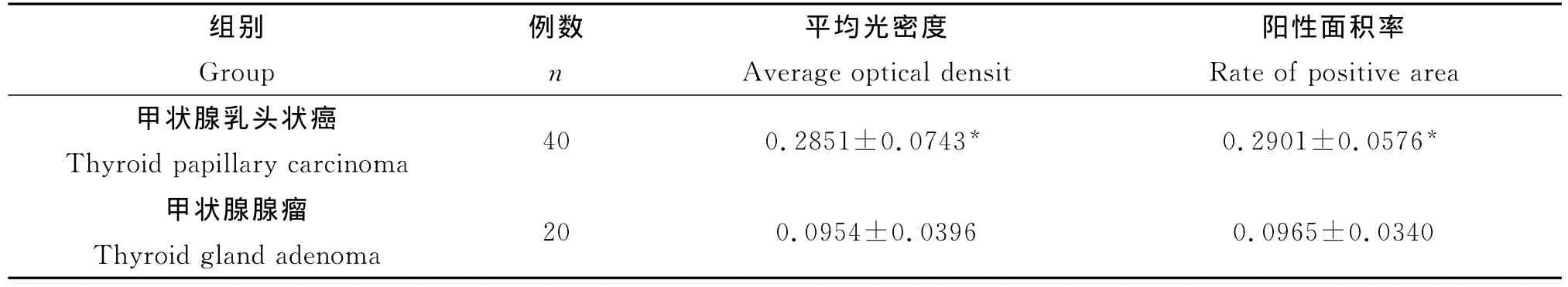
表1 甲状腺乳头状癌与甲状腺腺瘤组织ABCG2表达的平均光密度和阳性面积率(¯x±s)Table 1 The average optical density and t he rate of positive area of ABCG2 in thyroid papillary carcino ma and in thyroid gland adeno ma tissue(¯x±s)
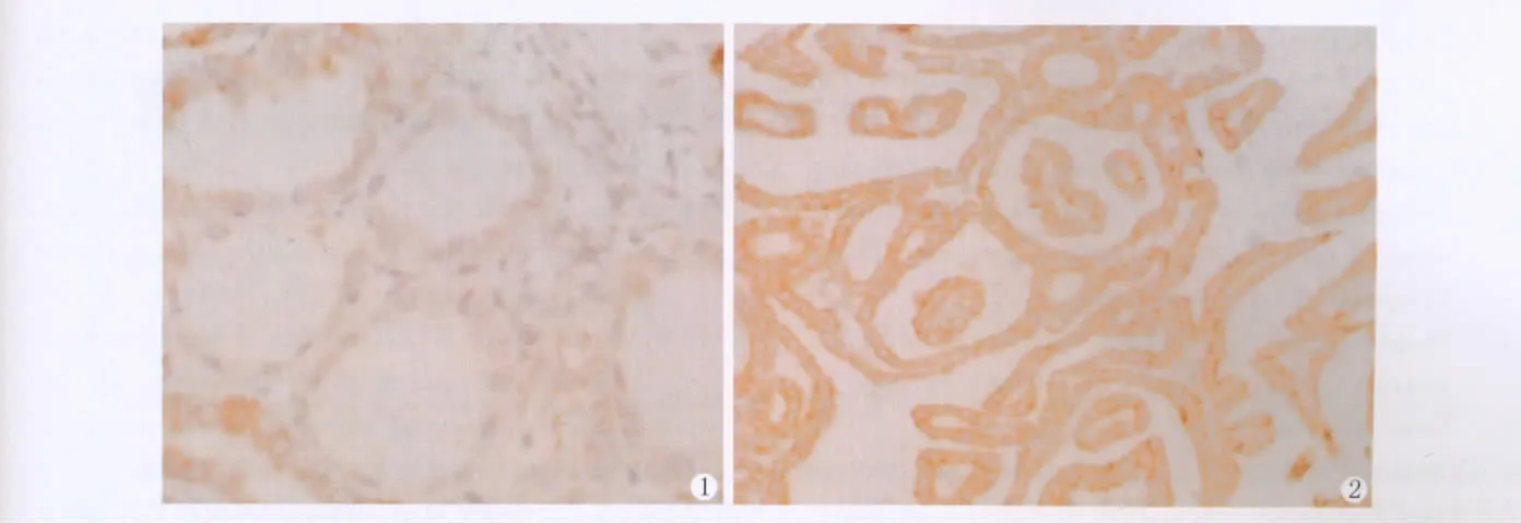
图1 ABCG2在甲状腺腺瘤中的表达。可见少量的棕黄色颗粒,ABCG2表达较弱(SP×200)图2 ABCG2在甲状腺乳头状癌组织中的表达。可见密集分布的棕黄色颗粒,ABCG2表达呈强阳性(SP×200)Fig.1 Expression of ABCG2 in thyroid gland adeno ma.There were few brown particles in the cytoplas m of cancer side tissue,ABCG2 expression was weak.SP×200Fig.2 Expression of ABCG2 in the cytoplas m of thyroid papillary carcinoma There were plenty of brown particles in the cytoplas m of thyr oid papillar y carcino ma.ABCG2 was str ongly expressed.SP×200
讨 论
甲状腺癌(TC)在头颈部肿瘤中居首位。其组织学类型主要包括乳头状癌(PTC),滤泡状癌(FTC),未分化癌(ATC)和髓样癌(MTC),以PTC所占比例最大[5-6]。乳头状癌是甲状腺癌中最常见的类型,属于一种低度恶性肿瘤[7-10],多发于20-50岁的成年人,男女比例约为1∶3-4。尽管15岁以下的儿童和青少年很少发生PTC,但PTC仍然是最常见的儿童型甲状腺癌。有文献报导PTC患者五年存活率女性为94%,男性为90%[11-13]。早期诊断和及时正确的治疗是提高生存率的关键。深入认识甲状腺癌的发病机制,将有可能为治疗提供新的线索和途径。
三磷酸腺苷(ATP)结合转运蛋白G超家族成员2(ABCG2)基因是近年来发现的ABC(ATP-binding cassette)超家族成员之一,是一种新发现的耐药相关半转运蛋白,与P糖蛋白和多药耐药相关蛋白同属ABC转运蛋白超家族[14-16]。目前研究较多的是,ABCG2通过泵出常见的化疗药物,如米托蒽醌、托普替康、伊立替康、甲氨碟呤等,使肿瘤细胞产生多药耐药,从而导致化学治疗的失败[17-18]。
ABCG2的另一重要功能是与干细胞的关系,有研究显示,ABCG2在干细胞中高表达,随着细胞的分化成熟其表达量迅速下降,成熟细胞中表达量非常低,推测可能肿瘤干细胞通过ABCG2的主动泵出功能,将抗癌药大多泵出细胞外,使得肿瘤干细胞逃脱了抗癌药的杀伤作用,为肿瘤的复发和转移奠定了基础 。我们的实验结果显示:甲状腺腺瘤组织中ABCG2呈低表达;甲状腺乳头状癌组织中ABCG2呈高表达。图像分析结果显示:甲状腺腺瘤组织和甲状腺乳头状癌组织之间ABCG2表达的平均光密度及阳性面积率的差异有显著性意义(P<0.05)。实验结果提示:ABCG2的高水平表达可能参与了甲状腺乳头状癌的发生发展,而且其在癌组织中的高表达可能参与了甲状腺乳头状癌化疗过程中多药耐药形成。同时在甲状腺乳头状癌组织中检测到ABCG2的高表达也说明了ABCG2可能在甲状腺乳头状癌发生的早期起了一定的作用。
[1]Volante M,Papotti M.Poorly differentiated thyroid carcino ma:5years after the 2004 WHO classification of endocrine tu mours.Endocr Pat hol,2010,21:1-6
[2]Br uce B,Khanna G,Ren L,et al.Expressi on of the cytoskelet on linker protein ezrin in hu man cancers.Clin Exp Metastasis,2007,24(2):69-78
[3]Mallikarjuna K,Pushparaj V,Biswas J,et al.Expression of epider mal gr owth factor receptor,ezrin,hepatocyte growth factor,and c-Met in uveal melanoma:an i mmunohistochemical study.Curr Eye Res,2007,32:281-290
[4]Krishnamurthy P,Ross DD,Nakanishi T,et al.The stem cell mar ker Bcr p/ABCG2 enhances hypoxic cell sur vival through interactions with heme.J Biol Chem,2004,279:242l8-24225
[5]Br uce B,Khanna G,Ren L,et al.Expression of the cytoskeleton linker protein ezrin in hu man cancers.Clin Exp Metastasis,2007,24:69-78
[6]Bongiovanni M,Bloo m L,Krane JF,et al.Cyto morphologic feat ures of poorly differentiated t hyroid carcino ma:A multi-institutional analysis of 40 cases.Cancer Cytopathol,2009,117:185-194
[7]Bongiovanni M,Sadow PM,Faquin WC.Poorly differentiated thyroid carcino ma:A cytologic-histologic review.Adv Anat Patho,2009,16:283-289
[8]Sanders EM,Volsi VA,Brierley J,et al.An evidencebased review of poorly differentiated thyroid cancer.World J Sur g,2007,31:934-945
[9]Erkilic S,Kocer NE.Insular carcino ma of the thyroid with unco mmon cytologic feat ures:Anisokaryotic cells and microfollicles containing dense colloid.Pathol Res Pract,2006,202:389-393
[10]Jogai S,Jassar A,Adisena A.Fine needle aspiration cytology of thyroid lesions.Acta Cytol,2005,49:483-488
[11]Waldner M,Schi manski C C,Neurath M F.Colon cancer and the i mmune syste m:ther ole of tu mor in vading T cells.World J.Gastroenterol.2006,12,623,7233-7238
[12]Zarife M A,Reis E A,Car mo T M.Increased frequency of CD56 Bright NK-630cells,CD3-CD16+CD56-NK-cells and activated CD4 + T-cellsor B-cellsinparallel 631with CD4+ CDC25 high T-cells contro lpotentially viremia in blood donor swith 632 HCV.J.Med.Virol.2009,81,49-59
[13]Kodama Y,Asai N ,Kawai K,et al.The RET protooncogene :a molecular therapeutic target in thyroid cancer.Cancer Sci,2005 ,96(3):143-148
[14]Wa1sh N,Kennedy S,Larkin AM,et al.Membrane transport proteins in human melanoma:associations with tttmour aggressiveness and metastasis.BrJ Cancer,2010,102:1157-1162
[15]Chen Z,Liu F,Ren Q,et al.Suppression of ABCG2 inhibits cancer cell proliferation.Int J Cancer,2010,126:841-851
[16]Gupta N,Martin PM,Miyauchi S,et al.Down-regulation of BCRP/ABCG2 in colorectal an d cervical cancer.Biochem Biophys Res Co mmun,2006,343:57l-577
[17]Ki m YH,Ishii G,Goto K,et al.Expression of breast cancer resistance protein is associated with a poor clinical outco me in patients wit h s mall-cell lung cancer.Lung Cancer,2009,65:105-111
[18]Tsunoda S,Oku mura T,Ito T,et al.ABCG2 expression is an independent unfavorable prognostic factor in esophageal squa mous cell carcino ma.Oncology,2006,71(3-4):251-258
Expression and clinical significance of ABCG2 protein in thyroid gland papillifor m carcinoma
Tang Tian
(Depart ment of Oncology,Renmin Hospital of Wuhan University,Hubei 430060,China)
Objective To investigate the expression and clinical significance of t he ABCG2 pr otein in thyroid papillary carcino ma.Methods The expression of the ABCG2 protein was examined by S-P i mmunohistoche mical staining in 40 cases of t hyroid carcino ma and 20 cases of t hyr oid gland adeno ma.These sa mples were taken fr o m Ren min Hospital of Wuhan University.The HPIAS-2000 i mage analysis system was used to measure the average optical density and positive area.Results ABCG2 protein expression was high in t hyr oid carcino ma,but l ow in t hyr oid gland adeno ma.I mage analysis de monstrated t hat the expression of the ABCG2 protein in thyroid carcino ma was significantly higher than that in thyroid gland adeno ma(P< 0.05).Concl usion The high expression of ABCG2 may be invol ved in t he occurrence and devel op ment of t hyr oid papillar y carcino ma,and also in t he f or mation of multiple dr ug-resistance in t he che mot herapy process of thyroid papillary carcino ma.
Thyr oid carcino ma;ABCG2 pr otein;I mmunohistoche mistry
R735.7
A
10.3870/zgzzhx.2012.04.016
2012-04-20
2012-06-06
唐甜,女(1981年),汉族,博士。