实时剪切波弹性成像量化可评估闭合性跟腱的术前硬度
2024-11-01杨文娴王雨豪刘卫勇
摘要:目的" 探讨剪切波弹性成像(SWE)对闭合性跟腱完全断裂的硬度研究。方法" 选取2021年7月~2024年1月中国科学技术大学附属第一医院经超声诊断为闭合性跟腱完全断裂患者53例,超声测量跟腱断裂的分离长度、断裂处距离附着处长度及跟腱厚度,并测量跟腱断裂近端、远端及健侧跟腱剪切波速(SWV),同时测量患侧及健侧比目鱼肌SWV。结果" 53例闭合性跟腱完全断裂患者,62.26%(33/53)跟腱断裂发生于左侧,37.73%(20/53)跟腱断裂发生于右侧。患侧跟腱断裂厚度、健侧跟腱厚度、断裂处距离附着处长度及患侧跟腱分离长度的侧别比较均无统计学意义(Pgt;0.05);患侧跟腱断裂远端与健侧跟腱的厚度分别为8.36±3.07 mm、5.01±0.97 mm,差异有统计学意义(Plt;0.05),患侧跟腱断裂近端与健侧跟腱的SWV、患侧跟腱断裂近端与患侧跟腱断裂远端的SWV比较差异均有统计学意义(Plt;0.05),健侧跟腱比目鱼肌与患侧断裂跟腱比目鱼肌的SWV比较差异无统计学意义(Pgt;0.05)。结论" 闭合性跟腱完全断裂大多累及左侧跟腱,患侧跟腱的弹性变化发生在游离跟腱,并不累及比目鱼肌近端的肌腱结构,SWE技术可用于定量评估患侧跟腱的术前硬度。
关键词:超声;闭合性跟腱完全断裂;剪切波速
Real-time shear wave elastography for measuring complete rupture of the closed Achilles tendon
YANG Wenxian1, WANG Yuhao2, LIU Weiyong1, 2
1Graduate School of Bengbu Medical University, Bengbu 233000, China; 2Department of Ultrasound, The First Affiliated Hospital of University of Science and Technology of China, Hefei 230000, China
Abstract: Objective To explore the hardness study of shear wave elastography (SWE) on closed complete Achilles tendon rupture. Methods Fifty-three patients with closed complete Achilles tendon rupture diagnosed by ultrasound in the First Hospital of the University of Science and Technology of China from July 2021 to January 2024 were selected, and the separation length of the Achilles tendon rupture, the length of the rupture site from the attachment site and the thickness of the Achilles tendon were measured ultrasonographically, as well as the proximal and distal Achilles tendon rupture and the healthy Achilles tendon shear wave velocity (SWV), and the SWV of the flounder muscle of both the affected and the healthy side were measured simultaneously. Results The 53 cases of closed patients with complete Achilles tendon rupture, 62.26% (33/53) of the Achilles tendon ruptures occurred on the left side and 37.73% (20/53) of the Achilles tendon ruptures occurred on the right side. Side-by-side comparisons of the thickness of the ruptured Achilles tendon on the affected side, the thickness of the Achilles tendon on the healthy side, the length of the rupture from the attachment, and the length of the separation of the Achilles tendon on the affected side were not statistically significant (Pgt;0.05); the thicknesses of the distal part of the rupture of the Achilles tendon on the affected side and that of the Achilles tendon on the healthy side were 8.36±3.07 mm and 5.01±0.97 mm, respectively, with statistically significant differences (Plt;0.05), the thicknesses of the proximal part of the rupture of the Achilles tendon on the affected side and the Achilles tendon on the healthy side, the The differences in SWV between the proximal part of the affected Achilles tendon rupture and the proximal part of the affected Achilles tendon rupture and the distal part of the affected Achilles tendon rupture were statistically significant (Plt;0.05), and the differences in SWV between the healthy Achilles tendon flounder muscle and the affected ruptured Achilles tendon flounder muscle were not statistically significant (Pgt;0.05). Conclusion Closed complete rupture of the Achilles tendon mostly involves the left Achilles tendon, and the elastic changes of the affected Achilles tendon occur in the free Achilles tendon and do not involve the proximal tendon structures of the flounder muscle, and the SWE technique can be used to quantitatively assess the preoperative stiffness of the affected Achilles tendon.
Keywords: ultrasound; closed complete Achilles tendon rupture; shear wave velocity
跟腱断裂是指跟腱在跟骨基底部、肌腱区域或肌肉肌腱连接处完全连续性中断,发病率为11/10万~37/10万[1] ,以男性为主,位居肌腱断裂发病率的第3位[2] 。近年来,跟腱断裂发病率呈上升趋势,这可能与人口老龄化、肥胖人群增多及饮食结构改变有关。受到患者体格检查时疼痛的限制,临床上常难以充分评估闭合性跟腱断裂的情况,而超声可为跟腱断裂情况提供丰富的诊断信息。闭合性跟腱完全断裂术前超声检查除诊断跟腱断裂外,还应评估跟腱断裂后的硬度改变。随着超声技术的发展,超声弹性成像可对肌腱组织力学改变进行非侵入性评估,尤其是实时剪切波弹性成像(SWE)可通过测量感兴趣区域(ROI)的杨氏模量值定量来评估肌腱生物学弹性[3] 。SWE能够获取剪切波在肌腱内的传播速度用来反应其硬度,从而对闭合性跟腱完全断裂的早期诊断具有很大的适用性[4] 。由于肌肉骨骼具有各向异性的特性,《二维剪切波弹性成像检查肌骨组织操作规范指南》推荐SWE以剪切波速(SWV)(m/s)来表示,不宜采用杨氏模量或剪切模量表示[5]。动态SWE证明在评估闭合性跟腱完全断裂时结果更为客观,然而,也存在一些局限性,特别是ROI的大小和深度有限[7] 。研究表明[8] ,肌腱损伤后恢复情况与弹性值之间存呈正相关,SWV越低则肌腱的力学性能和功能越差,愈合越慢。既往研究大多集中于健康人群跟腱硬度测量,或者断裂的跟腱硬度测量[9] ,未将断裂的跟腱断端近端和远端加以区分测量。本研究采用SWE技术对闭合性跟腱损伤的断端近端及远端、健侧跟腱硬度进行定量分析,旨在为闭合性跟腱完全断裂的术前评估提供指导依据。
1" 资料与方法
1.1" 一般资料
回顾性分析2021年7月~2024年1月中国科学技术大学附属第一医院经超声诊断为闭合性跟腱完全断裂的患者53例,年龄19~75(37.16±9.59)岁,其中男性51例,女性2例。53例患者中62.26%(33/53)跟腱断裂发生于左侧,37.73%(20/53)跟腱断裂发生于右侧。所有患者均经手术证实。纳入标准:符合“急性跟腱断裂的循证医学诊疗指南”中的相关诊断标准[10] ;经手术证实为闭合性损伤;首次跟腱断裂。排除标准:开放性跟腱断裂;代谢性或内分泌疾病导致断裂者。本研究已通过中国科学技术大学附属第一医院(安徽省立医院)伦理审核(审批号:202401281151000172845)。
1.2" 仪器与方法
使用Mindray Resona R9S型彩色超声诊断仪,L14-5WU型线阵探头,探头频率5~15 MHz,仪器配备剪切波弹性成像功能,检查条件设置为肌肉骨骼检查预设条件。
患者取俯卧位,双足悬垂于检查床外,在足踝及小腿背侧中下1/4处涂抹适量的耦合剂,将超声探头轻轻置于足踝背侧,超声探头方向与小腿长轴方向平行。先检查健侧跟腱,跟腱声像图呈细线样连续均匀平行的强回声,轻微移动超声探头,待显示跟腱最厚处后冻结图像,于跟腱连于跟骨附着2~3 cm处测量跟腱厚度,测量3次,取平均值。然后切换至弹性成像模式,按下仪器面板上“Elasto”按钮后,在仪器显示屏触“STQ弹性”条件,仪器自动弹出ROI,将ROI调节至5 mm×5 mm后,把ROI区置于跟腱中央,仪器屏幕上“M-STB Index”显示5颗星后,按下“update”键,记录“Median”值,测量3次,取平均值。将ROI置于健侧跟腱后方的比目鱼肌内,测量健侧比目鱼肌的SWV。然后再对患侧跟腱进行测量,先将超声探头置于足踝背侧近跟骨侧,先测量断裂部位距离跟骨的长度,观察跟腱断端远端二维超声改变,对跟腱断端远端厚度及SWV进行测量。将超声探头向头侧平移,待跟腱断裂部位显示于屏幕中央后,观察跟腱断端声像图改变,并测量跟腱断裂分离长度。再次将超声探头向头侧平移,待图像即将离开跟腱断裂部位后,停止探头平移,待图像稳定后,观察跟腱断端近端二维超声改变,并测量跟腱断端近端厚度及SWV,之后将ROI移动至跟腱深部的比目鱼肌内并测量SWV值。
1.3nbsp; 统计学分析
采用SPSS27.0软件进行统计分析,计数资料以n(%)表示,服从正态分布的计量资料以均数±标准差表示,两组连续性变量的比较采用独立样本t检验,多组间比较采用单因素方差分析,根据是否方差齐性行LSD检验。以Plt;0.05为差异有统计学意义。
2" 结果
2.1" 健侧跟腱及患侧跟腱二维声像图表现
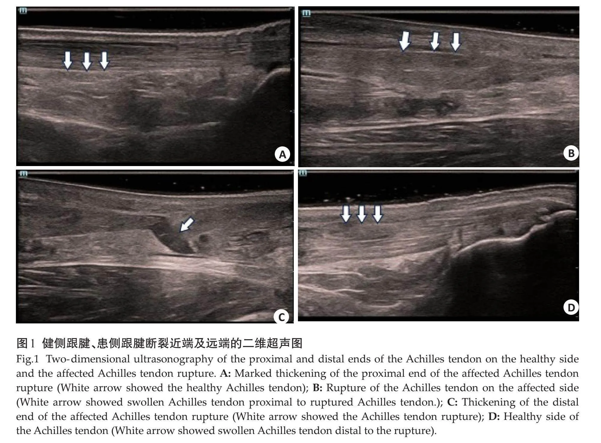
健侧跟腱纵向声像图表现为平行的束状结构,跟腱纤维呈细样连续均匀的强回声(图1A)。跟腱完全断裂时表现为肌腱纤维连续性中断,断端近端(图1B)及远端增厚(图1C),回声减低,断端不平整,跟腱断裂处被无回声或低回声的血肿充填(图1D),伴周围软组织肿胀。患侧断裂跟腱分离长度13.60±8.67 mm,断裂部位距离跟骨附着处47.71±11.09 mm。
2.2" 患侧跟腱断裂近端厚度、患侧跟腱断裂远端厚度、健侧跟腱的厚度、断裂处距离附着处长度及患侧跟腱分离长度的侧别比较
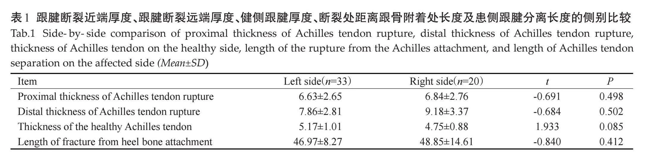
患侧跟腱断裂近端厚度、患侧跟腱断裂远端厚度、健侧跟腱的厚度、断裂处距离附着处长度及患侧跟腱分离长度的侧别差异均无统计学意义(Pgt;0.05,表1)。
2.3" 患侧跟腱断裂近端、患侧跟腱断裂远端及健侧跟腱厚度比较
53例患侧跟腱断裂近端、患侧跟腱断裂远端及健侧跟腱厚度分别为6.71±2.67、8.36±3.07、5.01±0.97 mm,差异有统计学意义(P=0.017)。两两比较显示,患侧跟腱断裂近端与患侧跟腱断裂远端的厚度、患侧跟腱断裂近端与健侧跟腱的厚度的差异无统计学意义(P=0.229、0.077);患侧跟腱断裂远端与健侧跟腱的厚度的差异有统计学意义(8.36±3.07 mm vs 5.01±0.97 mm,P=0.005)。
2.4" 患侧跟腱断裂近端、患侧跟腱断裂远端、健侧跟腱、患侧比目鱼肌、健侧比目鱼肌的侧别SWV比较
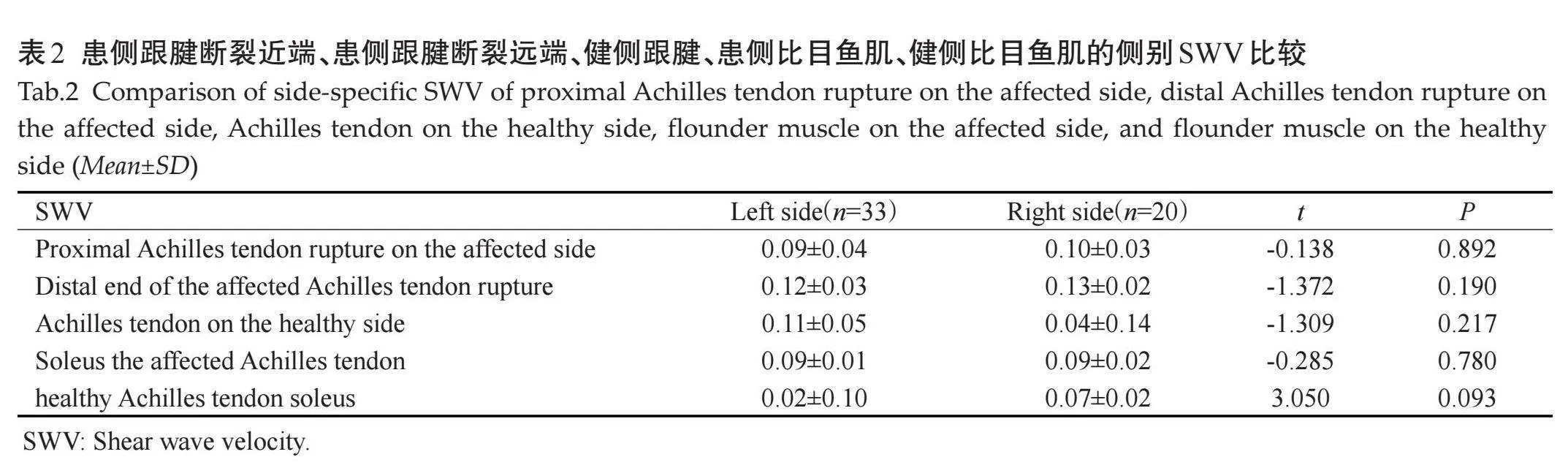
患侧跟腱断裂近端、患侧跟腱断裂远端、健侧跟腱、患侧比目鱼肌、健侧比目鱼肌的侧别SWV的差异均无统计学意义(Pgt;0.05,表2)。
2.5" 患侧跟腱断裂近端、患侧跟腱断裂远端与健侧跟腱SWV比较及健侧侧比目鱼肌与患侧比目鱼肌SWV比较
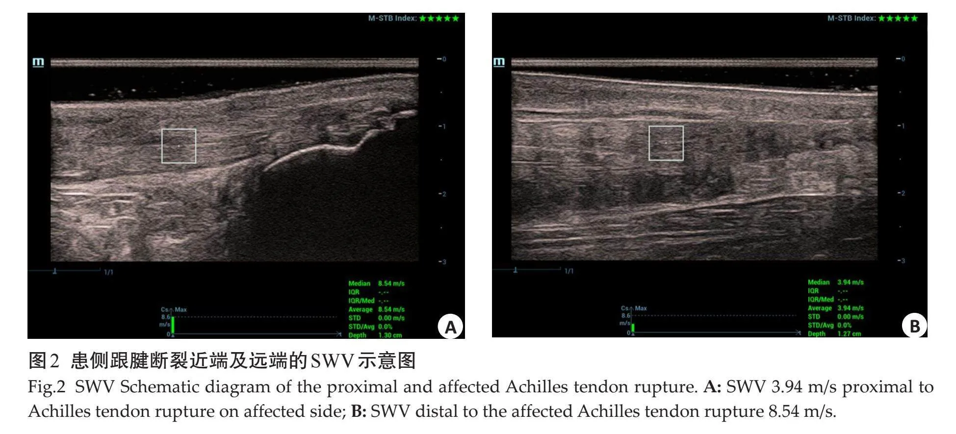
53例患侧跟腱断裂近端、患侧跟腱断裂远端及健侧跟腱SWV分别为3.11±1.11、3.99±1.13、4.06±1.38 m/s。患侧跟腱断裂近端、跟腱断裂远端与健侧跟腱的SWV比较有统计学意义(P=0.001)。两两比较显示,患侧跟腱断裂近端(图2A)与患侧跟腱断裂远端(图2B)的SWV、患侧跟腱断裂近端与健侧跟腱的SWV差异有统计学意义(3.11±1.11 m/s vs 3.99±1.13 m/s,Plt;0.001;3.11±1.11 m/s vs 4.06±1.38 m/s,P=0.003);患侧跟腱断裂远端与健侧跟腱的SWV差异无统计学意义(P=0.667);健侧比目鱼肌与患侧比目鱼肌的SWV差异无统计学意义(P=0.598)。
3" 讨论
跟腱断裂是一种常见的运动损伤,诊断主要依赖于临床体格检查,但受到患者疼痛及软组织肿胀的影响,体格检查常常出现假阴性诊断,单纯依赖临床检查误诊率高达20%~30%[11] 。超声因无创、无辐射及便捷等优势,已成为跟腱断裂首选的影像学检查方法。传统的二维超声评估跟腱断裂时无法深入了解跟腱的特性,其结果与预后跟腱的功能恢复没有明显的相关性。相比之下,SWE已被证实是肌腱[12] 和肌肉[13] 较为可靠的定量技术,在预测跟腱结构变化方面比肌骨超声更敏感,且具有更良好的可靠性和可重复性[14] ,甚至被运用于检测断裂跟腱的定量特性以及监测其治疗效果[15-17] 。由于跟腱内部缺乏血供,损伤后容易引起多种并发症并且不易修复和愈合[18] ,且跟腱断裂后跟腱的僵硬度影响到跟腱治疗后患者恢复程度,所以术前评估患侧跟腱断裂的硬度尤为重要。
跟腱断裂的男女比例为2:1~19:1[19] ,本研究中患者共53例,其中男性51例,女性2例,男性跟腱断裂的发生率明显高于女性,既往研究[20] 也证实了这一观点,可能原因是参与运动的男性多于女性[29] ,另外可能是性别生理差异,女性分泌的雌激素可以促进胶质蛋白的合成,维持跟腱的紧张状态,从而降低跟腱损伤的发生率。本研究中跟腱断裂位于左侧的发生率为62.26%(33/53),位于右侧的发生率为37.73%(20/53),与大多数人为右腿优势有关,肌肉的发达程度可以提高身体的稳定性和支持性,有助于减轻肌腱的负担,从而降低右侧跟腱断裂的发生率,与既往研究[21] 结果相符。
一般来说,患侧跟腱的SWV低于健侧跟腱[22] 。由于个体间SWV值的多样性且缺乏一般的参考值,到目前为止还未有普遍适用的健侧跟腱参考值,但是可以通过每位患者两侧个体内比较作为个体的“参考标准”。既往研究表明,跟腱断裂后健侧与患侧的剪切模量无差异[23] ,但目前关于跟腱侧别损伤引起厚度及SWV的比较尚未见报道,可能是由于SWE技术在测量跟腱硬度时有量程限制。本研究结果显示,跟腱断裂位于左侧或右侧,测得的厚度、SWV、患侧跟腱分离长度、距离跟骨附着处长度及比目鱼肌SWV差异均无统计学意义,表明跟腱侧别损伤引起的厚度及SWV与患侧跟腱分离长度、与跟骨附着处的距离及比目鱼肌的弹性无关。本研究结果表明跟腱发生断裂后患侧跟腱近端、远端均较健侧跟腱增厚,是由于跟骨结节上方2~6 cm处最窄,最为薄弱,由于跟腱长期慢性牵拉劳损,跟腱组织变脆,血运不丰富,因此跟腱发生断裂后组织水肿增厚较为明显,与以往研究[24] 结果一致。本研究结果还表明跟腱断裂远端较跟腱断裂近端增厚明显,可能是由于成人跟腱两端血供丰富,但中段血管数量较少,管径较小,存在血供差。因此,利用SWE技术可用于监测肌腱的恢复情况,避免早期肌腱负荷导致的再次损伤。
SWE是近年来超声领域的一项新技术,可从生物力学的角度评估病变信息。在二维超声图像的基础上显示组织硬度的信息,选择ROI精确定量评估组织硬度,分辨不同组织间以及同一组织不同病理状态的硬度差异[8] 。SWE可以提供肌腱力学性能信息,正常跟腱发生损伤时,跟腱的生物学会发生改变,硬度降低。SWE结果显示,跟腱断裂后,跟腱断裂近端及远端的SWV均减低,且跟腱断裂近端减低更为明显,说明跟腱发生断裂后,张力减低、组织血肿或积液会导致僵硬度降低,与以往研究[25-27] 结果一致;并且患侧比目鱼肌SWV与健侧比目鱼肌SWV的差异无统计学意义,表明跟腱断裂的弹性改变仅局限于游离跟腱,而不涉及比目鱼肌的近端肌腱结构,这与既往研究[28] 结果相同。本研究还发现患侧跟腱断裂远端的SWV高于患侧跟腱断裂近端的SWV,且两者有明显的差异性,此结果在以往的研究中尚未报道。可能的原因有:跟腱的远端有跟骨附着;跟腱的近端断裂后张力减低,局部凹陷,此处的跟腱横截面积小,偏心负荷大,血管少[29] 。
本研究存在一些局限性:本研究的样本量偏少,且为单中心研究;本研究纳入的均为需要手术的跟腱完全断裂的患者,跟腱不完全断裂未纳入;跟腱断裂后,由于局部组织水肿增厚,取样框与断裂跟腱之间无法保持完全垂直状态,测得的患侧跟腱SWV值稍有偏差。在未来的研究中应扩大样本量及多中心性研究来提供参考标准。
综上所述,闭合性跟腱完全断裂大多累及左侧跟腱,在跟腱发生断裂后会出现水肿增厚,且患侧断裂跟腱远端较患侧断裂跟腱近端增厚明显;利用SWE技术测得患侧断裂跟腱近端、远端的SWV均低于健侧跟腱,且跟腱断裂近端的SWV较跟腱断裂远端的SWV低,表明跟腱发生断裂后,跟腱的弹性变化发生在游离跟腱,对比目鱼肌近端肌腱的结构不产生影响。采用SWE技术测得的SWV可以评估患者术前跟腱的软硬程度,为手术治疗提供支持。
参考文献:
[1]" "Suchak AA, Bostick G, Reid D, et al. The incidence of Achilles tendon ruptures in Edmonton, Canada[J]. Foot Ankle Int, 2005, 26(11): 932-6.
[2]" "Park HG, Youn D, Baik JM, et al. Epidemiology of Achilles tendon rupture in South Korea: claims data of the national health insurance service from 2009 to 2017[J]. Clin Orthop Surg, 2021, 13(4): 539-48.
[3]" "郝云霞, 崔立刚, 孙" 洋. 剪切波弹性成像技术检测正常人髌腱弹性特征的初步研究[J]. 中国超声医学杂志, 2019, 35(1): 69-72.
[4]" "Ooi CC, Malliaras P, Schneider ME, et al. “Soft, hard, or just right?” Applications and limitations of axial-strain sonoelastography and shear-wave elastography in the assessment of tendon injuries[J]. Skeletal Radiol, 2014, 43(1): 1-12.
[5]" " 中国医师协会超声医师分会. 中国二维剪切波弹性成像检查肌骨组织操作规范指南[J]. 中华超声影像学杂志, 2024, 33(3): 193-200.
[6]" "Payne C, Watt P, Cercignani M, et al. Reproducibility of shear wave elastography measuresof the Achilles tendon[J]. Skeletal Radiol, 2018, 47(6): 779-84.
[7] Klauser AS, Miyamoto H, Bellmann‑Weiler R, et al. Sonoelastography: musculoskeletal applications[J]. Radiology, 2014, 272(3): 622-33.
[8]" Prado‑Costa R, Rebelo J, Monteiro-Barroso J, et al. Ultrasound elastography: compression elastography and shear‑wave elastography in the assessment of tendon injury[J]. Insights Imaging, 2018, 9(5): 791-814.
[9]" "Zhang LN, Wan WB, Wang YX, et al. Evaluation of elastic stiffness in healing Achilles tendon after surgical repair of a tendon rupture using in vivo ultrasound shear wave elastography[J]. Med Sci Monit, 2016, 22: 1186-91.
[10]" Ivanac G, Lemac D, Kosovic V, et al. Importance of shear-wave elastography in prediction of Achilles tendon rupture[J]. Int Orthop, 2021, 45(4): 1043-7.
[11] Breda SJ, van der Vlist A, de Vos RJ, et al. The association between patellar tendon stiffness measured with shear-wave elastography and patellar tendinopathy‑a case‑control study[J]. Eur Radiol, 2020, 30(11): 5942-51.
[12]Corrigan P, Zellers JA, Balascio P, et al. Quantification of mechanical properties in healthy Achilles tendon using continuous shear wave elastography: a reliability and validation study[J]. Ultrasound Med Biol, 2019, 45(7): 1574-85.
[13]" Snoj Ž, Wu CH, Taljanovic MS, et al. Ultrasound elastography in musculoskeletal radiology: past, present, and future[J]. Semin Musculoskelet Radiol, 2020, 24(2): 156-66.
[14]" Dirrichs T, Quack V, Gatz M, et al. Shear wave elastography (SWE) for monitoring of treatment of tendinopathies: a double-blinded, longitudinal clinical study[J]. Acad Radiol, 2018, 25(3): 265-72.
[15] Dirrichs T, Schrading S, Gatz M, et al. Shear wave elastography (SWE) of asymptomatic Achilles tendons: a comparison between semiprofessional athletes and the nonathletic general population[J]. Acad Radiol, 2019, 26(10): 1345-51.
[16]" Mendes B, Firmino T, Oliveira R, et al. Hamstring stiffness pattern during contraction in healthy individuals: analysis by ultrasound-based shear wave elastography[J]. Eur J Appl Physiol, 2018, 118(11): 2403-15.
[17]" Shane AM, Reeves CL, Nguyen GB, et al. Revision surgery for the Achilles tendon[J]. Clin Podiatr Med Surg, 2020, 37(3): 553-68.
[18]Zhang XN, Deng LQ, Xiao SL, et al. Sex differences in the morphological and mechanical properties of the Achilles tendon[J]. Int J Environ Res Public Health, 2021, 18(17): 8974.
[19]" Lantto I, Heikkinen J, Flinkkilä T, et al. Epidemiology of Achilles tendon ruptures: increasing incidence over a 33-year period[J]. Scandinavian Med Sci Sports, 2015, 25(1): e133-8.
[20] Barrios-Cárdenas AL, Lazo-Vera JO. Epidemiological, clinical and therapeutic characteristics of Achilles tendon rupture[J]. Acta Ortop Mex, 2021, 35(3): 252-6.
[21] Dirrichs T, Quack V, Gatz M, et al. Shear wave elastography (SWE) for the evaluation of patients with tendinopathies[J]. Acad Radiol, 2016, 23(10): 1204-13.
[22]" Zellers JA, Cortes DH, Corrigan P, et al. Side-to-side differences in Achilles tendon geometry and mechanical properties following Achilles tendon rupture[J]. Muscles Ligaments Tendons J, 2017, 7(3): 541-7.
[23] Gatz M, Betsch M, Bode D, et al. Intra individual comparison of unilateral Achilles tendinopathy using B-mode, power Doppler, ultrasound tissue characterization and shear wave elastography[J]. J Sports Med Phys Fitness, 2020, 60(11): 1462-9.
[24]" 刘" 芳, 李殿城, 朱家安, 等. 剪切波弹性成像定量评估大鼠坐骨神经卡压的实验研究[J]. 中国超声医学杂志, 2022, 38(8): 924-7.
[25]" 丁常伟, 张迎春, 宋" 馨, 等. 实时剪切波弹性成像评估强直型帕金森病患者早期上肢肌肉硬度的变化[J]. 中国医学影像学杂志, 2021, 29(8): 826-30.
[26] Alfuraih AM, O'Connor P, Tan AL, et al. Muscle shear wave elastography in idiopathic inflammatory myopathies: a case-control study with MRI correlation[J]. Skeletal Radiol, 2019, 48(8): 1209-19.
[27] Li QR, Zhang Q, Cai YH, et al. Patients with Achilles tendon rupture have a degenerated contralateral Achilles tendon: an elastography study[J]. Biomed Res Int, 2018, 2018: 2367615.
[28] Crawford SK, Thelen D, Yakey JM, et al. Regional shear wave elastography of Achilles tendinopathy in symptomatic versus contralateral Achilles tendons[J]. Eur Radiol, 2023, 33(1): 720-9.
[29] Hess GW. Achilles tendon rupture: a review of etiology, population, anatomy, risk factors, and injury prevention[J]. Foot Ankle Spec, 2010, 3(1): 29-32.
(编辑:林" 萍)