Effect of long-period stacking ordered structure on very high cycle fatigue properties of Mg-Gd-Y-Zn-Zr alloys
2023-11-18XingyuWANGChoHEXueLiLngLIYongjieLIUQingyunWANG
Xingyu WANG, Cho HE,b,∗, Xue Li, Lng LI,b, Yongjie LIU,b, Qingyun WANG,c,∗
a Key Laboratory of Deep Earth Science and Engineering, Ministry of Education, College of Architecture and Environment, Sichuan University, Chengdu 610065, China
b Failure Mechanics & Engineering Disaster Prevention and Mitigation, Key Laboratory of Sichuan Province, Sichuan University, Chengdu 610065, China
c School of Architecture and Civil Engineering, Chengdu University, Chengdu 610106, China
Received 26 May 2021; received in revised form 25 September 2021; accepted 10 November 2021
Available online 15 December 2021
Abstract Magnesium alloys with a long-period stacking ordered(LPSO)structure usually possess excellent static strength,but their fatigue behaviors are poorly understood. This work presents the effect of the LPSO structure on the crack behaviors of Mg alloys in a very high cycle fatigue(VHCF) regime. The LPSO lamellas lead to a facet-like cracking process along the basal planes at the crack initiation site and strongly prohibit the early crack propagation by deflecting the growth direction. The stress intensity factor at the periphery of the faceted area is much higher than the conventional LPSO-free Mg alloys, contributing higher fatigue crack propagation threshold of LPSO-containing Mg alloys. Microstructure observation at the facets reveals a layer of ultrafine grains at the fracture surface due to the cyclic contact of the crack surface, which supports the numerous cyclic pressing model describing the VHCF crack initiation behavior.
Keywords: Fatigue crack initiation; Long-period stacking ordered structure; Mg alloys; Ultrafine grains; Very high cycle fatigue.
1. Introduction
Magnesium alloy is a promising lightweight alloy for structural applications due to its low density,high specific strength,excellent thermal and electrical conductivity, high biocompatibility, and recyclability. It has broad application prospects in aerospace, transportation, biomedicine, and energy industries[1–3]. However, its hexagonal crystal structure with insufficient slip systems results in poor ductility [4,5]. The static strength of conventional Mg alloys is also insufficient. These deficiencies limit their further applications as structural materials. By adding some rare earth elements, Mg alloys containing a long-period stacking ordered (LPSO) structure are emerging as particularly attractive structural materials. The LPSO structure improves the static mechanical properties and ductility at room temperature [6–9]. In the past few years,considerable research effort has been devoted to the performance of the LPSO-containing Mg alloy under static loading conditions [10,11]. However, the knowledge about its fatigue behavior is limited. Applications of the Mg-LPSO alloys in structural engineering would result in large numbers of cyclic loadings, thus it is critical to fully investigate its fatigue behavior and related mechanisms.
It has been reported that in high-cycle fatigue (HCF, fatigue life ranges in 105∼107cycles) and very-high-cycle fatigue regimes (VHCF, fatigue life ranges in 107∼109cycles),crack initiation and early propagation usually account for more than 90% of the total fatigue life [12–14]. Its fatigue cracking behaviors have been reported to be closely related to its cyclic deformation mechanisms. At high amplitudes of cyclic strain, microstructural cracking generally starts at the twinning boundaries [15–17]. However, at low stresses below the yield strength, deformation twinning is nearly absent, and fatigue crack nucleation is mainly caused by slip bands [18–20]. For LPSO-strengthened Mg alloys, the LPSO lamella can effectively inhibit non-basal slip and deformation twinning because hard lamellas are distributed along the basal plane. However, the development of basal slip bands cannot be restrained effectively, because the motion of basal dislocation does not transverse the LPSO lamellas. Therefore, it is questionable whether the LPSO structure could enhance the fatigue crack initiation resistance of the material. Recently,Shao et al.[8]were the first to correlate the VHCF crack path of an LPSO-containing GWZ1031K-T4 alloy with the local microstructure. They reported that the unique microstructure of LPSO structure and stacking faults accounted for the enhanced fatigue resistance of the material. However, the observation of crack path is not fully representative for the crack initiation and early propagation, which plays a decisive role on the VHCF strength. It is very important to understand how the LPSO structure influences the fatigue behaviors of the material, especially on the crack initiation and propagation behavior. So far, very little information about the concerns mentioned above is available, and the underlying mechanism remains to be explored.
In the present work, we aimed to experimentally investigate the VHCF fatigue behaviors of the LPSO-strengthened Mg alloys.Combined use of the focused ion beam(FIB)technique and the transmission electron microscope (TEM) was applied to precisely reveal the effect of the LPSO structure on the crack initiation and propagation behaviors. The results may extend our knowledge of the formation mechanism of the microstructural crack behaviors of Mg-LPSO alloys and potentially provide a way to improve their fatigue properties.
2. Material and experimental procedure
The material in this work is Mg-10Gd-3Y-0.1Zn-0.4Zr(wt%) alloys. We prepared pure magnesium and zinc, Mg-30Y, Mg-30Gd, Mg-30Zr (wt%) master alloys in a vacuum electrical resistance furnace under a gas atmosphere comprising CO2and SO2. Then, a semicontinuous casting process and a solution treatment at 520 °C for 24 h (T4) were carried out. The ultimate tensile strength, yield strength, elongation, and modulus of elasticity are 260 MPa, 203.5 MPa,13%, and 45 GPa, respectively. Fig. 1a shows the microstructure of this alloy observed by a scanning electron microscope(SEM). It is characterized by equiaxed grains with an average size of approximately 37.5 μm by the linear section method. The LPSO structures in the material includes the block-shaped LPSO structures gathered at the grain boundaries and the thin LPSO lamellas (which is embedded in the interior of the grain). Fig. 1b shows the high magnification of the grain interior obtained by high-angle annular dark-field scanning transmission electron microscopy (HADDF-STEM).It is shown that abundant fine LPSO lamellas of nanometerscale thicknesses and stacking faults(SFs)are spread over the grain interior along the basal planes.

Fig. 1. Microstructure of Mg-10Gd-3Y-1.0Zn-0.4Zr alloy. (a) SEM image showing the LPSO lamellas indicated by arrows. (b) HADDF-STEM image showing stripe-shaped LPSO structures and SFs in the α-Mg matrix.
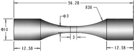
Fig. 2. Dimensions of fatigue specimen (units: mm).
Fatigue tests were conducted on an ultrasonic fatigue system (Shimadzu USF-2000) at a constant stress ratio ofR= -1 (symmetrical tensile and compressing loading) and a cyclic frequency off= 20 kHz. Fig. 2 shows the detailed dimensions of the testing specimens. Before testing, the specimens were mechanically polished by abrasive papers in various grades and cloths, then etched with a mixed solution of 4% nitric acid and 96% ethanol (vol%). Polishing and etching in advance make the LPSO lamella visible, which helps to quickly identify the position and orientation of the basal plane ofα-Mg matrix. Besides, a compressed air-cooling system was applied to eliminate the self-heating effect during ultrasonic fatigue tests, and the increase of temperature during testing was less than 5°C based on the results measured by an infrared camera (NEC R500EX). Thus the effect of the temperature rising on fatigue testing was neglected in this work.
After fatigue failure, the fractographic analysis of failed specimens was characterized by an SEM (Zeiss ULTRA-55),focusing on crack initiation and propagation zones. The secondary fatigue cracks at the side surfaces of specimens were also detected to reveal the relationship between the local microstructure and the crack path. To avoid the effect of the primary crack propagation on the local cracking behaviors,the observation location of the secondary cracks was at least 1 mm away from the fracture surface. Furthermore, the FIBassisted lift-out technique (FEI dual-beam Quanta 3D 200i))was applied to prepare TEM samples of the cross-sections exactly at slip bands, crack initiation site and crack propagation zone, respectively. The microstructure observations were carried out on an atomic resolution analytical TEM equipped with a spherical aberration corrector (JEM-ARM200F). The accelerating voltage was 200 kV. To associate crack surfaces with the LPSO structure, we used two imaging modes of high-resolution transmission electron microscopy (HRTEM)and high-angle annular dark-field scanning transmission electron microscopy (HAADF-STEM) to gain more insight into the microstructure evolution of slip bands and crack surfaces.All micrographs were recorded along with the(11¯20)axis of theα-Mg matrix.Note that the contrast of the HAADF-STEM image is highly sensitive to the atomic number (Z contrast)[21], so the LPSO structures segregated with Gd and Y elements generally show much higher brightness relative to that of the Mg matrix. Therefore, it helps to distinguish the LPSO lamella from theα-Mg matrix easily, as shown in Fig. 1b.
3. Results
3.1. Very high-cycle fatigue strength
Fig. 3 shows the S-N curve with the fatigue lives ranging from 104to 109cycles, in which the arrows indicate the runout specimens (with a fatigue life of more than 109cycles). The curve shows a decreasing trend with the increase of the stress amplitude. The fatigue strength of the LPSO-strengthened Mg alloys at 109cycles is approximately 110 MPa, which is higher than the widely-used Mg alloys of the AZ31 (88 MPa [22]), ZK60 (95 MPa [23]), and AM60(50 MPa [24]), but slightly lower than the AZ80 (135 MPa[25]) and GW123K (117 MPa [20]). However, the ultimate strengths of the reported AZ80 and GW123K are 332 MPa and 335 MPa respectively, which are much higher than that of the Mg-10Gd-3Y-0.1Zn-0.4Zr in this test (260 MPa). The ratio of the VHCF strength to the ultimate tensile strength of the LPSO-containing Mg alloys (0.42) is still higher than previously reported LPSO-free Mg alloys. Clearly, the effect of the LPSO lamellas on the improvement of the VHCF toughness is significant compared with the LPSO-free Mg alloys.This is in accordance with the result obtained by Shao et al.[8], who recently reported that the LPSO containing Mg alloys exhibits relatively good VHCF fatigue resistance. Since the microstructure of the materials generally exerts a very sensitive influence on their VHCF strength, the enhancement of the VHCF strength in Mg-10Gd-3Y-0.1Zn-0.4Zr alloys should be related to the profuse LPSO structure in theα-Mg matrix.The strengthening mechanisms of the LPSO-containing Mg alloy need to be further investigated.

Fig. 3. VHCF strength of the Mg-10Gd-3Y-0.1Zn-0.4Zr alloys.
3.2. Fracture morphology
In this work, fatigue cracks are all initiated from the specimen surface. Fig. 4 shows a typical fracture surface of a failed specimen at the stress amplitudes of 130 MPa. The fracture surface contains two fatigue cracks origination sites,as indicated by white arrows in Fig. 4a. The morphology is relatively rough around the crack initiation sites, and boundaries between the rough zone around the initiation site and the smooth zone at the stable crack propagation region can be identified as plotted by two curved lines. The rough morphologies around the crack initiation site have also been reported at the fracture surface in the VHCF regimes [16]. It is believed that its formation is closely associated with the local microstructural heterogeneities. Therefore, the early crack behavior within the rough area is referred to as the microstructurally small crack propagation, while the relatively smooth area can be regarded as a result of the stable crack propagation process.

Fig. 4. Typical fracture surface morphology of fatigue crack initiation (σa = 130 MPa, Nf = 3.45 × 10⁷ cycles). (a) Low magnification image of the fracture surface showing the location of crack initiation sites at specimen surface. (b) Enlarged view of the facets at crack initiation sites framed as b’ in (a). (c)Enlarged view of the facets at crack initiation sites framed as c’ in (a). (d) Observation on the side surface and fracture surface showing the positional relationship between the facet and LPSO lamellas.
Figs. 4b and 4c show enlarged views of the crack initiation site as framed by “b’” and “c’” in Fig. 4a, respectively.Two kinds of morphologies, isolated facets with smooth appearance and rough surfaces with numerous stripes, can be identified near the crack initiation sites. The stripes in the rough region should be a result of the transgranular cracking process, in which the crack tip changes its growth direction frequently because of microstructural heterogeneities.The LPSO lamellas should greatly influence the crack behaviors, and this will be revealed in the following sections. As for the faceted morphology, the size of the facets is approximately the same as the average grain size, in a range of 30 to 40 μm. Similar faceted morphologies were also observed in previous works. It is believed that the formation of the facets at the crack initiation site is a result of the transgranular cracking process along specific crystallographic planes [8,26]. To confirmed this, the specimen in the SEM chamber was tilted slightly so that both the side surface and fracture surface can be observed simultaneously. Fig. 4d presents the correlation of fracture surface and local microstructure at the framed location “d’” marked in Fig. 4c. The fine lamellar structures in the interior of the grains are LPSO lamellas. According to the observation on the LPSO orientation at the side surface,a cluster of three grains with almost the same orientations of the basal plane can be characterized next to the facet at the fracture surface. Even though the precise determination of the positional relationship between the LPSO lamellas and facet is impossible in the two-dimensional plane view, the facets can still be estimated to be parallel to the LPSO lamellas.Therefore, the LPSO structure should have a vital influence on crack initiation and early propagation behaviors.

Fig. 5. High magnification of flat subregions at stable crack propagation zone.
At the stable crack propagation stage, a higher magnification view of the typical smooth area is shown in Fig. 5,indicating that there are lots of grain-sized flat subzones as plotted by dash lines in the propagation region as well. However, in contrast to the facets in crack initiation sites, the flat subzones exhibit a relatively rough surface with numerous striations. Given that the difference in the fracture morphologies between crack initiation sites, early crack propagation zones, and stable crack propagation regions, the formation of the fracture surface should be a result of different interactions between the local microstructure and the crack behaviors. In the following sections,we will carry out detailed observations on the crack initiation sites and propagation zones to correlate the local microstructure to crack behaviors.
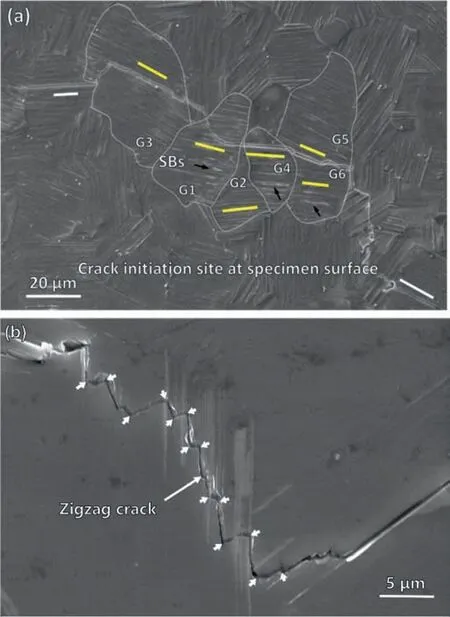
Fig. 6. (a) A typical SEM image of a secondary fatigue crack at the etched surface. Several segments of the crack path are parallel to the LPSO lamella.(b) Enlarged image of a typical “zigzag” crack path during transverse propagation across the LPSO lamella.
3.3. Interaction between fatigue crack and LPSO lamella
Fig. 6a shows an SEM image of a secondary fatigue crack at the side surface of a specimen (130 MPa, 7.94 × 10⁶cycles). For better interpretation, some of the grains are numbered as G1∼G6, and their boundaries have been marked out by the dash lines. It can be seen that slip markings, as indicated by black arrows, were sparsely distributed within G1∼G3 at the relatively low stress. Most of the grains are undeformed macroscopically even near the crack path. As indicated by the black arrows, all the slip bands are parallel to their neighboring LPSO lamellas (indicating the basal plane of theα-Mg matrix), so the observed slip bands are located at the basal planes. No obvious non-basal slip bands or deformation twinning can be seen at the surface. Therefore, the basal slip is the predominant plastic deformation mode during cyclic deformation in the HCF and VHCF regimes.As for the fatigue crack, it was initiated from the basal slip bands,showing a straight section parallel to the slip bands and LPSO lamellas. This observation of slip-induced crack initiation is quite similar to previous investigations on the crack initiation mechanism of Mg alloys at low-stress amplitudes [27],because the slippage on the basal plane is the easiest deformation available to Mg alloys. Furthermore, the directions of the LPSO lamellas within G1∼G6 are plotted by yellow lines.It is interesting that the LPSO lamellas at different grains around the crack initiation site have almost the same orientations. This observation agrees with the microstructure-crack relation in Fig. 4d and suggests that the clusters of favorably oriented grains are apt to localize plastic deformation and initiate fatigue cracks.
As for the effect of the LPSO lamellas on the small crack propagation, Fig. 6b presents the appearance of the fatigue crack path as it penetrates the LPSO structures. The crack path that intersects the LPSO structures exhibits a clear zigzag manner. The growth direction of the crack changes more than ten times within a straight-line length of approximately 20 μm. As compared with the straight transgranular cracks parallel to the LPSO structures in Fig. 6a, the frequent changes of crack growth direction led to the formation of the rough fracture surface containing numerous strips observed around the crack initiation site, as shown in Figs. 4b and 4c. In this case, the LPSO lamellas strongly prohibit the small crack propagation in the interior of unfavorably oriented grains. As a result, the resistance to the short fatigue crack propagation can be enhanced accordingly.
3.4. Positional relationship between slip bands and LPSO lamellas
There is a clear interaction between the LPSO lamellas and the fatigue cracks.We first focused on the development of slip bands that directly results in the initiation of fatigue cracks.Fig. 7a shows an SEM image of several slip bands (numbered as SB1∼SB5) that were activated in the interior of a grain.To deeply understand the positional relationship between the slip bands and the LPSO lamellas, a FIB technique was applied to slice a TEM sample exactly along the cross-section of the slip bands. Figs. 7b and 7c show the HAADF-STEM and the HRTEM images of the obtained cross-section,respectively. The direction of the (0001) basal planes is provided.It should be noted that the slip bands are actually the paths for the extensive dislocation motion and interaction. It is reported that the dislocation intensity zones of the slip bands are generally brighter in HRTEM, but darker in HADDFSTEM [28]. Therefore, the dislocation intensity zone with low brightness can act as an indicator to determine the location of the slip bands. In Figs. 7a and 7b, the relative position of the slip bands (SB1∼SB5) has been figured out based on the comparison with that in the SEM image. The crosssection clearly shows that all the slip bands are parallel to the LPSO lamellas (with brighter contrast in the HADDFSTEM image). They were all oriented along the basal plane since the direction of LPSO structures is consistent with the(0001) plane ofα-Mg grains. This observation further verifies the previous SEM observation in Section 3.2, revealing that only basal slip was activated for plastic deformation during the fatigue testing. The result also agrees with the previous investigations on the cyclic deformation of Mg alloys at lowstress amplitudes [19,20,26,29]. For detailed observation of the slip bands, Fig. 7d shows an amplified HRTEM image of the microstructure in the interior of the slip bands (SB2).Clearly, the SB2 is located between the LPSO lamella and theα-Mg matrix, which might be associated with the local strain concentration at the interface between the hard LPSO structure and the softα-Mg matrix. Meanwhile, the relatively low brightness of the slip bands in the HADDF-STEM image indicates that there exist intensive dislocation activities inside them. The slip bands can be regarded as a concentration path of dislocation motion to accommodate cyclic shear deformation. Based on the above observation, it is evidenced that the LPSO structure strongly prohibits the deformation along the c-axis of theα-Mg matrix. The deformation was strongly localized within the slip bands, and only basal slip is available for the development of the plasticity along the LPSO lamellas.

Fig. 7. Observation on the cross-section of the surface slip bands. (a) Location of FIB section at the slip bands. (b) Enlarged HAADF-STEM image showing the location of slip bands and LPSO structures. (c) HRTEM image showing the same location of (b). (d) HRTEM image of the slip band (SB2) showing the interior microstructure.
3.5. Microstructure of the facet at fatigue crack initiation site
The previous section demonstrated the strain localization at the basal slip bands due to the restriction of the LPSO structure,but the crack initiation behavior is still unclear.Since the faceted morphology is the representative fracture mode at the crack initiation area, a TEM sample exactly at the facet was prepared to characterize its microstructure evolution caused by the crack initiation process. Fig. 8a shows an SEM image of a facet located at the crack initiation site. The sliced film, indicated by a short line, is located at the center of the facet. The overall view of the section is provided in the inset, in which the inclined angle between the loading direction and crack surface of the facet is also plotted. The facet was roughly oriented along the plane of maximum shear stress,demonstrating that the facet formation is ascribed to the development of slip bands accommodating shear deformation.Fig. 8b shows the HADDF-STEM image of the cross-section of the facet. Note that the inclined surface of the facet (crack surface) was replaced horizontally for better observation. Particularly, the direction of the facet is consistent with the basal plane of the Mg matrix. A deformation layer can be characterized at the facet surface (∼1 μm in thickness). In the surface deformation layer, the traces of the slip bands containing intensive dislocation previously observed in Fig. 7d can be identified based on the contrast differences. Note that the cross-section contains no block-like LPSO structures, but several thin LPSO lamellas. The location of the slip bands is next to the thin LPSO lamellas. The shape of the slip bands changed from band to strip accordingly, as shown in Fig. 8b.Interestingly, the Mg matrix seems to be recrystallized into ultrafine grains after the fatigue cracks initiation process. To confirm this,the HADDF-STEM observation at a higher magnification is provided in Fig. 8d, in which the inset shows the diffraction pattern of the observed region. The grain boundaries (GB) can be easily identified due to the segregation of heavier solute elements. Besides, the deformation at the top of the section seems to be more intensive because the upper vestige of basal slip bands was tilted slightly compared with the initial (0001) plane of the Mg matrix. The enlarged view of the deformation layer is present in Figs. 8c.The recrystallization process should be the inducing factor to smash and redistribute the slip bands near the fracture surface.
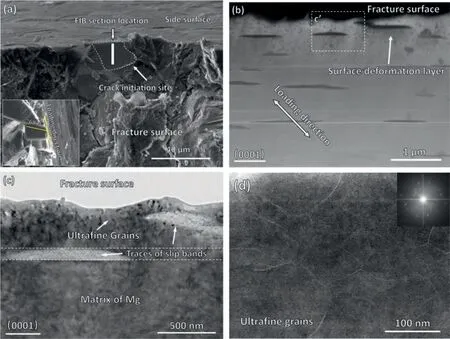
Fig. 8. Microstructure of facets at fracture surface. (a) Location of FIB-sectioned film at the facet. (b) HADDF-STEM image of the cross-section. (c) HRTEM image of the cross-section of the facet showing a deformation layer at the crack surface.(d)High magnification of the ultrafine microstructure in HADDF-STEM mode.
3.6. Surface microstructure at stable crack propagation zone
The fracture surface of the stable crack propagation zone is different from the crack initiation and early crack propagation region. For comparison, the grain-sized flat subzone at stable crack propagation region was also characterized by the FIB-assisted TEM observation. Fig. 9a is the SEM image of the slicing location at the flat subzone. The microstructure beneath the crack surface was obtained, as shown in Fig. 9b.The grain size varies significantly from the matrix to the fracture surface, and the grains present a gradient microstructure.The microstructure in the matrix possesses a relatively larger grain size of 1∼2 μm. While as getting closer to the fracture surface, the grain size decreases significantly. Magnified images of grains beneath the fracture surface (Figs. 9b and 9c)illustrate that there are lots of ultrafine grains here as well,with a grain size of about 100 nm. Furthermore, it should be noted that the fracture surface at the crack initiation region is approximately parallel to the basal plane (Fig. 8b). On the contrary, the fracture surface in the stable crack propagation zone is almost perpendicular to the loading direction, which is independent of the orientation of local grains. At the fatigue crack initiation site, the facets are results of cracking along slip bands. It is well known that the slip bands are generally located in the plane with the relatively large shear stress, so shear is the dominant deformation that drives the initiation and growth of the crack. At the stable propagating stage of the fatigue crack, the size of the plastic zone at the crack tip would increase accordingly, and the effect of local microstructure on the crack path is weakened. Fatigue crack prefers to propagate along the direction perpendicular to the loading stress. As a result, the normal stress becomes the decisive factor in controlling the direction of crack growth,which explains that the crack path is irrelevant to the grain orientation in the stable propagation zone.
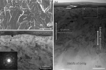
Fig.9. Microstructure of the facet in crack propagation zone:(a)location of FIB section on the crack surface;(b)Enlarged HRTEM image of the microstructure beneath the facet-like crack surface, showing a gradient microstructure of grains; (c) High magnification view of framed rectangular “c” in (b), presenting ultrafine grains near the fracture surface.
4. Discussion
4.1. Effect of the lpso structure on micro-plasticity and crack initiation
Observations on the slip bands in Fig. 6a indicated that plasticity is only visible at limited numbers of grains with favorable orientation for slipping. In the VHCF regime, the cyclic deformation is macroscopically elastic but microscopically plastic at isolated grains. More detailly, micro-plasticity in the form of slip bands only occurs along the basal plane and parallel to the LPSO lamellas, as shown in Fig. 7. Therefore, the Mg-10Gd-3Y-0.1Zn-0.4Zr alloys in this test deform heterogeneously,which only basal slip can be activated within favorably oriented grains. This inhomogeneous deformation at the microscale can be ascribed to the following two important factors. Firstly, the close-packed hexagonal structure of Mg alloys makes its critical resolved shear stress (CRSS)value of basal slip much lower than the non-basal slip and deformation twinning. The deformations along the c-axis of Mg alloys are generally absent at very low amplitudes of cyclic stress, as reported in the previous investigation on the HCF of LPSO-free Mg alloys [26]. Secondly, the LPSO lamellas are embedded into the Mg matrix as a strengthening phase, and they are parallelly distributed along the basal plane. As a result of the specific position, the LPSO lamella almost does not influence the gliding of basal dislocation that moves along the basal plane, but strongly prohibits the activities of non-basal slip and deformation twinning, which have to destroy the LPSO lamellas during cyclic deformation.Based on the above two reasons,the basal slip is the predominant cyclic deformation mechanism in the VHCF regime.
As for the effect of the LPSO structure on the crack initiation, the slip bands are always the preferable sites for fatigue crack initiation, which are demonstrated in Fig. 7. Since the neighboring LPSO structure constrains the development of slip bands, the crack path initiated from the slip bands was restricted by the orientation of the LPSO lamellas. Generally,the morphology of the slip-induced fatigue cracking presents a straight appearance at the specimen surface (Fig. 6a) and a facet at the fracture surface (Fig. 4). This is further confirmed by the observations on the cross-section of slip bands (Fig. 7)and the facets at the initiation site (Fig. 8), demonstrating the positional relationship between the slip bands,crack path,and LPSO lamella. Besides, Figs. 4a and 6a also demonstrate that the cluster of favorably oriented grains would become the vulnerable site for crack initiation. The localization of the grains with similar orientations can act as a relatively soft region for shear deformation, which is similar to the effect of micro texture on the crack behaviors in titanium alloys [30]. In this case, the LPSO lamellas seem to have a limited strengthening effect on the crack initiation process because the crack path does not penetrate them.
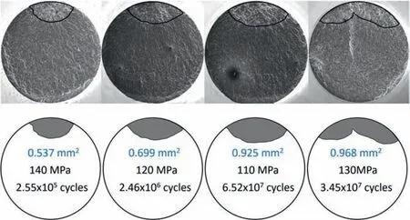
Fig. 10. The projected area of the rough zones around the crack initiation sites.
4.2. Effect of LPSO structure on short crack propagation behaviors
The effect of the LPSO lamellas on the small crack propagation depends on the directional relationship between the crack growth and the LPSO lamellas, as shown in Fig. 6b.The LPSO lamella tends to restrain the crack path along with its orientation, resulting in the straight appearance of crack paths. Once the crack path has to pass through the LPSO lamella, crack propagation is greatly hindered, as evidenced by the observation on the zigzagging in Fig. 6b. This phenomenon of crack growth traversing the LPSO lamellas was widespread at the early stage of crack propagation, so the fatigue performance of the Mg-LPSO alloys would be improved to a certain extent. Besides, the intense interaction between the LPSO structure and small crack results in the formation of the rough area near the crack initiation sites, which should be reflected by the shape and size of the rough zones. Considering the effect of the LPSO lamellas on small crack behaviors, quantitative fractography analysis was carried out on the rough areas in the following.
The rough zone around the crack initiation site has been characterized as shown in Fig. 4, and more typical examples can be found in Fig. 10. Similar rough morphology in the VHCF studies has also been reported in the VHCF researches on titanium and magnesium alloys [16,31,32]. It is believed that multiple fatigue cracks can initiate at the slip bands concurrently. The growth of these small cracks is strongly influenced by the neighboring local microstructures. Therefore,the rough area is a result of the early propagation and coalescence of multiple small microstructural cracks. The projection area of the rough zone on the plane perpendicular to the loading stress was measured for each failed specimen. For surface crack initiation, the stress intensity factor (SIF) at the periphery of the rough zones can be calculated by using the following equation proposed by Murakami et al. [33].

Fig. 11. Quantitative calculation of the stress intensity factor at the rough zone.
Whereσmaxis the amplitude of applied stress, andAreainiis the projected area of the rough zone around the crack initiation site. The obtained SIF values are presented in Fig. 11.It can be seen that the SIF varies within a range of 3.93∼5.00 MPa•m1/2, which is independent of fatigue life. Generally, it was well known that the SIFs at rough zones are approximately a constant, and their average value corresponds to the threshold for stable crack propagation [34,35]. Based on the values of the SIFs in Fig. 11, the fatigue crack propagation threshold of the Mg-10Gd-3Y-1.0Zn-0.4Zr alloys can be estimated to be 4.51 MPa•m1/2on average. For conventional LPSO-free Mg alloys, the fatigue crack behaviors have been experimentally investigated, and the crack propagation thresholds are about 1.25–1.45 MPa•m1/2for As21[36],2.59–3.45 MPa•m1/2for ZK60 [16] and 2.91–3.87 MPa•m1/2for AZ91 [37]. The average value of the SIFs at rough zones in this work is obviously higher than the crack propagation thresholds of LPSO-free Mg alloys. That is to say, the LPSO lamellas retarded the beginning of the stable crack propagation stage.In fact,the rough zone is the place where the LPSO lamellas strongly influence the small crack behaviors by restraining and deflecting its growth direction. The increased crack propagation threshold of the LPSO-containing Mg alloys indicates that the crack initiation and early propagation at the rough zones would experience an increased number of cyclic loadings.
At the crack initiation stage, basal slip is found to be the predominant deformation mechanism at the crack initiation site. Since the LPSO lamellas are paralleled to the path of basal slipping, they cannot impede the basal dislocation activities effectively in the interior of the primary grain. It is hard to say that the LPSO lamellas can improve fatigue crack initiation resistance effectively. However, at the stage of the early crack propagation, the misorientation of the LPSO lamellas to the crack growth direction significantly enhanced the resistance to the crack propagation, which brings a remarkable improvement of fatigue crack propagation resistance. Therefore,it is believed that the effect of the LPSO structure on the fatigue strength enhancement is mainly ascribed to the blocking of crack growth at the early stage of crack propagation.This provides another explanation for the enhanced fatigue resistance of the LPSO-containing Mg alloys compared with the LPSO-free Mg alloys.
4.3. Formation mechanism of ultrafine grains at the crack surface
Fatigue crack initiation is essential for the VHCF behaviors of materials because many studies show that crack initiation usually accounts for most of the total fatigue life [38,39].In this work, facet-like cracking is the representative failure mode, especially at the initiation site (Fig. 4). Based on the observation of the slip-cracking relationship, it is believed that these facets were caused by slip-induced crack initiation along the basal plane. Moreover, it is further revealed that the surface of the facets consists of a layer of ultrafine microstructure. In general, the characteristics of this fine-grained layer are often observed at the origination site of interior crack and named as a fine granular area (FGA) in the HCF and VHCF regimes [31,40]. It is fascinating that ultrafine grains both exist at the facet around the crack initiation site and the flat subzones at the crack propagation region in this work, which has not been reported previously.
At present, many research works have explained the formation mechanism of refined grains at the crack surface, but it is still in dispute. Some researchers [41,42] suggested that the microstructural stress risers, such as second-phase inclusion,micro-textured region,and soft phase of duplex material,led to local recrystallization of the material due to intense plastic deformation occurred in the local high-stress region.Fatigue cracks then initiate from the recrystallized region, so the formation of fine-grained regions occurs earlier than the initiation of fatigue cracks. On the other hand, some other researchers [43,44] found that the fine-grained layer at the fatigue crack initiation site was absent in fatigue tests with positive stress ratios. The fine-grained layer should be caused by numerous compressing of the crack surfaces under cyclic loading, named as the numerous cyclic pressing (NCP) model proposed by Hong et al. [44]. The NCP model believes that fatigue crack initiation is a prerequisite for the formation of fine-grained regions. The focus of the dispute between the two views is the sequence of fatigue cracks and fine-grained layers. Due to the lack of direct experimental evidence, it is difficult to arrive at a definite conclusion.
In this study, fatigue cracks originate from the basal slip bands, as shown in Figs. 6a. Further observation on the microstructure of these slip bands (Fig. 7) found severe local plastic deformation on the specific crystal plane of the Mg alloys, which led to the development of the slip bands. Note that there is no fine-grained structure inside the matrix before the initiation of fatigue cracks. However, the observation of the microstructure of the crack surface after the fatigue test (Fig. 8 and 9) clearly shows the existence of ultra-fined grains at the fracture surface. That is to say, the ultra-fined layer observed in this work should be formed after the fatigue crack initiation, which supports the NCP model very well [44]. The formation of ultrafine grains should be associated with the cyclic compression of the crack surface after the crack initiation from the slip bands. Although it is currently quite difficult to obtain direct evidence of the grain refinement process caused by cyclic compression of crack surfaces, the microstructural analysis of the slip bands and the crack surface in this study indirectly demonstrates that the fine-grained layer is formed after the initiation of fatigue cracks.
The ultrafine grains can be observed at the crack initiation and stable propagation zones, but their grain size and distribution differ significantly. At the crack initiation site, the thickness of the ultrafine-grained layer is about 1 μm, and the equiaxed microstructure with uniform grain size is characterized in the layer. At the stable crack propagation area,the thickness of the fine-grained layer reaches about 8–10 μm, and there is a gradient structure in the layer. Based on the previous discussion, it is believed that the fine-grained structure of the two regions is formed by the alternating interaction of the crack surfaces,but the mode of contacting and compressing of crack surfaces is very different, which is the reason for the formation of two different appearances of finegrained layers. At the fatigue crack initiation site, the maximum shear stress plane within the favorably oriented grains becomes the preferential crack growth path. Under the combined effects of decomposed normal stress and shear stress,both the compressed and sheared contact between the two crack surfaces contributes to the grain refinement. At the stable crack propagation stage,fatigue crack prefers to propagate along the direction perpendicular to the loading stress. Since the shear stress disappears along the crack surface, only normal stress is applied on the crack surface, making the crack surface compressively deformed with much higher stress amplitudes. Therefore, the deformation layer at the stable propagation zone has a much deeper thickness and present gradient microstructure, as shown in Fig. 9.
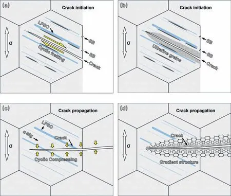
Fig. 12. Schematic model for ultrafine grains formation mechanisms. (a) Fatigue crack initiation along with the basal slip bands. (b) Formation of ultrafine grains at crack initiation site due to cyclic compressing and shearing.(c)Crack propagation path in the interior of grains.(d)formation of gradient microstructure caused by cyclic compressing.
Based on the observations and discussions above, the fatigue crack initiation and propagation processes in the LPSOcontaining Mg alloys are schematically illustrated in Fig. 12.In the VHCF regime, the cyclic deformation is highly inhomogeneous, and plasticity only accumulated at basal slip bands due to the restriction of the LPSO lamellas, as shown in Fig. 12a. In this case, the evolution of slip bands is only visible at the favorably oriented grains. Since the localization of the cyclic plastic strain contributes to the formation of the surface relief in the form of extrusions and intrusions,which plays a decisive role in the initiation of fatigue cracks[45]. As a result, fatigue cracks are apt to initiate from the basal slip bands next to the LSPO lamellas (Fig. 12b). After the crack initiation, the early crack propagation is strongly influenced by the LPSO lamellas. The LPSO lamellas can strongly prohibit the propagation of microstructurally small cracks by deflecting their growth direction frequently, leading to a higher crack propagation threshold. At the stable crack propagation stage, the direction of the applied stress is overall perpendicular to the crack path(Fig.12c).The LPSO lamellas cannot influence the propagation direction of fatigue cracks due to the increase of plasticity at the crack tip. The normal stress is much higher than that at the crack initiation stage,which contributes to the formation of gradient structure in the surface deformation layer (Fig. 12d).
It should be noted that the fatigue crack initiation and early propagation are very important to the VHCF behaviors of metallic materials. Based on the above discussion, the LPSO lamellas are incapable of impeding the slipping of basaldislocations at the basal planes, their effect on the fatigue crack initiation resistance at the primary grains seems to be limited. The frequent change of microstructural crack path might be the main reason for the excellent VHCF resistance of Mg alloys with the LPSO lamellas.
5. Conclusions
The VHCF failure mechanisms of the LPSO-containing Mg alloys were investigated with a focus on the effect of the LPSO lamellas on the micro-plasticity evolution and fatigue crack behaviors. The following conclusions can be drawn:
1. The LPSO structure limits non-basal slip and deformation twinning at low stresses, and basal slip is the predominant deformation. Fatigue crack initiates from the basal slip bands.
2. Facet morphology can be observed around the crack initiation site due to the slip-induced cracking mechanism.The LPSO lamella strongly hinders the short crack growth behavior.
3. The surface layer of the crack surface consists of finegrained microstructure caused by the cyclic contact of the crack surfaces. It is evidenced that the fine grains are generated after crack initiation.
4. The LPSO results in a relatively high crack propagation threshold, and this is the main reason for the improvement of the VHCF strength of the LPSO-containing Mg alloys.
Acknowledgment
The authors gratefully acknowledge the financial support from the National Natural Science Foundation of China (Nos.12072212 and 11832007),the National Key Research and Development Program of China(No.2018YFE0307104),and the Applied Basic Research Programs of Sichuan Province (No.2021YJ0071). We also highly appreciate the help of Dr. Yan Li from the Department of Mechanics, Sichuan University.
杂志排行
Journal of Magnesium and Alloys的其它文章
- NaH doped TiO2 as a high-performance catalyst for Mg/MgH2 cycling stability and room temperature absorption
- Microstructure, mechanical properties and fracture behaviors of large-scale sand-cast Mg-3Y-2Gd-1Nd-0.4Zr alloy
- Enhanced initial biodegradation resistance of the biomedical Mg-Cu alloy by surface nanomodification
- Elucidating the evolution of long-period stacking ordered phase and its effect on deformation behavior in the as-cast Mg-6Gd-1Zn-0.6Zr alloy
- The mechanism for tuning the corrosion resistance and pore density of plasma electrolytic oxidation (PEO) coatings on Mg alloy with fluoride addition
- The role of melt cooling rate on the interface between 18R and Mg matrix in Mg97Zn1Y2 alloys
