Effect of curcumin on palmitic acid-induced apoptosis and steroid production of ovarian granulosa cells by inhibiting HMGB1-mediated autophagy
2021-12-20LiaoYuanWangKaijuLiHaohuiChenHuipingLiXuanyi
Liao Yuan,Wang Kaiju,Li Haohui,Chen Huiping,Li Xuanyi
Abstract Objective:To explore the effect of curcumin on the injury of ovarian granulosa cells induced by palmitic acid(PA) and its possible mechanism.Methods:Human ovarian granulosa cells were cultured in vitro.CCK-8 was used to detect cell viability.ELISA was used to detect estradiol(E2)content in the cell supernatant.RT-qPCR was used to detect the expression of HMGB1,CYP19A and CYP11A mRNA.Flow cytometry was used to detect cell apoptosis.Western blotting was used to detect the expression of HMGB1,Beclin1,LC3B and P62 protein.Results:200 μmol/L or 400 μmol/L PA significantly decreased the cell viability,E2 content,and the expression of CYP19A and CYP11A mRNA,while increased the apoptosis rate of cells (P<0.05).Compared with the control group,the cell viability,E2 content,and the expressions of CYP19A,CYP11A and P62 in the PA group were significantly reduced,while the cell apoptosis rate,the expressions of HMGB1 and Beclin1,and LC3II/LC3I ratio were significantly elevated (P<0.05).Compared with the PA group,the cell viability,E2 content,and the expressions of CYP19A,CYP11A and P62 in PA+curcumin (2.5 μmol/L or 5 μmol/L) group were increased,while the cell apoptosis rate,the expression of HMGB1 and Beclin1,and LC3II/LC3I ratio were evidently reduced (P<0.05).Overexpression of HMGB1 reversed the protective effects of curcumin on the PA induced injuries of ovarian granulosa cells and up-regulated the expressions of autophagy-related proteins (P<0.05).Conclusion:Curcumin can attenuate PA-induced ovarian granulosa cell injury by inhibiting autophagy mediated by HMGB1.
Keywords ovarian granulosa cells;palmitic acid;curcumin;high mobility group box protein 1;autophagy
Introduction
Fatty acids in follicular fluid are important nutrients for oocyte growth.Physiological conditions,such as obesity,have a profound effect on the level of non-esterified fatty acids (NEFA) in follicular fluid,which may lead to lower quality of oocytes [1].Palmitic acid (PA) is the main fatty acid in follicular fluid.High concentrations of PA and stearic acid in follicular fluid are related to human adverse pregnancy outcome[2].Therefore,improving the injury of ovarian granulosa cells induced by PA may be a potential therapeutic strategy to improve human adverse pregnancy outcome.Curcumin is a low molecular polyphenol with a wide range of biological and pharmacological properties,including anti-inflammatory,antioxidant and anti-cancer [3].Studies have shown that curcumin can improve the quality of oocytes.Curcumin can protect oxidative stress of human granulosa cells and promote the viability of granulosa cells[4].It can be concluded that curcumin may have a certain effect on the injury of ovarian granulosa cells induced by PA.High mobility group box protein 1 (HMGB1) is a DNAbinding protein,which is secreted by inflammatory cells and damaged cells,and is the main participant of inflammation[5].Studies have shown that the expression of HMGB1 is increased in granulosa cells treated with high concentration of insulin.Knocking down HMGB1 can improve granulosa cell injury by inhibiting the activation of inflammatory pathway in granulosa cells [6].In addition,the content of HMGB1 in follicular fluid and autophagy of granulosa cells were significantly increased in patients with polycystic ovary syndrome (PCOS).HMGB1 promotes the progression of PCOS by inducing autophagy of granulosa cells [7].Based on this,this study explored whether curcumin plays an effective role in PA-induced ovarian granulosa cell injury by regulating HMGB1-mediated autophagy,in order to provide new scientific basis for developing new therapeutic strategies to improve follicular quality and adverse pregnancy outcomes.
Materials and methods
Main materials
Human ovarian granulosa cells were purchased from Wuhan Procell Life Science &Technology Co.,Ltd.PA and curcumin were purchased from Sigma-Aldrich.LipofectamineTM2000 Transfection Reagent and eBioscienceTMAnnexin V-FITC Apoptosis Detection Kit were purchased from Thermo Fisher Scientific.HMGB1 overexpression vector (oe-HMGB1) and overexpression vector negative control (oe-NC) were purchased from Wuhan BioRun Biotechnology Co.,Ltd.CCK-8 kit was purchased from MedChemExpress.The human estradiol (E2) ELISA kit was purchased from Wuhan Elabscience Biotechnology Co.,Ltd.HMGB1,Beclin1,LC3B and P62 antibodies were purchased from Abcam.
Cell culture
Human ovarian granulosa cells were cultured in an atmosphere containing 5% CO2and 95% O2at 37 ℃with DMEM medium which supplemented with 10%fetal bovine serum (FBS) and 1% penicillin/streptomycin(100 U/mL,Thermo Fisher Scientific).
Cell grouping
The cells were divided into control group,PA group,PA+curcumin 2.5 μmol/L group and PA+curcumin 5 μmol/L group.PA (concentration of 100,200 or 400 μmol/L) and curcumin (concentration of 1.25,2.5,5,10 or 20 μmol/L) were added to the cells.The control group cells were added with the same amount of solvent.The cells were cultured for 24 h.In the cell transfection experiment,the cells were divided into PA+curcumin 5 μmol/L group,PA+curcumin 5 μmol/L+oe-NC group(transfected oe-NC)and PA+curcumin 5 μmol/L+oe-HMGB1 group (transfected oe-HMGB1).
Cell transfection
Human ovarian granulosa cells were cultured to a density of 90% the day of transfection.oe-NC and oe-HMGB1 were transfected into human ovarian granulosa cells using Lipofectamine 2000 following the manufactures’instructions.After 48 h of post-transfection,200 μmol/L PA and 5 μmol/L curcumin were added to the cells.
Cell viability assay
Human ovarian granulosa cells growing in logarithmic phase were inoculated into 96-well plate with 1×10⁴cells per well.After the cells were cultured for 24 h,the optical density (OD) of each well at 450 nm was detected according to the instructions of CCK-8 kit.In addition,the blank group (not inoculated with cells)was used to set the reading value to zero.
Detection of E2 content in cell supernatant
The supernatant of human ovarian granulosa cells after administration or cell transfection was collected,and the content of E2 in the supernatant was detected according to the instructions of ELISA kit.
RT-qPCR
Human ovarian granulosa cells were collected and the total RNA was extracted by Trizol reagent.The extracted RNA was reverse transcribed into cDNA.2×SYBR Green qPCR Master Mix was used for followup detection.RT-qPCR was performed using a 7500 Fast RT-PCR instrument (Applied Biosystems,Darmstadt,Germany).The reaction procedure was as follows:95 ℃,10 min;95 ℃,15 s;60 ℃,30 s;72 ℃,10 s,40 cycles.Taking GAPDH as the internal reference,the relative expressions of HMGB1,CYP19A and CYP11A mRNA were calculated by 2-ΔΔCtmethod.The primer sequences used were as follows (5’-3’):HMGB1-Forward:CTCAGAGAGGTGGAAGACCATGT,HMGB1-Reverse:GGGATGTAGGTTTTCATTTCTCTTTC;CYP19A-Forward:AGGTGCTATTGGTCATCTGCTC,CYP19A-Reverse:TGGTGGAATCGGGTCTTTATGG;CYP11A-Forward:GAGATGGCACGCAACCTGAAG,CYP11A-Reverse:CTTAGTGTCTCCTTGATGCTGGC;GAPDH-Forward:CATGAGAAGTATGACAACAGCCT,GAPDH-Reverse:AGTCCTTCCACGATACCAAAGT.
Flow cytometry analysis
Cell apoptosis analysis was performed using flow cytometry with an Annexin V/PI apoptotic detection kit.Briefly,differently treated human ovarian granulosa cells were washed in PBS and re-suspended in binding buffer containing 10 μL of Annexin V and 5 μL PI according to the manufacturer’s instructions.The apoptotic cells were analyzed using a flow cytometry(BD Biosciences) equipped with FlowJo software(Tree Star).
Western blotting
Human ovarian granulosa cells were collected and lysed by RIPA lysate.Total protein obtained from the cells was separated by electrophoresis on SDS-PAGE and electrotransferred onto polyvinyl idene difluoride(PVDF)-plus membranes.After blocking with 5%skim milk for 2 h,the membranes were incubated overnight at 4 ℃with the following primary antibodies:HMGB1(1:1,000),Beclin1(1:2,000),LC3B(1:1,000),P62 (1:2,000) and GAPDH (1:5,000).Then,the membranes were washed three times in TBST and incubated with secondary antibodies conjugated with HRP at 37 ℃for 2 h.Relative densitometry was performed by using a computerized software package(NIH Image 1.63 software).
Statistical analysis
SPSS 21.0 software was used to analyze statistical data.All data were shown as mean±standard deviation(SD).One-way analysis of variance followed by LSDttest was used for statistical analysis.Pvalues less than 0.05 were considered statistically significant.
Results
Effect of PA on ovarian granulosa cells
There was no significant difference between PA 0 μmol/L group and PA 100 μmol/L group (P>0.05).The cell viability,E2 content,and CYP19A and CYP11A mRNA expressions in PA 200 μmol/L group and PA 400 μmol/L group were significantly lower than those in PA 0 μmol/L group,while the apoptosis rate of cells was significantly higher (P<0.05) (Figure 1).
Effect of curcumin on ovarian granulosa cells in⁃duced by PA
The cell viability of curcumin 10 μmol/L group and 20 μmol/L group was significantly lower than that of the control group (P<0.05),while there was no significant difference in cell viability among other groups (P>0.05).Therefore,in the follow-up experiment,the concentration of curcumin was 2.5 μmol/L and 5 μmol/L.
Compared with the control group,the cell viability,E2 content,and the mRNA expressions of CYP19A and CYP11A in PA group were significantly decreased,while the apoptosis rate was significantly increased (P<0.05).Compared with PA group,the cell viability,E2 content,and CYP19A and CYP11A mRNA expressions in PA+curcumin 2.5 μmol/L group and PA+curcumin 5 μmol/L group were significantly increased,while the apoptosis rate was significantly decreased(P<0.05)(Figure 2).
Effect of curcumin on HMGB1 expression and au⁃tophagy in ovarian granulosa cells induced by PA
Compared with the control group,the mRNA and protein expression of HMGB1,Beclin1 protein expression and LC3II/LC3I ratio in the PA group were significantly increased,while the P62 protein expression was significantly decreased (P<0.05).Compared with the PA group,the mRNA and protein expression of HMGB1,Beclin1 protein expression and LC3II/LC3I ratio in the PA+curcumin 2.5 μmol/L group and PA+curcumin 5 μmol/L group were significantly decreased,while the P62 protein expression was significantly increased(P<0.05)(Figure 3).
Overexpression of HMGB1 reversed the effects of curcumin on ovarian granulosa cells induced by PA
There were no significant differences in HMGB1 mRNA and protein expression levels,cell viability,E2 content,CYP19A and CYP11A mRNA levels,cell apoptosis rate,Beclin1 and P62 protein levels and LC3II/LC3I ratio between PA+curcumin 5 μmol/L group and PA+curcumin 5 μmol/L+oe-NC group(P>0.05).Compared with PA+curcumin 5 μmol/L+oe-NC group,the expression of HMGB1 mRNA and protein,cell apoptosis rate,Beclin1 protein level and LC3II/LC3I ratio in PA+curcumin 5 μmol/L+oe-HMGB1 group were increased,while the CYP19A and CYP11A mRNA levels and P62 protein level were markedly decreased(P<0.05)(Figure 4).
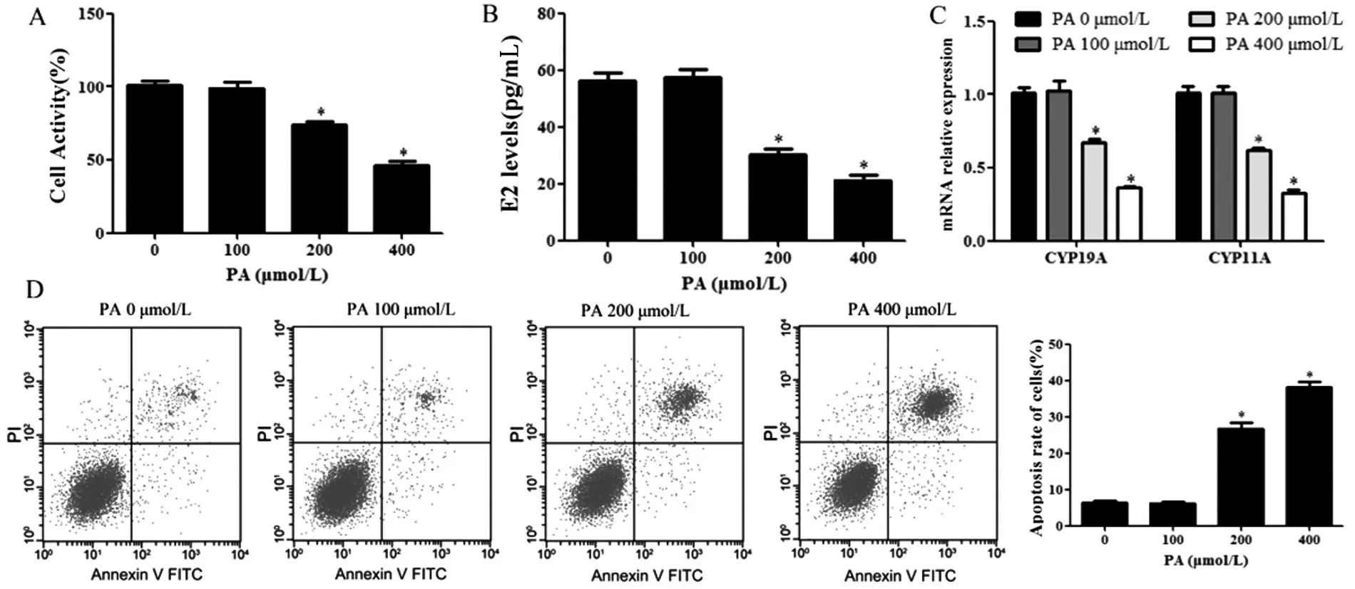
Figure 1 Effect of PA on ovarian granulosa cells.A:Detection of cell viability by CCK-8;B:Detection of E2 in cell supernatant by ELISA;C:Detection of CYP19A and CYP11A mRNA expression by RT-qPCR;D:Detection of apoptosis by flow cytometry.Compared with 0 μmol/L group,*P<0.05.
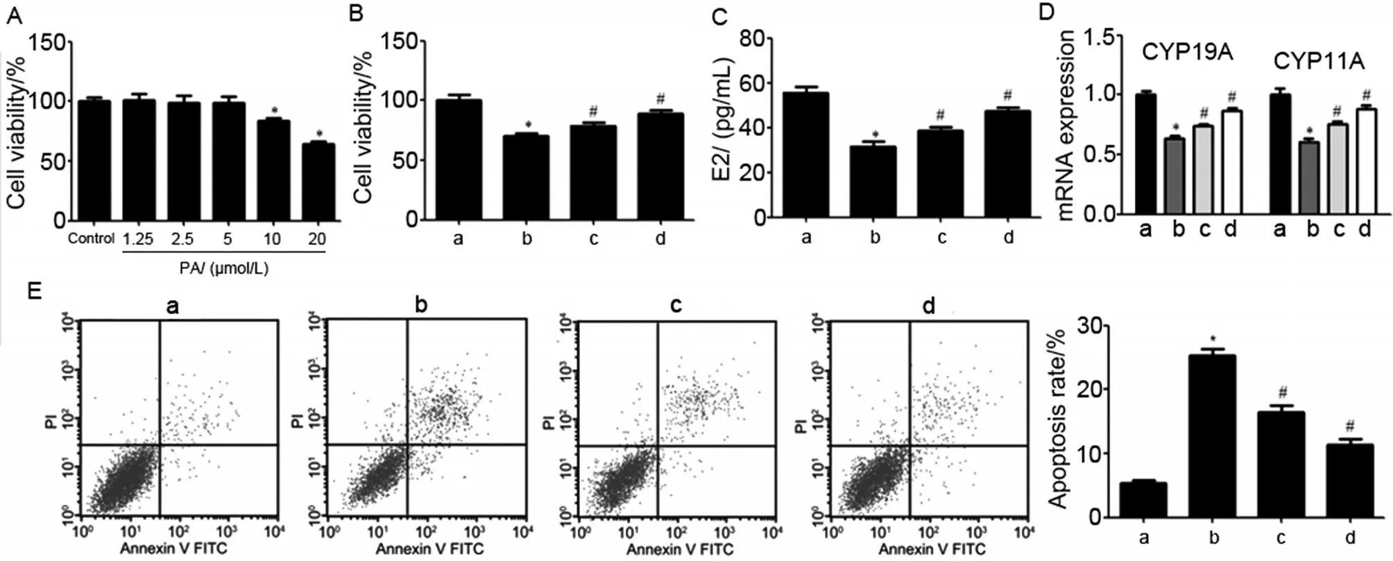
Figure 2 Effect of curcumin on ovarian granulosa cells induced by PA.a:control group;b:PA group;c:PA+curcumin 2.5 μmol/L group;d:PA+curcumin 5 μmol/L group.A-B:Detection of cell viability by CCK-8;C:Detection of E2 content in cell supernatant by ELISA;D:Detection of CYP19A and CYP11A mRNA expression in cells by RT-qPCR;E:Detection of apoptosis by flow cytometry.Compared with the control group,*P<0.05;Compared with the PA group,#P<0.05.
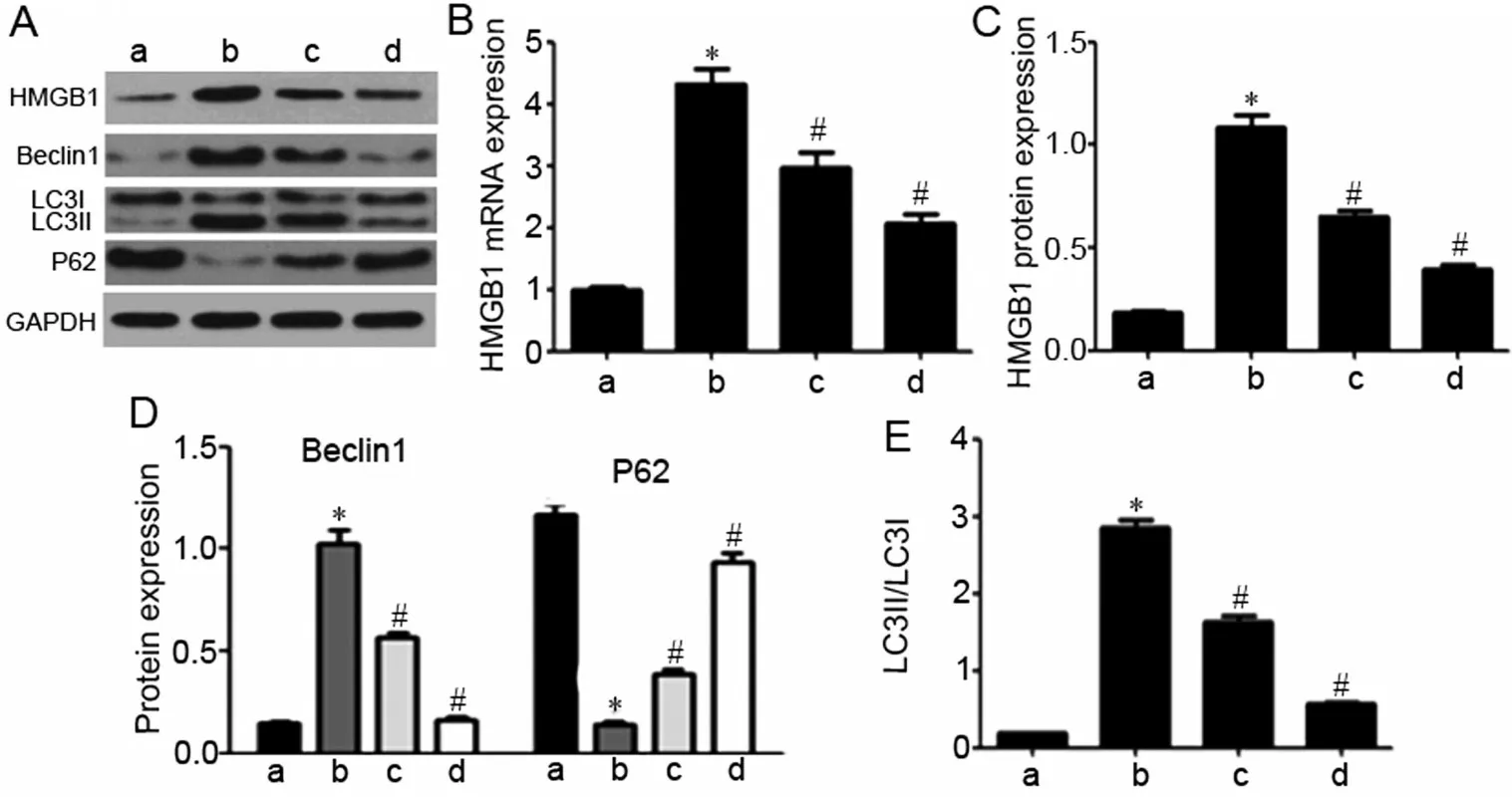
Figure 3 Effects of curcumin on HMGB1 expression and autophagy in PA-induced ovarian granulosa cells.a:control group;b:PA group;PA+curcumin 2.5 μmol/L group and PA+curcumin 5 μmol/L group.A:Representative western blots of HMGB1,Beclin1,LC3 and P62;B-C:Quantitative analysis demonstrated the mRNA and protein expression of HMGB1;D-E:Quantitative analysis demonstrated the protein expression of Beclin1 and LC3II/LC3I ratio.Compared with the control group,*P<0.05;Compared with the PA group,#P<0.05.
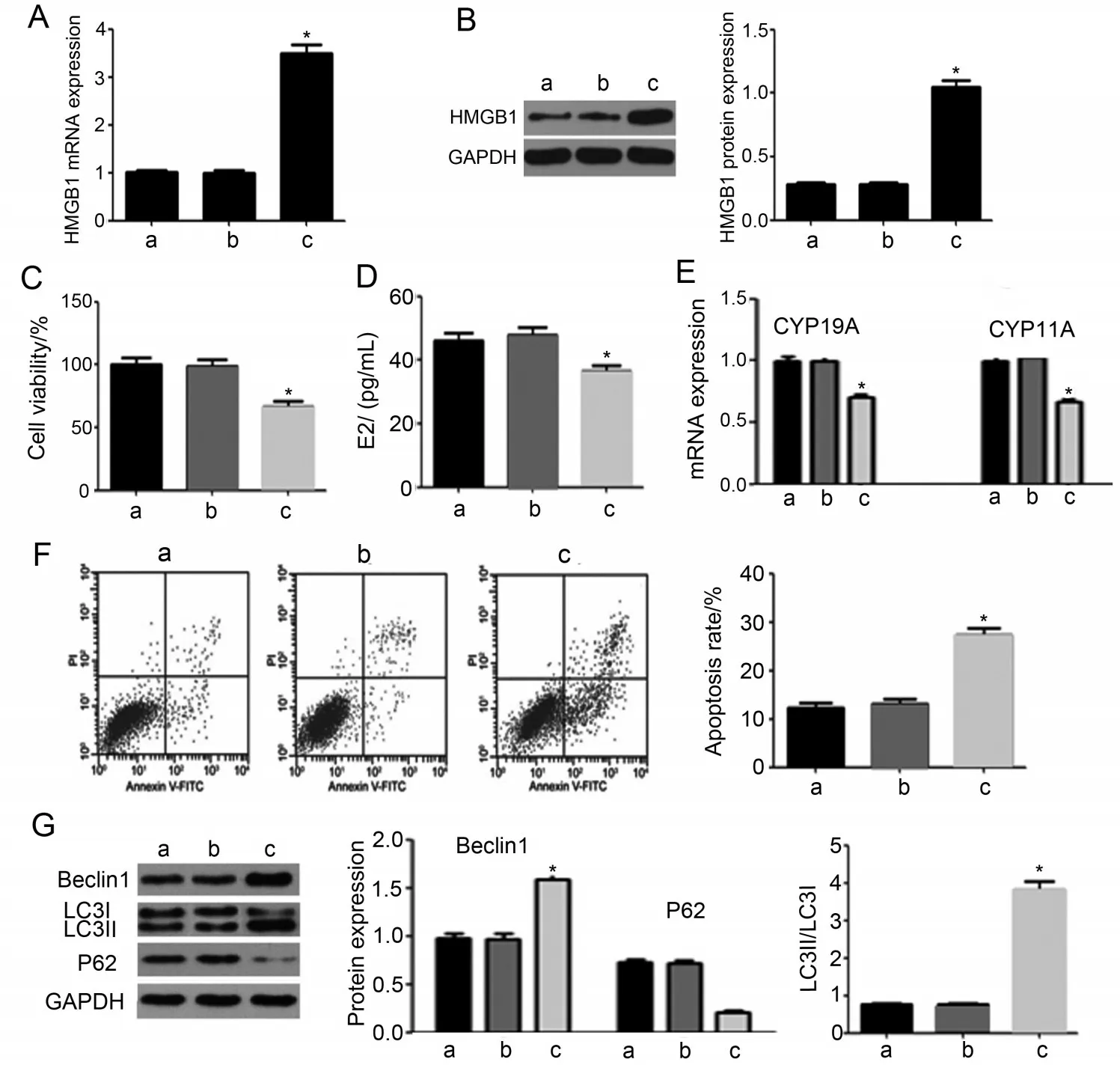
Figure 4 Overexpression of HMGB1 reversed the effects of curcumin on ovarian granulosa cells induced by PA.a:PA+curcumin 5 μmol/L group;b:PA+curcumin 5 μmol/L+oe-NC group;c:PA+curcumin 5 μmol/L+oe-HMGB1 group.A:Expression of HMGB1 mRNA was detected by RT-qPCR;B:The expression of HMGB1 protein was detected by Western blotting;C:The cell viability was evaluated by CCK-8 assay;D:The E2 content in cell supernatant was assessed by ELISA;E:CYP19A and CYP11A mRNA expression was measured by RT-qPCR.F:The cell apoptosis was determined by flow cytometry;G:The protein expressions of Beclin1,LC3II/LC3I and P62 were measured by Western blotting.Compared with PA+curcumin 5 μmol/L+oe-NC group,*P<0.05.
Discussion
Granulosa cells,as the main cells producing steroids,can produce aromatase,catalyze the conversion of androgen to estrogen,and promote follicular development through paracrine pathway [8].Granulosa cells may be closely related to the decline of female fertility caused by obesity [9].PA is a ubiquitous saturated fatty acid,which has been proved to be toxic to Sertoli cells and may lead to disorders of testicular development and spermatogenesis [10].Evidence has proved that PA also has cytotoxicity to granulosa cells and can induce endoplasmic reticulum stress and apoptosis in granulosa cells [11].The results in this study showed that high concentrations of PA (200 μmol/L and 400 μmol/L)could inhibit the viability of granulosa cells,the secretion of E2 and the mRNA expression of steroid synthase CYP19A and CYP11A,and promote apoptosis.
Curcumin can regulate multiple biological targets and cellular pathways to play a therapeutic role in a variety of diseases,such as cancer,cardiovascular disease,neurological and autoimmune diseases[12].Curcumin plays an significant role in protecting granulosa cells from external damage.For example,the results of Vashisht et al]13]showed that curcumin through exosome can reduce the dysfunction of granulosa cells caused by lipopolysaccharide,inhibit the expression of pro-inflammatory cytokines,and promote the expression of CYP19A gene and the production of E2.In addition,curcumin could promote the production of progesterone and E2 in premature ovarian failure mice,and significantly inhibit the expression of aging-related proteins,oxidative stress and apoptosis of granulosa cells in mice with premature ovarian failure [14].Curcumin also has a protective effect on mice with polycystic ovary syndrome.It can effectively inhibit ovarian injury and dehydroepiandrosteroneinduced granulosa cell apoptosis [15].Previous studies have confirmed that PA can inhibit the proliferation and survival rate of granulosa cells,induce apoptosis and the production of endoplasmic reticulum stress markers.In the presence of PA,the ability of oocytes to develop into blastula stage is lower [16].This study has also proved that high concentration of PA has cytotoxicity to granulosa cells.In this study,PA was used to treat granulosa cells to establish an injury model in order to explore the effect of curcumin on granulosa cell injury.The results show that both low and high doses of curcumin could promote PA-induced granulosa cell viability,E2 secretion and steroid synthase CYP19A and CYP11A mRNA expression,and inhibit apoptosis.The results show that curcumin has a protective effect on granulosa cell injury induced by PA.
HMGB1 is a small protein with cytokine activity in the nucleus,cytoplasm and extracellular cells,and exists in all types of cells [17].The evidence shows that the expression of HMGB1 in serum and follicular fluid of patients with polycystic ovary syndrome is significantly increased,and HMGB1 may be a new candidate biomarker of polycystic ovary syndrome [18].Knocking down HMGB1 could promote the proliferation of granulosa cells,increase the secretion of E2 and progesterone,and inhibit the apoptosis of ovarian granulosa cells [19].The results of Jiang et al [20]also proved that down-regulation of HMGB1 expression can promote granulosa cell proliferation,increase ovarian coefficient and inhibit granulosa cell apoptosis by inhibiting inflammation-related signal pathway.Evidence has proved that curcumin can play a protective role by inhibiting HMGB1-mediated cell injury [21].The results showed that PA treatment could induce the expression of HMGB1 in granulosa cells.Both low and high doses of curcumin could inhibit the expression of HMGB1 in PA-treated granulosa cells.In addition,overexpression of HMGB1 could partially reversed the effect of curcumin on PA-treated granulosa cells,resulting in decreased cell viability,E2 secretion and mRNA expression of steroid synthase CYP19A and CYP11A,and increased apoptosis rate.The results show that curcumin plays a protective role in PA-induced granulosa cell injury by inhibiting HMGB1.
Autophagy is essential for the natural decomposition of unnecessary or dysfunctional components in cells.Proper autophagy can circulate cell components and organelles,thereby maintaining cell function [22].However,abnormal autophagy may lead to cell dysfunction or even death.The results of Yang et al [23]showed that trio-methyl phosphate can cause damage to the female reproductive system,reduce the viability of ovarian granulosa cells and induce autophagy,while inhibition of autophagy can increase the viability of granulosa cells.In addition,4-hydroxycyclophosphamide can induce granulosa cell damage by promoting autophagy,while resveratrol can improve granulosa cell injury induced by 4-hydroxycyclophosphamide by inhibiting autophagy[24].At present,evidence has proved that HMGB1 is an important autophagy regulator,which can induce autophagy and promote cell injury [25].Similarly,HMGB1 can promote the progression of ovarian disease by inducing autophagy of granulosa cells [7].The results show that PA treatment could induce autophagy of granulosa cells.Both low and high doses of curcumin could inhibit autophagy of granulosa cells treated with PA.In addition,overexpression of HMGB1 could partially reverse the effect of curcumin on autophagy of PAtreated granulosa cells.The results suggest that curcumin plays a protective role in PA-induced granulosa cell injury by inhibiting autophagy induced by HMGB1.
To sum up,PA can inhibit the viability of granulosa cells and the secretion of steroids,and induce apoptosis.Curcumin can promote the survival and steroid production of PA-treated granulosa cells,and inhibit apoptosis by inhibiting the activation of autophagy mediated by HMGB1.The results of this study provide new scientific basis for developing new treatment strategies in order to improve follicular quality and adverse pregnancy outcomes.
AcknowledgmentsThis study was funded by Scientific Research Project of Health Industry in Hainan Province (No.20A200353) and The National Natural Science Foundation of China(81803277).