彩色多普勒超声与心电图诊断老年高血压性心脏病的价值
2021-03-24范海静
范海静

[摘要] 目的 分析彩色多普勒超聲、心电图诊断老年高血压性心脏病的价值。 方法 选择2017年12月至2020年3月我院收治的老年高血压性心脏病患者100例作为观察组,选择同期健康体检老年人30例作为对照组,比较对照组与观察组(高血压Ⅰ期、Ⅱ期、Ⅲ期)心脏超声指标;比较单独和联合使用心电图及彩色多普勒超声诊断心脏病的效能及其对心脏病类型的检出率。 结果 观察组与对照组、观察组各亚组间的E/A、LVEF、LVMM、LAD比较,差异均有统计学意义(P<0.05)。联合诊断的灵敏度、特异度分别为94.00%、100.00%,高于心电图的75.00%、96.67%及彩色多普勒超声的92.00%、90.00%,三组比较,差异有统计学意义(P<0.05);联合诊断心脏病的ROC曲线下灵敏度、特异度分别为93.18%、99.12%,高于心电图的73.98%、95.19%及彩色多普勒超声的89.91%、89.54%,三组比较,差异有统计学意义(P<0.05)。心电图的主动脉弹性减退检出率(0)、左心室肥厚检出率(18.00%)、左心房增大检出率(22.00%)均低于彩色多普勒超声(6.00%、30.00%、37.00%)和联合诊断(6.00%、32.00%、39.00%),差异有统计学意义(P<0.05)。 结论 彩色多普勒超声能有效诊断老年高血压性心脏病,且联合心电图能进一步提高诊断效能,提高对心脏病类型的检出率。
[关键词] 彩色多普勒超声;心电图;老年患者;高血压性心脏病
[Abstract] Objective To analyze the value of color doppler ultrasonography and electrocardiogram in diagnosing senile hypertensive heart disease. Methods A total of 100 elderly patients with hypertensive heart disease admitted to our hospital from December 2017 to March 2020 were selected as the observation group, and 30 healthy elderly patients who underwent physical examination during the same period were selected as the control group. Echocardiographic index was compared between the control group and the observation group (hypertension at stages Ⅰ to Ⅲ). The efficiency and detection rate on type diagnosis of heart disease by electrocardiogram or color doppler ultrasonography alone, electrocardiogram combined with color doppler ultrasonography were compared. Results Comparison of E/A, LVEF, LVMM and LAD between the observation groups and control group (hypertension at stages Ⅰ to Ⅲ) , the differences were statistically significant(P<0.05). The sensitivity and specificity in the diagnosis of heart disease were of electrocardiogram combined with color doppler ultrasonography were 94.00% and 100.00%, higher than 75.00% and 96.67% of electrocardiogram; higher than 92.00% and 90.00% of color doppler ultrasonography; the differences of comparison of the above indexes between the three diagnosis methods were statistically significant(P<0.05). The sensitivity and specificity of ROC curve in the diagnosis of heart disease of electrocardiogram combined with color doppler ultrasonography were 93.18% and 99.12%, higher than 73.98% and 95.19% of electrocardiogram; higher than 89.91% and 89.54% of color doppler ultrasonography; the differences of comparison of the above indexes between the three diagnosis methods were statistically significant(P<0.05). The detection rate of decreased aortic elasticity(0), the detection rate of left ventricular hypertrophy(18.00%), and the detection rate of left atrial enlargement(22.00%) were all lower than that of color Doppler ultrasound(6.00%, 30.00%, 37.00%) and combined diagnosis(6.00%, 32.00%, 39.00%), with statistically significant differences(P<0.05). Conclusion Color doppler ultrasonography can effectively diagnose senile hypertensive heart disease, but electrocardiogram combined with color doppler ultrasonography can further improve the efficiency and type on detection rate of heart disease diagnosis.
[Key words] Color doppler ultrasonography; Electrocardiogram; Elderly patients; Hypertensive heart disease
高血压患者在长期高血压水平作用下,导致左心房负荷增加,左心房肥大、扩张,引起高血压性心脏病(Hypertensive heart disease,HHD),可增加心源性猝死、心力衰竭等风险,患者早期缺乏明显症状,进展到一定程度,会出现右上腹疼痛、呼吸困难、全身水肿、少尿等症状,预后变差,因此早期确诊高血压性心脏病對改善患者预后具有重要意义。目前诊断HHD应用最广泛的是心电图,HHD患者的心肌细胞电生理异常在心电图中有明显表现,临床可通过心电图进行诊断,且能反复使用、操作简单,但也存在误诊、漏诊风险。心脏彩色多普勒超声操作简单、无创、分辨率高,逐渐用于心脏疾病检查中,其应用价值较高,近年来不断有研究以心电图为基础分析心脏彩色多普勒超声诊断HHD的价值,但结论并未形成共识,因此需要进一步的研究[1-2]。本研究以HHD患者100例和健康体检者30例为研究对象,分析心电图、心脏彩色多普勒超声及二者联合应用诊断HHD的价值,为提高该病的诊断准确性提供参考,现报道如下。
1 资料与方法
1.1 一般资料
选择2017年12月至2020年3月我院收治的老年高血压性心脏病患者100例作为观察组,选择同期健康体检老年人30例作为对照组。纳入标准[3]:年龄≥60岁;符合第7版《内科学》中HHD的诊断标准;进行心电图和心脏彩色多普勒超声检查;签署知情同意书。排除标准[4]:合并冠心病、心肌病、心脏瓣膜病;继发性高血压;躯体明显缺陷;心脏手术史;合并自身免疫疾病、严重炎症。观察组中,男58例,女42例;平均年龄(65.12±5.12)岁;高血压病程5~16年,平均(9.43±2.12)年;高血压Ⅰ期45例、Ⅱ期33例、Ⅲ期22例;合并高脂血症12例、糖尿病19例。对照组中,男16例,女14例;平均年龄(64.98±3.27)岁。两组性别、年龄比较,差异无统计学意义(P>0.05),具有可比性。本研究获得医院医学伦理委员会批准。
1.2 方法
采用美国西门子公司ACUSONSC2000彩色多普勒超声诊断仪,探头频率2.5 MHz。患者取左侧卧位,在胸骨旁左室长轴切面、短轴切面、心尖四腔心切面进行扫描,测量左心室射血分数(Left ventricular ejection fraction,LVEF)、左心室心肌质量(Left ventricular myocardial mass,LVMM)、左心房内径(Inside diameter of left atrium,LAD)、左室后壁厚度(Left ventricular posterior wall thickness,LVPW)、室间隔厚度(Interventricular septum thickness,IVS)、左心室舒张末期内径(Left ventricular end-diastolic diameter,LVDD);采用彩色多普勒超声的频谱多普勒测量二尖瓣血流舒张晚期左心室充盈峰速(A)、舒张早期左心室充盈峰速(E),计算峰值速度比(E/A)。使用光电ECG-2340心电图分析仪,患者取平卧位,测量P波(Ⅱ时间)、PR、Ptfvl及QRS波群电压。
1.3观察指标与评价标准
比较对照组与观察组(高血压Ⅰ期、Ⅱ期、Ⅲ期)的心脏超声指标(E/A、LVEF、LVMM、LAD、LVPW、IVS、LVDD)。彩色多普勒超声诊断标准[4]:①左室肥厚:左室后壁厚度及室间隔厚度均>12 mm;②心脏扩大:左室>56 mm,升主动脉内径>36 mm,同时左室长轴切面左房>36 mm。心电图确诊标准[5]:①左室肥厚:胸壁导联SV1+RV5男性>4.0 mV,女性>3.5 mV;②左房增大:P波宽度与P-R段的比值>1.6;PⅡ时长>0.11 s,Ptfv1绝对值>0.04 mm·s,Ptfv1负向波<0.04 mm·s;P波双峰且间距>0.04 s,在Ⅰ、Ⅱ、AVL、V4-V6导联更明显。比较单独和联合使用心电图及彩色多普勒超声诊断高血压性心脏病的效能(特异度、灵敏度)及对心脏病类型的检出率,灵敏度=真阳例数/(真阳例数+假阴例数)×100%,特异度=真阴例数/(真阴例数+假阳例数)×100%,根据灵敏度、特异度和ROC曲线下面积(AUC)值,分析三种方法诊断该病效能。
1.4 统计学方法
应用SPSS 18.0统计学软件进行数据分析,计量资料用(x±s)表示,采用t检验或F检验;计数资料用[n(%)]表示,采用χ2检验,P<0.05为差异有统计学意义。
2 结果
2.1 两组心脏超声指标比较
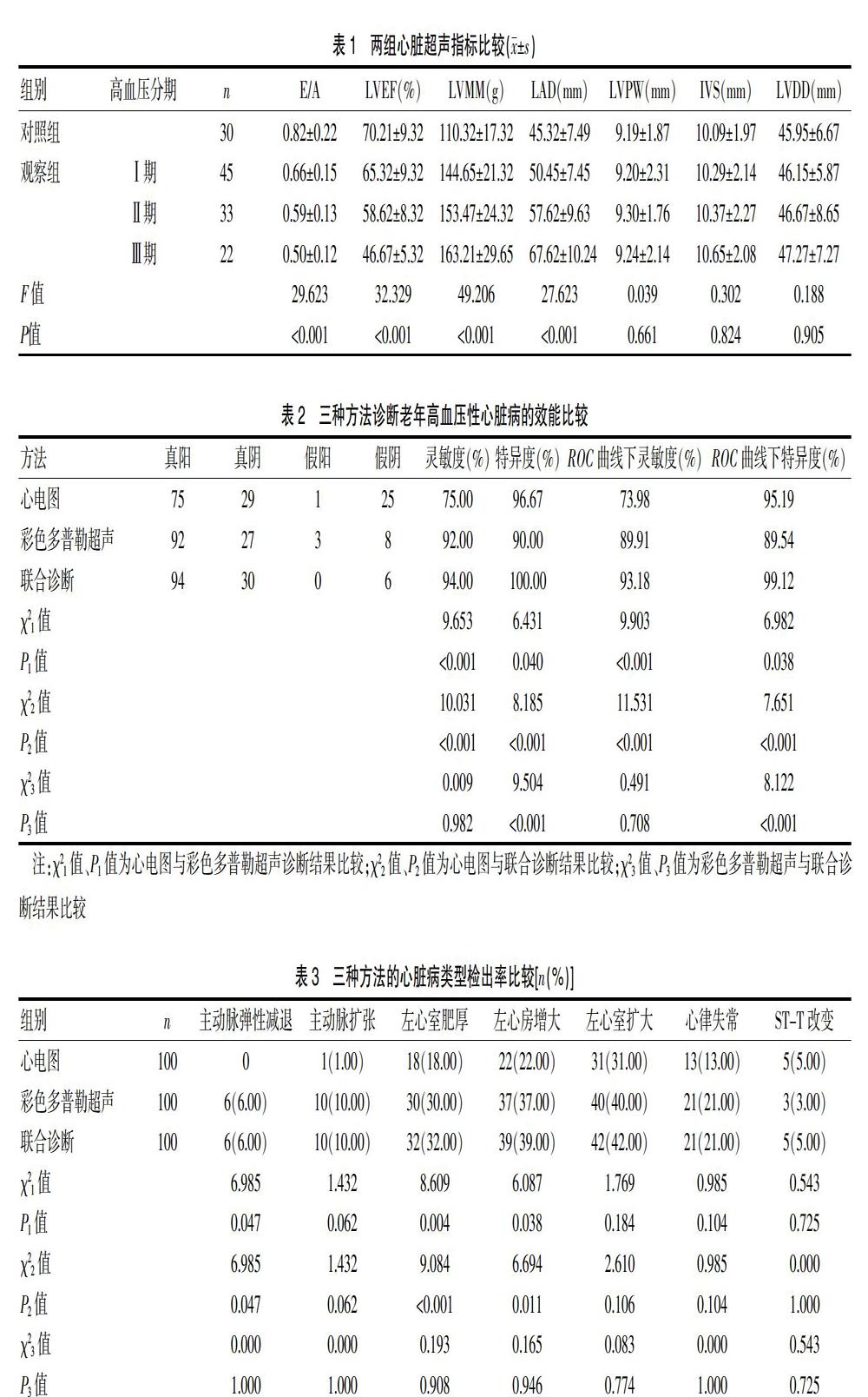
观察组与对照组、观察组各亚组间的E/A、LVEF、LVMM、LAD比较,差异均有统计学意义(P<0.05)。观察组与对照组、观察组各亚组间的LVPW、IVS、LVDD比较,差异无统计学意义(P>0.05)。见表1。
2.2 三种方法诊断老年高血压性心脏病的效能比较
联合诊断的灵敏度、特异度分别为94.00%、100.00%,高于心电图的75.00%、96.67%及彩色多普勒超声的92.00%、90.00%,三组比较,差异有统计学意义(P<0.05);联合诊断心脏病的ROC曲线下灵敏度、特异度分别为93.18%、99.12%,高于心电图的73.98%、95.19%及彩色多普勒超声的89.91%、89.54%,三组比较,差异有统计学意义(P<0.05)。见表2。心电图、彩色多普勒超声、联合诊断的AUC值分别为0.631、0.699、0.744。
