Hepatic isolated ectopic adrenocortical adenoma mimicking metastatic liver tumor
2021-03-05JingYiZhngYnLuoFeiLiuBoLi
Jing-Yi Zhng , Yn Luo , Fei Liu , , Bo Li
a Department of Ultrasound, West China Hospital, Sichuan University, Chengdu 610041, China
b Department of Liver Surgery, West China Hospital, Sichuan University, Chengdu 610041, China
A 55-year-old male had surgery for colon cancer in December 2017. A mass was found in the right posterior lobe of his liver in January 2019. There was no previous history of hepatitis, and all tumor markers were within normal ranges. Ultrasound examination found a 2.0 ×1.7 cm hypoechoic nodule with unclear boundaries and regular shape in hepatic segment VII. Contrast-enhanced ultrasound (CEUS) displayed hyperenhancement in the arterial phase and wash-out in the early portal phase, and the delayed phase was hypoenhanced ( Fig. 1 ). Contrast-enhanced computed tomography (CECT) presented a 1.5 cm slightly enhanced hepatic mass in the arterial phase and rim-enhancement in the portal phase ( Fig. 2 A-C). Magnetic resonance imaging (MRI) showed high intensity on T2W fat-suppressed scan and peripheral hyperintensity on contrast-enhanced T1W portal phase with no uptake of the contrast agent during the delayed phase ( Fig. 2 D-F), and diffusion weight was restricted in the right posterior upper hepatic segment. Preoperative diagnosis was metastatic carcinoma of the liver.
Laparoscopic local liver resection was performed. The incised hepatic specimen showed a gray-yellow and solid nodular mass adjacent to the liver capsule ( Fig. 3 A). The tumor was about 2.2 cm in diameter, and the boundaries were clear. After routine treatment of liver resection, the postoperative course was uneventful, and the patient was discharged at postoperative day 6. Immunohistochemical staining for melanoma antigen recognized by T-cells 1 (MART-1), synaptophysin (Syn), chromogranin A (CgA), calretinin (CR), and Ki-67 (<2%) was positive. Inhibin, pan-cytokeratin (PCK), epithelial membrane antigen (EMA), and hepatocytes were negative. Adrenal cortical adenoma originating from ectopic adrenal tissue in the liver was confirmed by postoperative histopathology ( Fig. 3 B). In addition, during imaging examinations at a 6-month follow-up appointment, the patient showed no manifestation of liver tumor recurrence.
Ectopic adrenal cortical adenoma rarely occurs in the liver. It can be divided into two types: functional and non-functional adenoma. The patient in this case had non-specific clinical symptoms, suggesting a high possibility of a non-functional tumor. At present, the image findings can only be used to distinguish intrahepatic adrenal cortical adenoma from the liver tumor [1]. A previous study suggested that imaging features of adrenal-gland-derived tumors are rooted by blood supply. The blood vessels are from the right adrenal vein to the inferior vena cava, and the tumor is connected to the medial limb of the right adrenal gland [2]. In this case, the possibility that the subcapsular tumor in the right posterior lobe was an ectopic adrenal tumor cannot be ruled out. It was near the adrenal vein or connected to the medial limb. A review of our images indicated that ultrasonography is not a reliable modality because only tumor-related images were obtained. It was not easy for CT and MRI to locate the right adrenal vein and medial limb. The connection between the tumor and the right adrenal gland was not initially detected.
Previous studies have reported that the ectopic adrenal cortical adenoma was misdiagnosed as hepatic malignancy [3–5]. Cases are often misdiagnosed as hepatocellular carcinoma when hepatic viral markers are positive, and the characteristic images show hyperarterial enhancement and wash-out in the portal vein phase on CECT or MRI [ 6 , 7 ]. Earlier reports [ 8 , 9 ]showed rim-enhancement on CECT and hypointensity with a subtle continuous perilesional rim in enhanced T1W which are the features of colorectal liver metastases. CEUS hyperenhancement in the arterial phase following a rapid wash-out indicates a high possibility of hepatic metastases [10]. The imaging findings of our case included wash-out in the early portal phase on CEUS, rim-enhancement on CECT, and peripheral hyperintensity on contrast-enhanced MRI. All of these images were the typical features of metastases in the liver. This patient had colon cancer one year earlier which also misled us to the diagnosis of a hepatic metastasis from colon cancer preoperatively.
In conclusion, when the lesion is adjacent to the right adrenal gland, the physician should keep “intrahepatic adrenocortical adenoma”in mind, even the images support the diagnosis of liver metastasis.

Fig. 1. The ultrasonic imaging features of ectopic adrenal cortical adenoma. A: Conventional ultrasound image showed a hypoechoic focal lesion with unclear boundary and regular shape (arrows). B: Arterial phase CEUS image showed hyperenhancement of the focal lesion (arrows). C: Early portal phase CEUS showed washed out of focal lesion(arrows). D: Delayed phase CEUS image showed hypoenhancement of the focal lesion (arrows). CEUS: contrast-enhanced ultrasound.

Fig. 2. The imaging features of ectopic adrenal cortical adenoma. A: Non-contrast enhanced CT showed a hypodense focal lesion (arrow). B: Arterial phase contrast-enhanced CT image demonstrated a slight enhancement of the focal lesion (arrow). C: Portal phase contrast-enhanced CT image showed rim-enhancement of the focal lesion (arrow).D: Fat-suppressed T2W MRI image showed a high intensity of focal lesion (arrow). E: Portal phase contrast-enhanced T1WI revealed peripheral hyperintensity of the focal lesion (arrow). F: Delayed phase contrast-enhanced T1WI showed a low signal intensity of focal lesion (arrow). CT: computed tomography; MRI: magnetic resonance imaging.
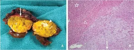
Fig. 3. Histopathology confirmed the mass to be an adrenal cortical adenoma. A: Specimen photograph showed a gray-yellow and solid nodular mass (arrowhead) with a complete tumor envelope (arrow). B: Photomicrograph (hematoxylin and eosin, ×200) showed a well-encapsulated nodule (arrow) surrounded by hepatocytes (star) and separated from adjacent perihepatic tissue by a fibrotic capsule (triangle) and hepatocytes.
Acknowledgments
None.
CRediT authorship contribution statement
Jing-Yi Zhang:Investigation, Resources, Software, Writing -original draft.Yan Luo:Conceptualization, Validation.Fei Liu:Funding acquisition, Resources, Software, Validation, Writing - review & editing.Bo Li:Conceptualization, Funding acquisition, Investigation, Writing - review & editing.
Funding
This study was supported by a grant from the Sichuan Science and Technology Program ( 2019YFS0370 ).
Ethical approval
This study was approved by the Ethics Committee of West China Hospital of Sichuan University and written informed consent was obtained from the patient.
Competing interest
No benefits in any form have been received or will be received from a commercial party related directly or indirectly to the subject of this article.
杂志排行
Hepatobiliary & Pancreatic Diseases International的其它文章
- Liver transplantation and BCLC classification: Limitations impede optimum treatment
- Do the existing staging systems for primary liver cancer apply to combined hepatocellular carcinoma-intrahepatic cholangiocarcinoma?
- Overlap of concurrent extrahepatic autoimmune diseases is associated with milder disease severity of newly diagnosed autoimmune hepatitis
- Aggressive surgical approach in patients with adrenal-only metastases from hepatocellular carcinoma enables higher survival rates than standard systemic therapy
- Integrating transcriptomes and somatic mutations to identify RNA methylation regulators as a prognostic marker in hepatocellular carcinoma
- The utility of two-dimensional shear wave elastography and texture analysis for monitoring liver fibrosis in rat model