Giant pyogenic granuloma on the glans penis
2020-12-28SheetnshuKumrTrunNrngBishnDssRdotrDipnkrDe
Sheetnshu Kumr , Trun Nrng ,*, Bishn Dss Rdotr ,Dipnkr De
a Department of Dermatology,Venereology and Leprology,Postgraduate Institute of Medical Education and Research, Chandigarh, India
b Department of Histopathology, Postgraduate Institute of Medical Education and Research,Chandigarh, India
A middle aged uncircumcised man presented with a painless growth on the glans penis which started 6 months back as a pinhead sized papule and rapidly progressed in size.The patient reported occasional bleeding from the lesion on minimal trauma.The patient was unable to perform sexual intercourse owing to the size of lesion and concern of bleeding.The patient was in monogamous relationship with his wife and denied any high risk sexual behaviour. On examination,there was a non-tender,oval,dark red,smooth,shiny, exophytic pedunculated growth of size 2.5 cm×2 cm×0.5 cm on the glans with a small but broad pedicle at the base of the lesion (Fig. 1). Histopathological examination of the mucosal biopsy from the lesion revealed lobular proliferation of capillaries with thinned out epithelium (Fig. 2). The diagnosis of penile pyogenic granuloma(PG)was reached on the basis of above findings.The lesion was referred to urology services and the lesion was surgically excised.
PG is an acquired benign vascular proliferation presenting as painless papule or nodule on skin or mucosa which sometimes bleeds spontaneously or on minimal trauma[1].The etio-pathogenesis of PG is largely unknown.It is considered to be a reactive phenomenon in which trauma, irritation or other unknown stimuli leads to angiogenesis and vascular proliferation [2]. Histopathological examination usually reveals lobules of small capillaries in a fibro-myxoid matrix with thinned out epidermis [2].
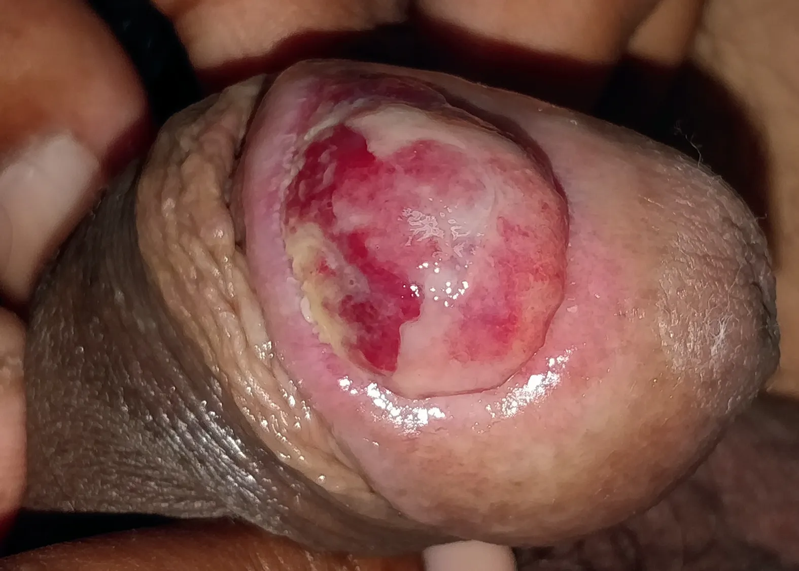
Figure 1 A dark red, smooth, shiny, oval, exophytic growth of size 3 cm × 2 cm × 0.5 cm on the glans penis with a small pedicle at the base of the lesion.
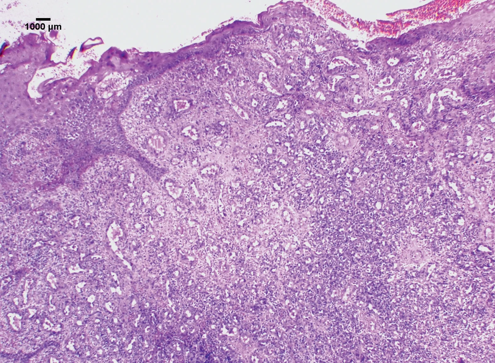
Figure 2 Histopathological examination(H&E,100×)of the mucosal biopsy from the lesion revealing lobular proliferation of capillaries with thinned out epithelium.
Penile PG is rare with only few reported cases in literature and the size of lesion rarely exceeds 1 cm[2-4].Although mostly asymptomatic, large penile PG may lead to significant concern among patients and partners owing to difficulty in performing sexual activity and episodes of bleeding. Although penile PG has usually been known to occur in adults, it has also been reported in pediatric age group [5]. Histopathology helps in differentiating penile PG from other differentials like genital warts and penile carcinoma. Early diagnosis, management and counselling are imperative. Awareness of this benign and easily manageable entity among clinicians will help in early diagnosis and management, thus sparing the patient of undue anxiety. Various treatment options are available for PG including surgical excision, cryotherapy, electrodessication, laser ablation and micro-embolization [1].Surgical excision and cryotherapy had been associated with low recurrence rates as compared to other modalities. Provision of histopathological evaluation and single step procedure make surgical excision the modality of choice especially for larger lesions [2].
To conclude, we present a case of giant penile PG managed successfully with surgical excision. Penile PG is a rare entity characterized usually asymptomatic papule or nodule which is associated with bleeding either spontaneously or minor trauma. Histopathology reveals lobules of small capillaries which helps in differentiating it from other differentials like genital warts and carcinoma. Surgical excision is treatment of choice owing to low risk of recurrence.Close and regular follow-up is imperative to monitor and manage recurrence at an earlier stage.
Author contributions
Study design:Sheetanshu Kumar, Tarun Narang, Dipankar De.
Data acquisition:Sheetanshu Kumar, Tarun Narang, Bishan Dass Radotra.
Drafting of manuscript:Sheetanshu Kumar, Tarun Narang.
Critical revision of the manuscript:BIshan Dass Radotra,Dipankar De.
Conflicts of interest
The authors declare no conflict of interest.
杂志排行
Asian Journal of Urology的其它文章
- Primary lymphomas of the genitourinary tract: A population-based study
- Xanthogranulomatous prostatitis:Impressive MRI appearance of a rare entity
- Huge urinoma caused by spontaneous ureteral rupture secondary to ureteral obstruction due to prostate cancer
- Cystoscopic extraction of an inadvertently placed ureteral stent in inferior vena cava
- Ambiguous clear cell carcinoma in medullary sponge kidney: A case report
- Investigation of confocal microscopy for differentiation of renal cell carcinoma versus benign tissue. Can an optical biopsy be performed?