Ambiguous clear cell carcinoma in medullary sponge kidney: A case report
2020-12-28YangChenTianyuLiJiwenCheng
Yang Chen, Tianyu Li,Jiwen Cheng
Department of Urology, The First Affiliated Hospital of Guangxi Medical University, Nanning, China
KEYWORDS Medullary sponge kidney;Renal clear cell carcinoma;Preoperative biopsy
Abstract Medullary sponge kidney (MSK) is a characteristic renal malformation, with a relatively low incidence. Radiologically, identification of MSK is sometimes ambiguous when compared to a renal mass. Here, we report a novel renal clear cell carcinoma in MSK, and discuss our approach to treatment. We recommended that a preoperative biopsy should be performed, followed by a comprehensive discussion regarding the appropriate perioperative preparations and careful surgical techniques that should be performed for this complex disease.
1. Introduction
Medullary sponge kidney (MSK), first described in 1939, is a renal malformation characterized by mineral deposits in the ectatic precalyceal papillary collecting ducts,including terminal ducts of Bellini and inner medullary collecting ducts [1-4]. Pathologically, there are two distinct features that are observed. Firstly, there are a large number of abnormal interstitial cells in the papillum with primitive characteristics, including a fibroblastic appearance. Secondly, there are cystic dilated medullary ducts with a single or multilayered epithelium, unlike normal medullary ducts which have a multilayered epithelium [4]. Although some cases of MSK have no obvious symptoms, generally there are certain common clinical manifestations, such as frequently recurring renal stones, nephrocalcinosis, hypercalciuria, hypocitraturia,microhematuria and macrohematuria, pyelonephritis,chronic kidney disease and abdominal pain[3,5-9].In the general population,the prevalence of MSK is thought to be 0.5%-1.0%, and may be more frequent in persons predisposed to form calcium stones [8,10,11]. Although computed tomography (CT) techniques with a high resolution can accurately diagnose MSK [12], there is sometimes confusion in the analysis of images which makes it hard to distinguish between MSK and renal tumors [13].Here, we present a clinical case of medullary sponge kidney without distinct clinical symptoms that was subsequently confirmed to also be a case of clear cell carcinoma.
2. Case report
A 56-year-old man presented with symptoms of renal colic for 1 month and was examined and determined to have a right renal mass. His medical history was significant for presentation of medullary sponge kidney, hyperuricemia,hypertension, and gouty arthritis. A physical examination presented with no obvious symptoms in the urinary system. There was however an obvious movement disorder,and swelling in the right ankle and knee. Moreover, his right index finger and middle finger tip joint presented with stiffness. This patient was first admitted to the local hospital 1 month ago for lumbago without gross hematuria. His radiological exam hinted at the presence of MSK(which mainly presented with renal calculi) and a suspicious right renal mass. Considering the complexity of his disease, the patient was admitted to our hospital for further treatment.
Upon admission, the plain film of the kidney, ureter,bladder (KUB) had the characteristic appearance of a“bouquet of flowers”. The CT scan in the excretory phase showed a medullary cyst in the right kidney (Fig. 1).Additionally, our CT scan revealed multiple kidney stones and a right renal mass (renal carcinoma) (Fig. 2). A ultrasound of the urinary system also identified a slightly echoic region in the right lower kidney. A routine blood examination presented moderate anemia (hemoglobin:86 g/L), while a renal function examination presented a serum creatinine level of 341 μmol/L and a uric acid level of 732 μmol/L.After treatment,serum creatinine and uric acid levels decreased to 288 μmol/L and 586 μmol/L,respectively. After discussion with other investigators, we concluded that the renal mass was nontypical with no apparent reinforcement in the CT scan (Fig. 2). To a certain extent, the possibility of renal malformations could not be eliminated, and thus we decided to initially perform a needle biopsy. The histopathology results identified as renal clear cell carcinoma. Considering the renal malformations, poor renal function, and history of kidney stones, we chose to perform robotic assisted nephron-sparing surgery (NSS) after discussion with the patient.The extracted tumor tissues are shown in Fig.3A.Also,histopathology confirmed the presence of clear renal cell cancer without adipose infiltration (Fig. 3B). Postoperatively, two double-J ureteral stents were placed for indwelling in the bilateral ureters to keep the ureter open. To address the renal colic and decline in renal function, we provided health education to the patient regarding blood pressure control, healthy diet, and uric acid control. Moreover, intake of potassium citrate was recommended for reduction in the risk of stones [8,14].Particularly, when the falling renal colic blocked the ureter, surgery was advised immediately.
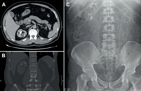
Figure 1 The radiological image in the computed tomography (CT) and kidney urinary bladder (KUB) radiograph. (A and B) Transverse section of the CT in excretory phase shows medullary cyst in the right kidney; (C) The Urinary tract plain X-ray has the characteristic appearance of a “bouquet of flowers” (We did not deal with the renal colic for without evident obstruction; postoperatively, two double-J ureteral stents were indwelling in bilateral ureters to keep the ureter open).
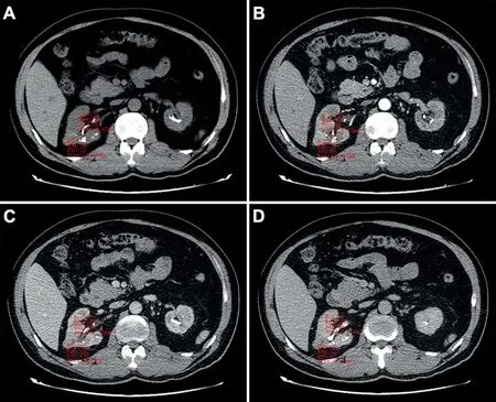
Figure 2 The figures of computed tomography in unenhanced phase (A), arterial phase (B), venous phase (C) and excretory phase (D) identified a mass in the right kidney. In four phases,the CT values were similar(unenhanced phase:29 for renal parenchyma,32 for mass;arterial phase:90 for renal parenchyma, 93 for mass; venous phase: 127 for renal parenchyma, 106 for mass; excretory phase: 69 for renal parenchyma, 67 for mass).
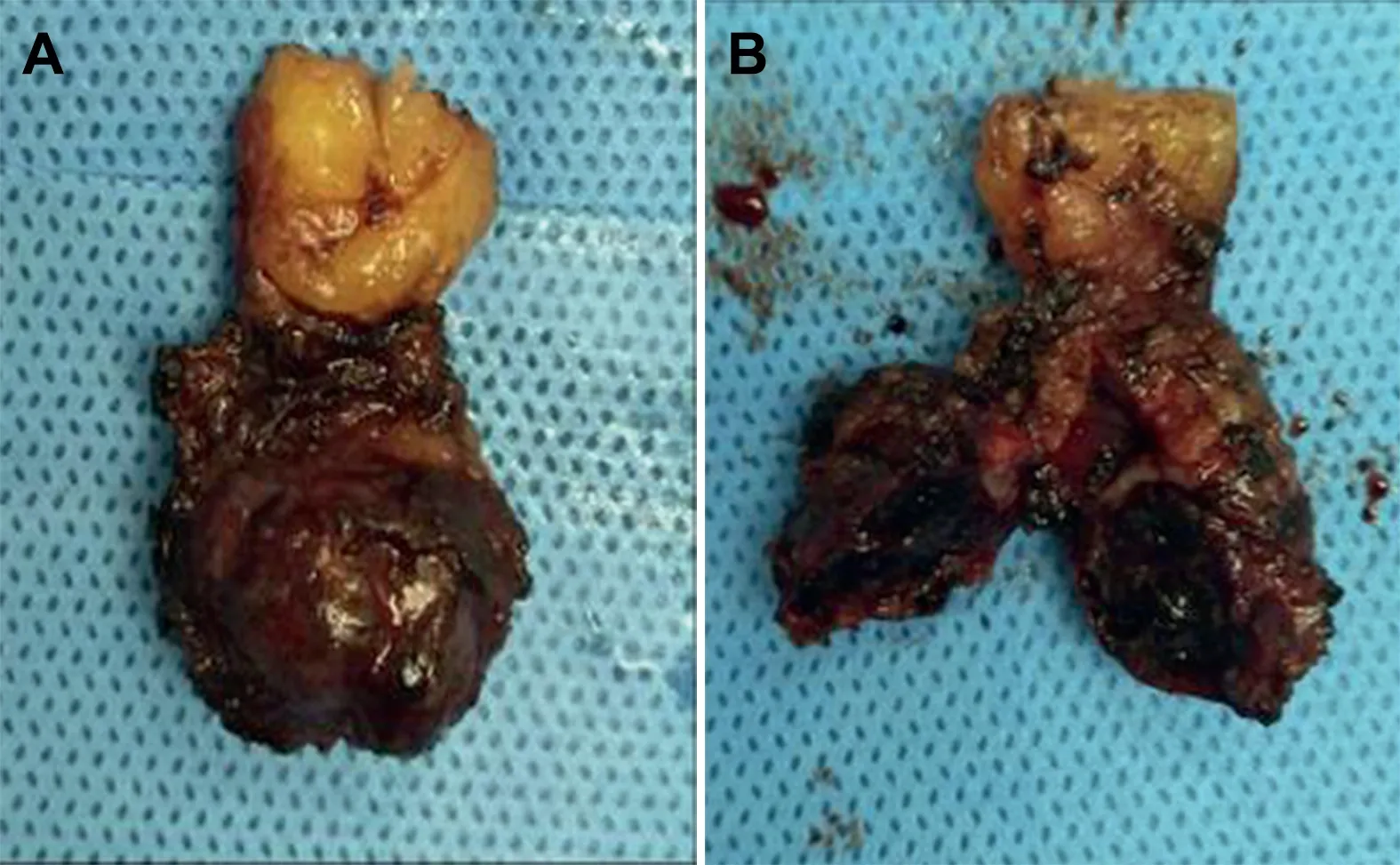
Figure 3 The gross pathology of tumor tissues. The tumor was removed integrally, without obvious capsule (A). When incising the tumor, the sarcodous tissues were shown (B).
3. Discussion
MSK is a specialized renal disease with an abnormal structure. In the past, MSK was diagnosed by intravenous pyelography (IVP). However, some cases might be misdiagnosed by this method due to poor image quality resulting from disturbances by bowel gas and stool, and non-detection of small stones[12,15].A better choice is the use of CT, which has recently been applied to uncover obvious “cystic” dilations of the prepapillary collecting ducts [3]. However, it is also thought that CT sometimes does not have enough spatial resolution to reveal the most common forms of MSK [3]. Thus, to a certain extent the diagnosis of MSK can be confusing.
In certain cases, atypical images with radiography can also make it hard to easily identify MSK and renal masses.In 1989, Kaver et al. [13] reported on a segmental MSK which mimicked a renal mass, while in 2005 Fellegara et al.[16]also encountered a case of MSK that was similar to a renal tumor. Interestingly, in 1991, Choong and Phillips [17] conversely observed a case of renal transitional cell carcinoma that mimicked MSK (Table 1). These reports suggest that the differential diagnosis between MSK and a renal tumor may be sometimes very difficult without characteristic radiographic findings.In this report,we also presented a renal clear cell carcinoma that had been previously diagnosed as MSK.Preoperatively,the observed radiological features were nontypical for confirmation of a renal mass(without obvious reinforcement in the contrast CT) (Fig. 2). Thus, after discussions with other investigators and the patient, we performed a puncture biopsy, which finally confirmed a diagnosis of renal clear cell carcinoma. This result gave us enough information to support the subsequent surgery. Renal cell carcinoma(RCC) is a widespread malignancy with 15 distinct histologic subtypes, among which renal clear cell carcinoma(RCCC) is the most common (over 70%) [18,19]. Many therapeutic options are available for RCCC [3,20]. In the case of RCCC with a localized status (localized RCCC and locally advanced RCCC), surgery is strongly recommended. For advanced/metastatic cases, cytoreductive nephrectomy and other methods such as immunotherapy and chemotherapy are suggested. In our case, there was no obvious evidence of metastatic disease. In addition,the presence of poor renal function preoperatively pushed us to perform the NSS to protect the residual nephrons and their function. Moreover, robotic assisted surgery guaranteed the precision of the process.

4. Conclusion
In summary,a preoperative biopsy,full communication and discussion with physicians and patients, perioperative preparation, and a careful operation are recommended for these complex diseases.
Conflicts of interest
The authors declare no conflict of interest.
Author contributions
Study concept and design:Jiwen Cheng, Tianyu Li.
Data acquisition:Yang Chen.
Drafting of manuscript:Yang Chen,Tianyu Li,Jiwen Cheng.
Critical revision of the manuscript:Jiwen Cheng.
杂志排行

Asian Journal of Urology的其它文章
- Primary lymphomas of the genitourinary tract: A population-based study
- Xanthogranulomatous prostatitis:Impressive MRI appearance of a rare entity
- Giant pyogenic granuloma on the glans penis
- Huge urinoma caused by spontaneous ureteral rupture secondary to ureteral obstruction due to prostate cancer
- Cystoscopic extraction of an inadvertently placed ureteral stent in inferior vena cava
- Investigation of confocal microscopy for differentiation of renal cell carcinoma versus benign tissue. Can an optical biopsy be performed?