Minimizing electrostatic interactions from piezoresponse force microscopy via capacitive excitation
2020-03-27QingfengZhuEhsanNasrEsfahaniShuhongXieJiangyuLi
Qingfeng Zhu, Ehsan Nasr Esfahani, Shuhong Xie*, Jiangyu Li,*
a Shenzhen Key Laboratory of Nanobiomechanics, Shenzhen Institutes of Advanced Technology, Chinese Academy of Sciences, Shenzhen 518055, China
b Hunan Provincial Key Laboratory of Thin Film Materials and Devices, Xiangtan University, Xiangtan 411105, China
c Department of Mechanical Engineering, University of Washington, Seattle 98195, WA, USA
d Key Laboratory of Low Dimensional Materials and Application Technology of Ministry of Education, and School of Materials Science and Engineering, Xiangtan University, Xiangtan 411105, China
Keywords:Piezoresponse force microscopy Electrostatic interactions Capacitive excitation
ABSTRACT Piezoresponse force microscopy (PFM) has emerged as one of the most powerful techniques to probe ferroelectric materials at the nanoscale, yet it has been increasingly recognized that piezoresponse measured by PFM is often influenced by electrostatic interactions. In this letter, we report a capacitive excitation PFM (ce-PFM) to minimize the electrostatic interactions. The effectiveness of ce-PFM in minimizing electrostatic interactions is demonstrated by comparing the piezoresponse and the effective piezoelectric coefficient measured by ce-PFM and conventional PFM. The effectiveness is further confirmed through the ferroelectric domain pattern imaged via ce-PFM and conventional PFM in vertical modes, with the corresponding domain contrast obtained by ce-PFM is sharper than conventional PFM. These results demonstrate ce-PFM as an effective tool to minimize the interference from electrostatic interactions and to image ferroelectric domain pattern, and it can be easily implemented in conventional atomic force microscope (AFM)setup to probe true piezoelectricity at the nanoscale.
Spontaneous polarization in ferroelectrics arises from the collective ordering of dipoles, and thus exhibits interfacial effects at the domain walls and size effects at the nanoscale when the long-range symmetry is broken [1-3]. Nanostructured ferroelectrics have thus attracted considerable interests for their exotic topological domains [4, 5] as well as potential device applications [6, 7]. Piezoresponse force microscopy (PFM) is one of the most established tools to study ferroelectrics at the nanoscale,yet it has recently been realized that apparent piezoresponse measured by PFM can be effected by the electrostatic interactions between the atomic force microscope (AFM) tip/cantilever and the sample surface [8-15]. This makes it difficult to probe the true ferroelectricity and piezoelectricity at the nanoscale,and it is highly desirable to minimize electrostatic contributions to the piezoresponse signals measured by PFM. In the past few years, there has been various attempts to address this issue,though significant challenges remain [16-21]. In this work, we report a capacitive excitation PFM technique, termed as ce-PFM,that is effective in minimizing electrostatic contributions to the piezoresponse signals and can be easily implemented in a conventional AFM setup.
PFM is a contact mode AFM technique that is based on converse piezoelectric effect. In a conventional PFM setup, a periodic bias V =VACcos(ωt) with frequency ω and amplitude VACis applied to the sample through a conductive cantilever tip, and the local oscillation of electric field under the tip induces piezoelectric vibration of the sample that is measured by the first harmonic component Aωof cantilever deflection A =Aωcosdriven by the sample, where the phase φ is dependent on polarization direction. Beside this “true” piezoelectric response, there exists other non-piezoelectric contributions to the deflection signal [22]. Since the tip is conductive, there is inevitably electrostatic interactions between the probe and the sample, resulting in artifacts in measured piezoresponse, and very often the adsorbents on the sample surface make the problem even worse [8,23, 24].
To overcome these difficulties, we developed ce-PFM as schematically shown in Fig. 1a, wherein the sample on an insulating substrate is put on top of a metal disc subjected to an alternating current (AC) voltage that serves as excitation source.The setup is similar with a capacitor structure and thus be named as capacitive excitation PFM. The metal disc induces an AC electric field according to Gauss's law, as confirmed by our finite element method (FEM) simulation in Fig. 1b. This electric field in turn induces piezoelectric vibration of the sample that can be measured locally by the cantilever deflection. Indeed,when the excitation frequency sweeps around the tip-sample contact resonance frequency, the deflection response exhibits a clear resonance peak for a LiNbO3crystal as shown in Fig. 1c.Note that the cantilever is non-conductive and is only used for deflection detection here. Hence electrostatic interactions can be minimized in ce-PFM. Importantly, the method is distinct from background-free PFM reported by Wang et al. [18], which excites the piezoresponse from substrate with a grounded conductive tip.
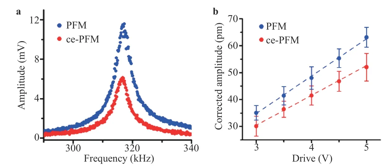
Will the proposed method minimize electrostatic contributions to piezoresponse measured? To answer this question, we compare piezoresponse measured on a classical ferroelectric material LiNbO3using conventional PFM and ce-PFM, as shown in Fig. 2. As seen in Fig. 2a, LiNbO3exhibits a much larger piezoresponse peak measured by conventional PFM than ce-PFM. Since the strength of electric field generated by the tip in conventional PFM and the disc in ce-PFM is different, the smaller piezoresponse measured by ce-PFM can be the result of combination of less concentrated electric field and minimized electrostatic interactions.
The effectiveness of ce-PFM is made more evident by comparing the intrinsic PFM and ce-PFM responses as a function of the applied excitation voltage averaged over 9 randomly chosen points, obtained by fitting the measured deflection response to a simple harmonic oscillator model [25-27], as show in Fig. 2b. It is observed that for LiNbO3, the conventional PFM and ce-PFM yield similar trend, showing clear linear behavior as expected from piezoelectricity, and the effective piezoelectric coefficient estimated from measured slopes are comparable at 13.90±0.32 and 10.87±0.28 pm/V, respectively. This establishes without ambiguity that the electrostatic contribution is indeed minimized by ce-PFM.

Fig. 1. Principle of ce-PFM. a Schematic of ce-PFM set-up; b finite element simulation of electric potential distribution under ce-PFM configuration with a positive applied bias; c piezoresponse of LiNbO3 versus excitation frequency measured by resonance-enhanced ce-PFM and analyzed by simple harmonic oscillator (SHO) model, with excitation .

Fig. 2. Comparison of vertical piezoresponses obtained from resonance-enhanced a conventional PFM and ce-PFM on LiNbO3, b intrinsic piezoresponse versus the excitation voltage for conventional PFM and ce-PFM. (1 pm = 1×10-12 m)

Fig. 3. Comparison of LiNbO3 domain pattern imaged by vertical PFM and ce-PFM. a PFM amplitude; b PFM phase; c ce-PFM amplitude; d ce-PFM phase mappings. The excitation voltage is 10 V for all the mappings.

Fig. 4. Domain pattern imaged via lateral ce-PFM. a FEM simulation of lateral displacement; b lateral ce-PFM amplitude; c lateral ce-PFM phase; d corresponding line scan profiles across the regions of simulation (blue line) and experiment (red line); the cantilever was aligned parallel to y-axis when measuring the.
One of the key applications of PFM is to image domain structure of ferroelectric materials, and this can be accomplished by ce-PFM as well. To demonstrate this capability, we choose periodically poled LiNbO3as an example, which exhibits lamellar 180° domains as revealed by conventional PFM mappings of amplitude and phase in Fig. 3a and 3b measured at non-resonant frequency. Note that the PFM amplitude is not perfectly symmetric across the domain walls, probably due to long-range electrostatic interactions between the conductive tip and the sample[8, 28]. The ce-PFM, on the other hand, reveals a clear domain pattern with more symmetric amplitude response and 180°phase reversal across the domain walls (Fig. 3c and 3d)), and the contrast in ce-PFM amplitude mapping is sharper than that of conventional PFM, especially near domain walls, since no electrical interferences exist between the sample and the cantilever.This proves that ce-PFM can image ferroelectric domain structures well. An interesting observation from Fig. 3b and 3d is that there is a 180° phase flip for the same domain imaged by PFM and ce-PFM due to opposite electric polarity between tip-biased PFM and disc-biased ce-PFM (Fig. 1). Note that both PFM and ce-PFM mappings were obtained using a low excitation frequency away from resonance, and thus phase values are physically meaningful [29].
It is also interesting to note that the same domain pattern can be visualized by lateral ce-PFM response, as shown in Fig. 4.Simulation by FEM in Fig. 4a reveals that even though the polarization is vertical, there is lateral displacement resulting from shear strain due to mechanical constraints between domains,and the shear strain has alternating signs at domain walls, positive at a ⊗|⊙ wall and negative at a ⊙|⊗ wall. This prediction is indeed confirmed by our lateral ce-PFM imaging, as shown in Fig.4b and 4c for the amplitude and phase mappings. The corresponding line scan in Fig. 4d shows qualitative agreement between simulation and experiment, wherein the experimental data is obtained by averaging 8 line-scans around the red line in Fig. 4b and 4c. Similar trend was also observed via conventional PFM [30, 31].
In summary, we have developed ce-PFM to probe and image true piezoresponse at the nanoscale with minimized artifacts and cross-talks, overcoming one of the main difficulties in conventional PFM that the PFM signal is often influenced by electrostatic interaction. We accomplish these by using nonconductive probe to detect the piezoelectric vibration of the sample, while the excitation is realized through capacitive effect using a metal disc underneath. This provides an effective tool to minimize electrostatic interaction, and it can be easily implemented in conventional AFM setup to probe true piezoelectric materials at the nanoscale.
Supplementary Material
See supplementary material for FEM simulations, PFM, and ce-PFM technique details.
Acknowledgement
We acknowledge the National Key Research and Development Program of China (Grant 2016YFA0201001), the National Natural Science Foundation of China (Grants 11372268,11627801, and 1472236), Unite State National Science Foundation (Grant CBET-1435968), the Leading Talents Program of Guangdong Province (Grant 2016LJ06C372), and Shenzhen Science and Technology Innovation Committee (Grant KQJSCX20170331162214306).
杂志排行
Theoretical & Applied Mechanics Letters的其它文章
- Micromechanical analysis on tensile properties prediction of discontinuous randomized zalacca fibre/high-density polyethylene composites under critical fibre length
- Neurodynamics analysis of cochlear hair cell activity
- Prolonged simulation of near-free surface underwater explosion based on Eulerian finite element method
- Spatial artificial neural network model for subgrid-scale stress and heat flux of compressible turbulence
- An analytical model to predict diffusion induced intermetallic compounds growth in Cu-Sn-Cu sandwich structures
- Molecular investigation on the compatibility of epoxy resin with liquid oxygen
