高血压性脑出血患者发病24h内血肿扩大的危险因素分析:一项单中心256例回顾性研究
2015-06-28王大永徐翔郭建文
王大永,徐翔,郭建文
高血压性脑出血患者发病24h内血肿扩大的危险因素分析:一项单中心256例回顾性研究
王大永,徐翔,郭建文
目的研究发病24h内的急性高血压性脑出血(AHCH)患者发生血肿扩大的危险因素。方法回顾性分析2008年3月-2013年3月广东省中医院符合纳入排除标准的AHCH患者256例的病例资料。收集患者的一般情况、既往病史、个人史、临床特点、CT检查结果、实验室检查指标、活血化瘀类中药使用情况。根据血肿扩大与否分为血肿扩大组与非血肿扩大组。首先对各项指标进行单因素分析,然后将经过单因素分析有统计学意义的因素作为自变量,血肿扩大结果作为因变量,采用多因素logistic回归分析法分析脑出血患者早期血肿扩大的独立相关因素。将各危险因素和血肿扩大作为自变量,3个月随访时以mRS(modified Rankin Scale)量表作为因变量,采用logistic回归分析急性期血肿扩大是否影响患者3个月的预后。mRS 0~2级为恢复良好,3~6级为严重致残或死亡。检验水准取α=0.05。结果256例患者中,有43例发生了血肿扩大,血肿扩大发生率为16.80%。单因素分析结果显示,男性、入院GCS评分、入院NIHSS评分、病程、天冬氨酸转氨酶(AST)是导致血肿扩大的危险因素,而多因素分析则显示,仅男性、病程是导致血肿扩大的独立危险因素。此外,logistic回归分析结果显示血肿扩大是影响患者结局的独立危险因素,入院时血肿体积、病程、NIHSS评分也是影响患者3个月随访结局的独立危险因素。结论男性、发病到入院时间较短(2h内)的患者应警惕血肿扩大的风险,而出血量大、血肿再次扩大、高NIHSS评分、发病时间短则提示患者预后不良。
脑出血;血肿扩大;相关因素
脑出血(intracerebral hemorrhage,ICH)即原发性脑实质出血(spontaneous intracerebral hemorrhage,SICH)具有高发病率,高致残率,高病死率的特点,危害极大[1]。ICH病死率在所有卒中类型中最高,其30d死亡发生率高达30%~50%,75% ICH患者1年后会遗留严重残疾甚至死亡[2]。脑出血超急性期血肿扩大的发生率为12.3%~58.9%[3-6],大部分发生在24h内。血肿扩大会使ICH患者预后变差[7-8],神经功能恶化,病死率增高[9]。
对于急性脑出血于发病24h内发生血肿扩大的影响因素,由于不同研究者的研究方法不同,得到的结论也不尽相同[10]。临床实际工作中,患者往往由多种危险因素导致脑血肿扩大。本研究采用多因素logistic回归分析方法,综合分析256例急性高血压性脑出血患者于发病24h内发生血肿扩大的影响因素及其预后,以期为临床工作提供帮助。
1 资料与方法
1.1 研究对象 回顾性分析2008年3月-2013年3月广东省中医院神经科确诊为高血压性脑出血的住院患者。高血压性脑出血诊断标准采用1995年全国第四届脑血管病会议制定的关于高血压性脑出血的诊断标准[11],所有病例均经头颅CT平扫确诊。纳入标准为:①符合第四届全国脑血管病学术会议制订的诊断标准,并经头颅CT证实为脑出血;②发病在24h内;③入院后24h内行第2次头颅CT检查。排除标准为:①发病时间大于24h;②因脑外伤、肿瘤、动脉瘤、动静脉畸形、烟雾病等所致的脑出血;③因手术或死亡等原因未能行第2次CT检查。剔除标准为诊断不明确、资料不全等影响结果判定者。
1.2 研究方法 查阅收集患者的一般情况、既往病史、个人史、临床特点、CT检查结果、实验室检查结果、活血化瘀类中药使用情况。头颅CT血肿大小的测量采用多田公式法,即血肿体积=血肿的最大长径(A)×宽(B)×层数(C)/2。脑出血早期血肿扩大标准采用Brott标准[12],即脑出血患者第2次CT检查血肿体积比首次CT检查>33%。由两位资深神经影像医师在CT扫描工作站独立分析。根据首次CT检查后血肿扩大与否分为血肿扩大组与非血肿扩大组。首先对各研究指标进行单因素分析,然后将经过单因素分析有统计学意义的因素及早期使用活血化瘀中药情况作为自变量,血肿扩大作为因变量,采用多因素logistic回归分析法分析高血压脑出血患者早期血肿扩大的独立相关因素;进一步以发病3个月随访的mRS(modified Rankin Scale)为因变量,其他危险因素和血肿扩大为自变量,采用多因素logistic回归分析法分析急性期血肿扩大是否影响患者3个月的预后。
1.3 统计学处理 采用SPSS 19.0软件进行统计分析。首先进行单因素分析,计量资料符合正态分布、方差齐者,以表示,组间比较采用t检验,方差不齐者采用校正t'检验;非正态分布或方差不齐,以Md±QR表示,采用秩和检验;计数资料以构成比或率表示,组间比较用χ2检验或Fisher法检验;等级资料采用秩和检验。在多因素分析中,将经过单因素分析有统计学意义的因素作为自变量,血肿扩大作为因变量,采用二分类非条件logistic回归分析法分析脑出血患者早期血肿扩大的独立危险因素。另外,将上述危险因素和血肿扩大作为自变量,3个月mRS为因变量,logistic回归分析法分析急性期血肿扩大是否影响患者3个月的预后。mRS 0~2级为恢复良好,3~6为严重致残或死亡。检验水准取α=0.05。
2 结 果
2.1 病例资料 研究人员检索病历,纳入2008年3月-2013年3月符合诊断标准的脑出血病例共901例,根据纳入标准、排除标准、剔除标准,去除出血性脑梗死31例,脑动静脉畸形43例,脑动脉瘤19例,脑肿瘤9例,脑外伤8例,因医生未开CT检查项目、患者经济原因或入院后死亡等原因而未能在24h后行第2次头颅CT检查者共70例,入院24h内行手术治疗108例,病程为24h~2周的患者95例,恢复期(2周<病程<6个月)患者96例,后遗症期(病程>6个月)患者166例,最终筛选出符合研究方案要求的病例256例(图1)。
2.2 血肿扩大的发生率 纳入患者256例,年龄21~91岁,其中男173例,年龄64.0±22.0岁;女83例,年龄69.0±22.0岁。纳入的256例患者中,43例发生了血肿扩大,非血肿扩大组213例。血肿扩大发生率为16.8%。

图1 901例脑出血患者纳入流程图Fig. 1 Flow chart of 901 ICH patients
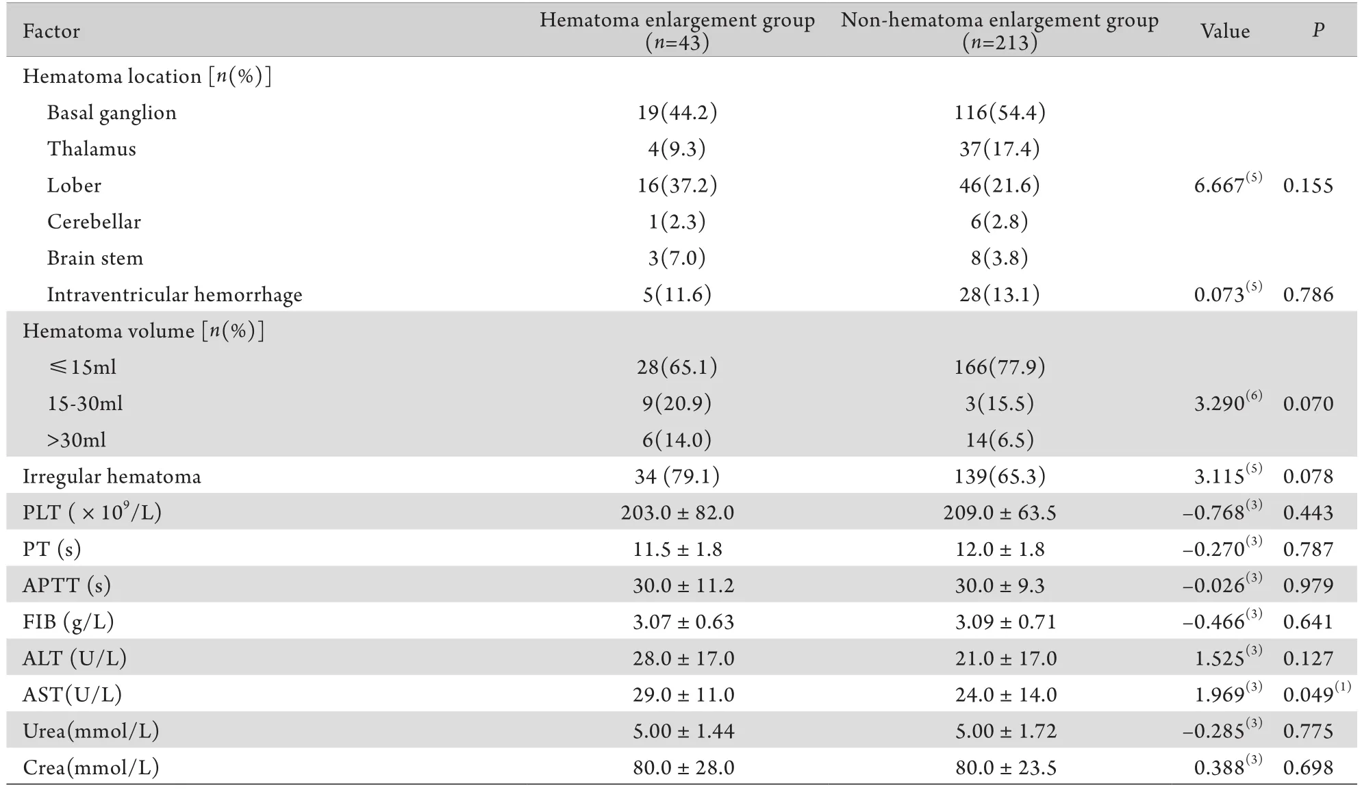
2.3 血肿扩大各研究指标的单因素分析 单因素分析发现,差异有统计学意义的研究指标包括男性、入院GCS评分、入院NIHSS评分、病程、天冬氨酸转氨酶(AST)等(表1)。
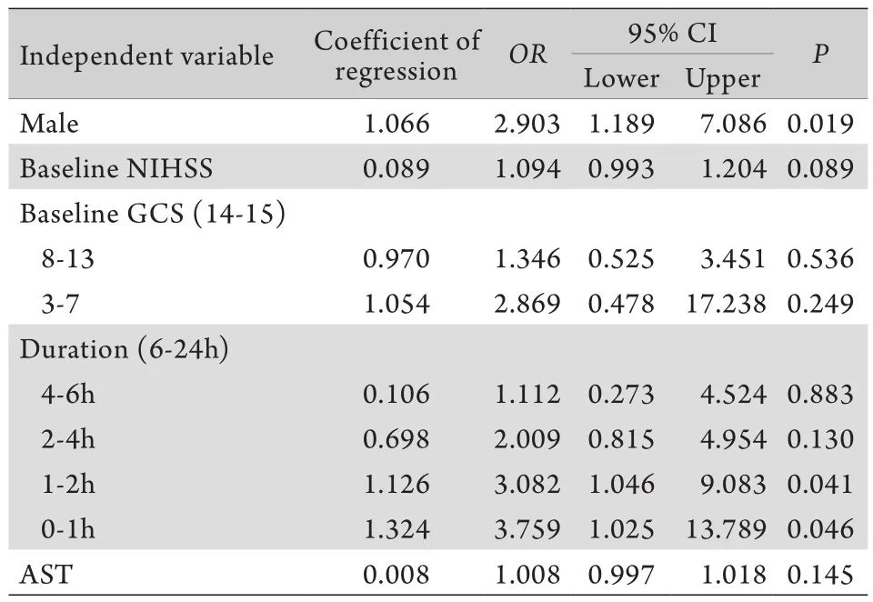
2.4 血肿扩大的多因素分析 将单因素分析有统计学意义的因素(男性、入院GCS评分、入院NIHSS评分、病程、AST)作为自变量,血肿扩大作为因变量,采用多因素Logistic回归分析脑出血患者早期血肿扩大的独立相关因素(表2)。结果显示有2个血肿扩大的独立危险因素:男性(OR=2.903,95%CI 1.189~7.086,P=0.019),发病距离首次CT检查时间[0~1h(OR=3.759,95%CI 1.025~13.789,P=0.046),1~2h(OR=3.082,95%CI 1.046~9.083,P=0.041)]。
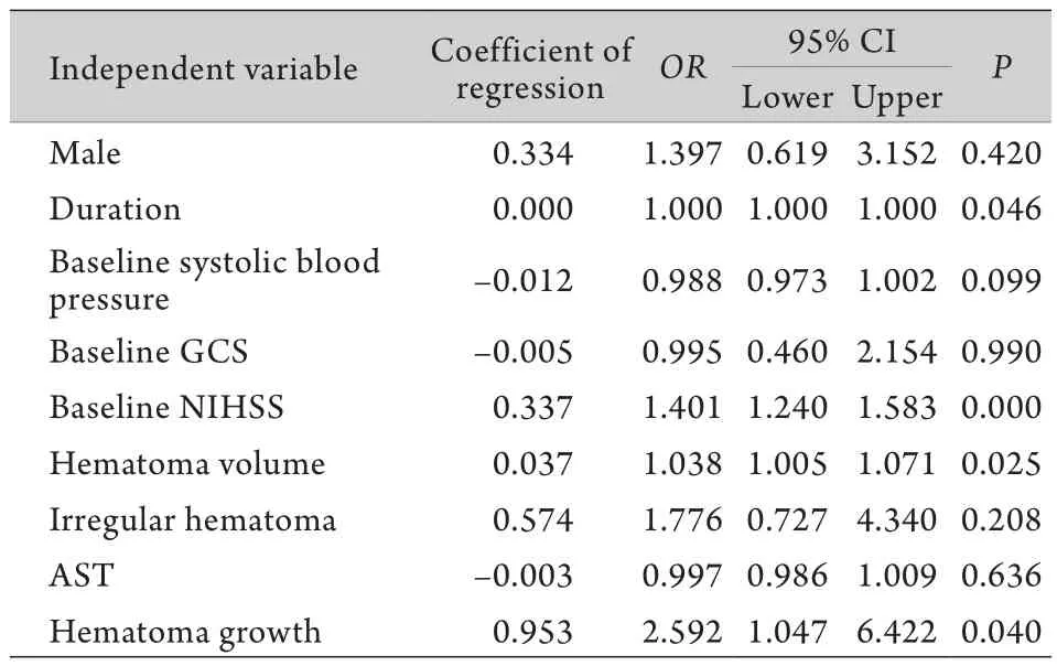
2.5 影响脑出血患者3个月随访mRS独立危险因素的多因素分析 将单因素分析有统计学意义的因素(男性、入院GCS评分、入院NIHSS评分、病程、AST)和血肿扩大作为自变量,3个月随访结局作为因变量。根据mRS评分,mRS 0~2分为生活独立,3~6分为生活依赖或死亡。采用多因素logistic回归分析影响脑出血患者3个月随访mRS的独立危险因素(表3)。结果显示有4个血肿扩大的独立危险因素,分别是发病到入院时间(OR=1.000,95%CI 1.000~1.000,P=0.046)、入院时NIHSS评分(OR=1.401,95%CI 1.240~1.583,P=0.000),首次CT显示的脑出血量(OR=1.038,95%CI 1.005~1.071,P=0.025),血肿是否扩大(OR=2.593,95%CI 1.047~6.422,P=0.040)。

表1 脑出血早期血肿扩大各指标的单因素分析结果Tab. 1 The univariate analysis on the hematoma enlargement

(续表)

表2 256例脑出血患者早期血肿扩大独立危险因素的logistic分析Tab. 2 Multivariate logistic regression analysis on the independent risk factors of hematoma enlargement in 256 patients

表3 影响脑出血患者3个月随访mRS独立危险因素的多因素logistic分析Tab. 3 Multivariate logistic regression analysis on the independent risk factors of 3 months outcome
3 讨 论
本研究纳入256例患者,其中43例在第1次头颅CT检查后发生了血肿扩大,血肿扩大发生率为16.8%,接近Fujii等[13]报道的14.0%,比Kazui等[14]报道的36%稍偏低,原因可能有:①采用的血肿扩大标准不一致:本研究采用的是Brott标准[12],即脑出血患者第2次CT检查血肿体积比首次CT检查>33%即可定义为血肿扩大,而Kazui等[14]研究采用的标准为两次CT检查血肿体积变化量V2-V1≥12.5cm3或者两次CT检查血肿体积比V2/ V1≥1.4。②纳入脑出血患者的病程有所区别:Kazui等纳入的脑出血患者病程在48h内,本研究纳入的脑出血患者为发病24h内。③观察时间长短不同:Kazui等[14]在入院后72h复查CT,本研究在入院后24h复查CT。④有部分患者入院后因死亡等原因而未能在24h后行第2次头颅CT检查,而有部分患者入院24h内行手术治疗,这些死亡或者手术的患者有发生血肿扩大的可能性,本研究为设计的严谨未能纳入这部分患者,有可能导致一部分血肿扩大的患者漏诊,因此实际的血肿扩大发生率要比研究结果16.8%高得多。
本研究单因素分析结果显示,男性、入院GCS评分、入院NIHSS评分、病程、肝功能(AST)是导致血肿扩大的危险因素,而多因素分析结果则显示,仅男性、病程是导致血肿扩大的独立危险因素。血肿扩大是影响患者结局的独立危险因素,此外,logistic分析显示除血肿扩大之外,入院时血肿体积、病程、NIHSS评分也是影响患者3个月随访结局的独立危险因素。本研究结果提示,男性、发病到入院时间较短(2h内)的患者应警惕血肿扩大的风险,而出血量大,血肿再次扩大、高NIHSS评分、发病时间短则提示患者预后不良。
目前关于高血压性脑出血血肿扩大危险因素的研究很多,研究指标主要有血肿体积、血肿形态、血肿部位、CT斑点征、高NIHSS评分、低GCS评分、首次CT检查距发病时间、血压水平、肝功能、酗酒、血糖等[3,7-9]。研究方法大都局限于采用单因素分析,较少使用多因素回归等统计方法。现实的情况是,某个患者具有多种危险因素,单因素分析往往难以反映临床实际。多因素logistic回归分析可以真实的反映临床患者多重因素的作用,更加贴近临床。
本研究的局限在于是回顾性研究,缺失病例偏多,某些量表评分缺如,只能用回顾性测评代替,样本数少且为单中心样本,在后续的研究中有待完善。
[1] Qureshi AI, Mendelow AD, Hanley DF. Intracerebral haemorrhage[J]. Lancet, 2009, 373(9675): 1632-1644.
[2] van Asch CJ, Luitse MJ, Rinkel GJ,et al. Incidence, case fatality, and functional outcome of intracerebral haemorrhage over time, according to age, sex, and ethnic origin: a systematic review and meta-analysis[J]. Lancet Neurol, 2010, 9(2): 167-176.
[3] Lim JK, Hwang HS, Cho BM,et al. Multivariate analysis of risk factors of hematoma expansion in spontaneous intracerebral hemorrhage[J]. Surg Neueol, 2008, 69(1): 40-45.
[4] Qin HX. The clinical analysis of risk factors on early hematoma enlargement incerebral hemorrhage[J]. Med J West Chin, 2013, 25(1): 93-95. [覃惠洵. 脑出血早期血肿扩大危险因素临床分析[J]. 西部医学, 2013, 25(1): 93-95. ].
[5] Li ZX, Chu XF, Dou RX,et al. Dangerous factors of hematoma enlargement in spontaneous intracerebral hemorrhage after the evaluation of the irregular shape of hematoma[J]. J Apop Nerv Dis, 2011, 28(2): 151-155. [李卓星, 褚晓凡, 窦汝香, 等. 量化评价血肿形态对脑出血血肿扩大危险因素分析[J]. 中风与神经疾病杂志, 2011, 28(2): 151-155.]
[6] Li Q, Liu ZB, Pan XC,et al. The analysis of the clinical features and risk factors on hematoma enlargement inpatients with intracerebral hemorrhage[J]. J Brain Nerv Dis, 2011, 19(6): 453-456. [李旗, 刘振宝, 潘晓春, 等. 脑出血早期血肿扩大的临床特点及相关因素分析[J]. 脑与神经疾病杂志, 2011, 19(6): 453-456. ]
[7] Davis SM, Broderick J, Hennerici M,et al. Hematoma growth is a determinant of mortality and poor outcome after intracerebral hemorrhage[J]. Neurology, 2006, 66(8): 1175-1181.
[8] Tuhrim S, Horowitz DR, Sacher M,et al. Volume of ventricular blood is an important determinant of outcome in supratentorial intracerebral hemorrhage[J]. Crit Care Med, 1999, 27(3): 617-621.
[9] Leira R, Davalos A, Silva Y,et al. Early neurologic deterioration in intracerebral hemorrhage: predictors and associated factors[J]. Neurology, 2004, 63(3): 461-467.
[10] Wang GQ, Li SQ, Zhang WW,et al. Can minimal invasive puncture and drainage for hypertension spontaneous basal ganglia intracerebral hemorrhage improve patient outcome: A prospective non-randomized comparative study[J]. Med J Chin PLA, 2014, 39(7): 531-541. [王国强, 李世强, 张微微, 等. 微创穿刺引流对高血压自发基底神经节区脑出血预后的影响——前瞻性非随机对照研究[J]. 解放军医学杂志, 2014, 39(7): 531-541.]
[11] Chinese Society for Neuroscience, CSN Chin J Neurol. Key points of cerebrovascular diseases diagnosis[J]. Chin J Neurol, 1996, 29(6): 379-380. [中华神经科学会, 中华神经外科学会.各类脑血管疾病诊断要点[J]. 中华神经科杂志, 1996, 29(6): 379-380.]
[12] Brott T, Broderick J, Kothari R,et al. Early hemorrhage growth in patients with intracerebral hemorrhage[J]. Stroke, 1997, 28(1): 1-5.
[13] Fujii Y, Tanaka R, Takeuchi S,et al. Hematoma enlargement in spontaneous intracerebral hemorrhage[J]. J Neurosurg, 1994, 80(1): 51-57.
[14] Kazui S, Naritomi H, Yamamoto H,et al. Enlargement of spontaneous intracerebral hemorrhage: Incidence and time course[J]. Stroke, 1996, 27(10): 1783-1787.
Multivariate logistic regression analysis of risk factors of hematoma enlargement in patients of hypertensive intracerebral hemorrhage within 24hrs of onset: A retrospective study of 265 cases from a single center in China
WANG Da-yong1, XU Xiang1, GUO Jian-wen2*1Department of Neurosurgery, Tangshan Industry Hospital, Tangshan, Hebei 063000, China
2Department of Neurology, Guangdong Province Hospital of TCM, Guangzhou 510120, China
ObjectiveTo study the risk factors of hematoma enlargement in acute hypertensive cerebral hemorrhage (AHCH) patients within 24 hours after onset.MethodsA retrospective review of clinical data of consecutive patients with AHCH who met the inclusion and exclusion criteria and admitted from Mar. 2008 to Mar. 2013 was performed. The patients' data included patients' demography, previous medical history, clinical features, findings of CT, results of laboratory examinations, and the use of traditional medicines for promoting blood circulation. Patients were divided into hematoma enlargement group and non-hematomaenlargement group. Univariate analysis was performed on the above factors first, and then with the statistically significant factors used as independent variables, hematoma growth as dependent variables, logistic regression analysis was performed to investigate the possible independent relevant factors for the early enlargement of hematoma in AHCH patients. The risk factors and enlargement of hematoma served as independent variables, the data of mRS scale obtained from 3-month follow up as dependent variables, logistic regression was then performed to investigate the influence of acute hematoma enlargement during 3-month follow up in AHCH patients. mRS 0–2 was assigned as good recovery, and mRS 3–6 as serious disability or death. The inspection level was α=0.05.ResultsAmong 256 patients, 43 (16.8%) were found to have hematoma enlargement. Univariate analysis showed the risk factors led to hematoma enlargement in AHCH patients were gender (male), Glasgow coma scale at admission, NIHSS (National Institute of Health Stroke Scale) at admission, course of disease, and liver function (AST). However, only two factors, namelygender (male) and course of disease, were the independent risk factors of the hematoma enlargement in AHCH patients according to the multivariate regression analysis. In addition, logistic regression revealed that the hematoma enlargement was the independent risk factor influencing the final outcome of AHCH patients, and the hematoma volume, NIHSS, and course of disease were the independent risk factors influencing the outcome of 3 month follow up.ConclusionsA male AHCH patient with shorter duration from onset to admission (within 2 hours) should alert attending physician there would be a risk of hematoma expansion. Larger amount of bleeding, enlarged hematoma, higher NIHSS and shorter duration from onset to admission herald a poor prognosis.
hypertensive intracerebral hemorrhage, hematoma enlargement, Logistic regression analysis
R743.34
A
0577-7402(2015)02-0151-05
10.11855/j.issn.0577-7402.2015.02.13
2014-10-30;
2014-11-30)
(责任编辑:沈宁)
王大永,医学硕士,副主任医师。主要从事神经外科与神经介入方面的工作
063000 河北唐山 河北唐山工人医院神经外科(王大永、徐翔);510120 广州 广东省中医院神经一科(郭建文)
郭建文,E-mail:jianwen_guo@me.com
*Corresponding author, E-mail: jianwen_guo@me.com
