趋化因子及其受体在肝细胞癌微环境中的作用
2015-01-09谢渭芬
陈 升 刘 娇 张 新 谢渭芬
随着对肿瘤研究的不断深入,研究人员发现肿瘤细胞并非“孤军奋战”,它们可以招募周围的间质细胞形成一个新的整体,称之为肿瘤微环境(TME)。肿瘤微环境是由肿瘤细胞、多种间质细胞及其分泌的蛋白、细胞外基质等组成。肝细胞癌(以下简称肝癌)是中国常见恶性肿瘤之一,研究人员发现在肝癌的微环境中,趋化因子及其受体在促进肝癌细胞增殖、招募免疫细胞进入肿瘤微环境、重塑细胞外基质及肿瘤细胞转移等方面都发挥着重要的作用[1]。
1 趋化因子及其受体的结构和分类
趋化因子是一类小分子蛋白,根据蛋白序列中保守性半胱氨酸(C)的数目及前两个半胱氨酸之间的距离可分为CXC亚家族、CC亚家族、C亚家族和CX3C亚家族[2]。趋化因子的受体属于G蛋白偶联受体家族,根据与不同配体的结合分为CC类受体(CCR)、CXC类受体(CXCR)、C受体(CR)和CX3C受体(CX3CR)。趋化因子通过与相应受体结合来发挥其生物学功能,但趋化因子与受体结合存在冗余的现象,即一种趋化因子可以结合多种受体,而一种受体也可以结合多种趋化因子。当趋化因子与相应的受体结合后,G蛋白亚基Gα-1和Gβ-γ分离并激活磷脂酰肌醇激酶和鸟苷三磷酸酶Rho下游通路,最终导致细胞外的钙离子内流,触发下游信号通路,引起多种生物学效应。趋化因子根据其生物学功能又可分为维持稳态的趋化因子和炎性反应相关的趋化因子。维持稳态的趋化因子在细胞的定植、次级淋巴器官的抗原递呈和免疫监视等生理情况下起着重要的作用。炎性反应相关的趋化因子在组织炎性反应情况下招募中性粒细胞、单核细胞、自然杀伤细胞进入炎性组织,从而启动机体固有免疫反应[3-4]。
2 趋化因子在肝癌微环境中的来源
在肝癌中,肿瘤细胞本身为趋化因子的重要来源之一,其通过分泌多种趋化因子来招募间质细胞进入肿瘤内,形成肝癌微环境。Zhou等[5]的研究发现,转移能力较强的肝癌细胞(MHCC-97L、MHCC-97H、LM3)比转移能力较弱的肝癌细胞(HepG2、PLC、Huh7)分泌更多的 CXCL5,并且在肝癌患者样本中发现,CXCL5主要表达于肝癌实质细胞内,还证实了肝癌细胞中CXCL5表达的强弱与瘤内中性粒细胞浸润程度及预后相关。
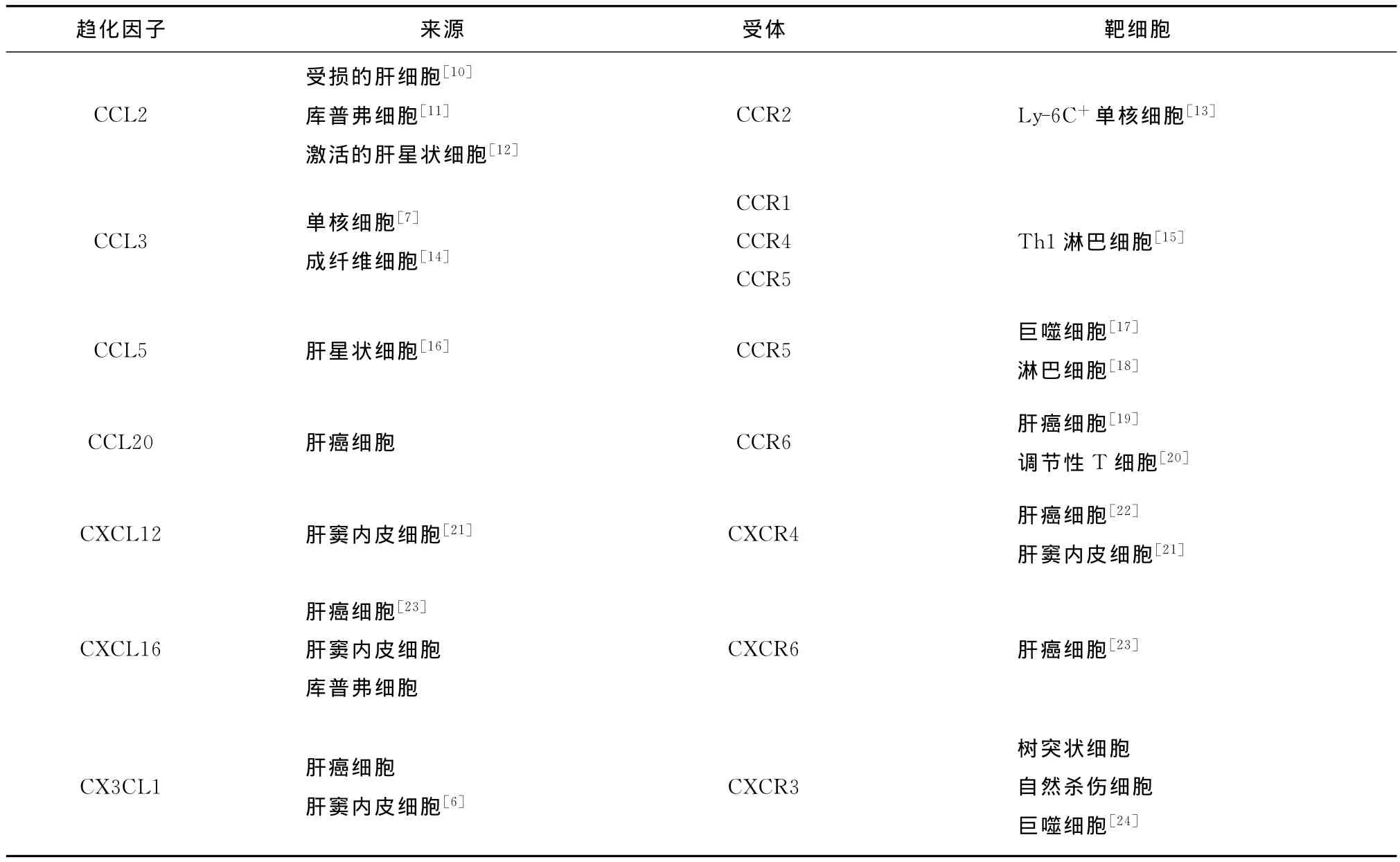
除了肿瘤细胞外,与肿瘤相关的巨噬细胞、库普弗细胞、树突状细胞、激活的星状细胞及肝窦内皮细胞等均可合成和分泌趋化因子(见表1)。Matsubara等[6]等报道了CX3CL1不仅表达于肝癌细胞,还表达于肝窦内皮细胞和胆管细胞。Piqueras等[7]和 Zimmermann等[8]研究发现,通过CCR1和CCR5招募的CD14+CD16+单核细胞可以分泌CCL3、CCL4等多种细胞因子。Bai等[9]研究发现,骨髓间质来源的HS-5细胞通过分泌CCL5来激活肝癌细胞(Huh7)PI3K/AKT通路和上调基质金属蛋白酶-2(MMP-2)的表达,从而促进肝癌细胞增殖、迁移和侵袭并抑制其凋亡。

表1 肝癌微环境中重要的趋化因子和受体
3 趋化因子与肝癌细胞的增殖
迄今为止,已发现多种趋化因子在促进肿瘤细胞增殖方面发挥着重要的作用。Cao等[25]检测了48对肝癌与癌旁标本中CXCL1的mRNA含量,发现Ⅰ~Ⅱ期患者中有51.4%的癌组织较癌旁组织表达升高,Ⅲ~Ⅳ期达72.7%;将肝癌细胞(7721、7402)过表达CXCL1或外源性加入CXCL1,均可明显增强肝癌细胞的增殖能力。Du等[26]研究发现,激活CCL20/CCR6趋化因子轴可以增强肝癌细胞的增殖能力,下调CCR6则可抑制肝癌细胞的增殖。Shi等[27]报道了下调肝癌细胞(97L、LM3)趋化因子CC受体样蛋白1(CCRL1)的表达后,可以抑制肝癌细胞的增殖并减慢皮下成瘤小鼠的肿瘤生长速度;同时肿瘤细胞分泌的CCL19和CCL21也随之减少,抑制了其共同受体CCR7的活性和下游Akt/GSK-3β和 Wnt通路的激活;这说明CCRL1表达上升可以抑制具有促瘤能力的CCR7的活性,从而减慢了肿瘤的增殖速度。Ochoa-Callejero等[28]研究发现,CCR5拮抗剂maraviroc不仅可抑制CCL5/p38/ERK通路的激活,从而缓解乙硫氨酸诱导的小鼠肝癌细胞增殖,而且肝癌细胞的凋亡数量明显增多。
4 趋化因子与肝癌的转移和侵袭
趋化因子对肝癌细胞的转移和侵袭同样起着重要的作用。Liu等[29]研究发现,CXCL12-CXCR4的激活可以促进肝癌细胞的转移。Nagamine等[30]研究发现,岩藻多糖通过下调Huh7中CXCL12来达到抗肿瘤活性的作用。Uchida等[31]报道CCL20可以促进CCR6高表达肝癌细胞(HepG2)形成伪足,肝癌组织中CCR6表达升高与癌细胞肝内转移及预后较差相关。Tian等[32]研究发现,缺氧诱导因子-1α(HIF-1α)可以结合在CXCL16转录活性区,启动CXCL16的表达,从而促进肿瘤细胞的转移和侵袭,生存分析也显示CXCL16表达高的患者预后较差。
趋化因子还可以诱导肝癌细胞分泌MMP,对细胞外基质和基底膜进行降解,增强肿瘤细胞的侵袭能力。Zhang等[33]研究发现,骨桥蛋白可以刺激肝癌细胞(SMMC7721、HepG2)合成 CXCR4和MMP-2,从而增强肝癌细胞的侵袭能力。若下调CXCR4,则MMP-2表达下降,肝癌细胞的侵袭能力受到抑制。Shih等[34]报道了肝癌成血管细胞分泌的CCL2可以激活肝癌细胞的CCR2/JNK信号通路,使miR-21表达上调,从而诱导肝癌细胞发生上皮-间质转化(EMT)和MMP-9的释放,最终使肝癌细胞侵袭和转移能力增强。
在转移性肝癌的研究中,Itatani等[35]报道了在人肝转移的结肠癌细胞中,Smad4的缺失可以上调CCL15表达,CCL15可以招募能分泌 MMP-9的CCR1+髓细胞进入肝癌组织,从而促进了癌细胞在肝内的转移。Zhao等[36]在小鼠结肠癌肝转移模型中发现,肿瘤细胞分泌的CCL2可以结合到骨髓来源且具有促进肝癌增殖的CD11b/Gr1mid细胞表面的CCR2受体,从而招募其进入肝癌组织内,抑制CCL2分泌或沉默CD11b/Gr1mid细胞上的CCR2均可抑制肿瘤增殖,这说明CCL2-CCR2轴在招募这类特殊的细胞进入肝癌组织内起着重要的作用。然而对以上这些趋化因子及受体是否参与了肝癌器官特异性转移尚无定论,因此在这方面进一步研究将丰富肝癌器官特异性转移的理论。
5 趋化因子与肝癌的血管生成
新生血管形成往往是肿瘤形成的标志之一,新生血管为肿瘤的增殖、转移和侵袭创造了有利条件。肝癌是一种血管丰富的肿瘤,它的增殖、转移很大程度上依赖血供范围,趋化因子和受体在其中发挥了重要的作用。在CXC趋化因子家族中,含有谷氨酸-亮氨酸-精氨酸模序(ELR)的趋化因子(如CXCL8、CXCL1、CXCL5和 CXCL12)是有效的成血管因子,而缺少ELR模序的CXC类趋化因子(如CXCL4、CXCL9等)可以抑制血管生成,两类趋化因子的平衡关系决定了瘤内血管的内皮细胞增生程度,进而影响肿瘤的生长和转移[21,37]。
目前,许多研究围绕CXCL12与血管生成的关系而展开。Li等[38]报道CXCL12和CXCR4在肝癌组织血管内皮细胞中的表达均明显上调,表明肝癌可以通过自分泌方式促进血管生成;Siegel等[39]研究发现用bevacizumab治疗肝癌后,患者循环血液中血管内皮生长因子-A(VEGF-A)和CXCL12下降。Xue 等[40]用 CXCL12 刺 激 肝 癌 细 胞SMMC-7721,发现VEGF分泌增加;若沉默CXCR7则VEGF分泌减少,且裸鼠皮下成瘤的瘤体明显缩小,血管指标CD31表达也减弱;结果间接证实了CXCR7可能是CXCL12受体之一,激活CXCR7会促进血管生成。
小鼠肝癌模型研究发现,通过尾静脉注射胸腺瘤细胞后,CCR1基因敲除小鼠(CCR1-/-)肝转移瘤的微血管密度明显低于对照组[41]。Choi等[23]研究发现,若下调肝癌细胞CXCL8的表达,则VEGF分泌明显减少,血管出芽能力减弱,小鼠瘤内CD31、CD34、血管内皮生长因子受体2(VEGFR2)及血管内皮细胞钙黏蛋白(E-cadherin)等血管标志物表达下降。
6 趋化因子与肝癌的炎性反应
肝脏的慢性炎性反应常伴随着持续的肝损伤,不断的损伤和再生会导致肝脏纤维化、硬化,直至肝癌。趋化因子与炎性反应的关系密切,明确趋化因子在慢性肝损伤到肝癌发展过程中的作用,以及寻找能预测慢性肝脏炎性反应恶变的趋化因子作为诊断依据,对肝癌的诊治具有重要的意义。中性粒细胞作为肿瘤炎性微环境中典型的炎性细胞,可以分泌多种促炎介质,参与抗炎反应及随后的组织重建等过程。Gao等[42]研究发现,在肝癌组织标本中的CXCR6表达明显高于癌旁组织,CXCR6缺失会抑制Gr-1+中性粒细胞浸润和促炎因子白细胞介素-1β(IL-1β)、IL-6和IL-8产生,联合检测肝癌标本中CXCR6与中性粒细胞的表达可以提高对生存和预后判断的准确性。Kuang等[43]研究发现,癌旁间质中有促炎作用的IL-17+细胞数量较癌组织明显增多,用IL-17处理过的肝细胞或肝癌细胞可以明显提高中性粒细胞的迁移能力,并且IL-17可以刺激肝脏血管内皮细胞CXCL1、CXCL5等数种CXC类趋化因子表达明显上调,但CC类趋化因子表达并未上调。如果抑制内皮细胞表面的CXCR1和CXCR2,则中性粒细胞迁移能力明显下降。以上结果说明IL-17可以通过血管内皮细胞表达的CXC类趋化因子来促进中性粒细胞进入肝癌组织发挥抗炎作用。
7 趋化因子与肝癌的免疫
目前有研究发现,与肝癌相关的免疫细胞包括库普弗细胞、自然杀伤细胞和树突状细胞,肝脏特有的肝窦内皮细胞、肝星状细胞和肝细胞都可以作为抗原递呈细胞,这种特殊的微环境也影响了由趋化因子介导的免疫细胞的招募过程[44]。Tang等[45]用过表达CX3CL1的小鼠肝癌细胞对小鼠进行皮下成瘤,发现CX3CL1可诱导机体产生肿瘤特异性的CD4+CD8+细胞毒性T细胞,其分泌IL-2可明显减慢肿瘤的生长速度。有研究已证实,CCL21可以招募树突状细胞和T细胞以减弱肿瘤细胞介导的免疫抑制作用[46]。在此基础上,Zhou等[47]在肝癌小鼠腹腔内注射抗CD25鼠单克隆抗体(PC61)以去除小鼠体内的Treg细胞,并联合注射CCL21,发现肿瘤的血管生成和癌细胞增殖受到抑制,且瘤内的CD4+CD8+细胞毒性 T细胞和CD11C+树突状细胞数量增加,具有免疫抑制功能的IL-10和转化生长因子-β(TGF-β)释放减少,最终小鼠的生存时间延长,说明去除小鼠体内Treg细胞可以增强CCL21的抗肿瘤作用。Marukawa等[48]用过表达CCL2的肝癌细胞在小鼠体内进行皮下成瘤,发现瘤块内的 Mac-1+巨噬细胞、CD4+CD8+T淋巴细胞数量增加,肿瘤坏死因子-α(TNF-α)表达明显升高,瘤体较对照组明显缩小,说明CCL2可以招募并激活巨噬细胞和T淋巴细胞以达到抗肿瘤增殖的目的。
8 结语
肝癌的预后不良且缺乏有效的治疗手段,学者们致力于寻找新的治疗靶点和治疗手段。以往的研究表明了趋化因子及其受体在肝癌的发病机制中起着举足轻重的作用。目前针对肝脏疾病的趋化因子拮抗剂已在动物实验中初见成效,但令人遗憾的是,趋化因子对肝癌的发生发展既有抑制也有促进,基于肝癌趋化因子及其受体的拮抗剂研究目前尚处于起步阶段。其中CXCL12-CXCR4轴的抑制剂研究得较多,特别是在防止肿瘤转移和复发方面的研究前景较为光明。肝癌趋化因子或受体的拮抗剂应用于临床需注意:(1)趋化因子拮抗剂可能会延迟人体正常炎性反应;(2)由于趋化因子及其受体存在冗余现象,抑制其中一对趋化因子及受体可能会反馈增强其他受体的活性;(3)明确合适的靶趋化因子轴进行治疗以及药物剂量至关重要。相信随着对趋化因子及其受体作用机制的研究和对肝癌发病机制和发展过程了解的不断深入,必将为新药的研发提供新的方向。
1 Zimmermann HW,Tacke F. Modification of chemokine pathways and immune cell infiltration as a novel therapeutic approach in liver inflammation and fibrosis.Inflamm Allergy Drug Targets,2011,10:509-536.
2 Charo IF,Ransohoff RM.The many roles of chemokines and chemokine receptors in inflammation.N Engl J Med,2006,354:610-621.
3 Oo YH,Shetty S,Adams DH.The role of chemokines in the recruitment of lymphocytes to the liver.Dig Dis,2010,28:31-44.
4 Proudfoot AE, Handel TM, Johnson Z, et al.Glycosaminoglycan binding and oligomerization are essential for the in vivo activity of certain chemokines.Proc Natl Acad Sci U S A,2003,100:1885-1890.
5 Zhou SL,Dai Z,Zhou ZJ,et al.Overexpression of CXCL5 mediates neutrophil infiltration and indicates poor prognosis for hepatocellular carcinoma.Hepatology,2012,56:2242-2254.
6 Matsubara T,Ono T,Yamanoi A,et al.Fractalkine-CX3CR1 axis regulates tumor cell cycle and deteriorates prognosis after radical resection for hepatocellular carcinoma.J Surg Oncol,2007,95:241-249.
7 Piqueras B,Connolly J,Freitas H,et al.Upon viral exposure,myeloid and plasmacytoid dendritic cells produce 3 waves of distinct chemokines to recruit immune effectors.Blood,2006,107:2613-2618.
8 Zimmermann HW,Seidler S,Nattermann J,et al.Functional contribution of elevated circulating and hepatic non-classical CD14CD16 monocytes to inflammation and human liver fibrosis.PLoS One,2010,5:e11049.
9 Bai H,Weng Y,Bai S,et al.CCL5 secreted from bone marrow stromal cells stimulates the migration and invasion of Huh7 hepatocellular carcinoma cells via the PI3K-Akt pathway.Int J Oncol,2014,45:333-343.
10 Ramm GA.Chemokine(C-C motif)receptors in fibrogenesis and hepatic regeneration followingacute and chronic liver disease.Hepatology,2009,50:1664-1668.
11 Heymann F, Trautwein C, Tacke F. Monocytes and macrophages as cellular targets in liver fibrosis.Inflamm Allergy Drug Targets,2009,8:307-318.
12 Marra F,Valente AJ,Pinzani M,et al.Cultured human liver fat-storing cells produce monocyte chemotactic protein-1.Regulation by proinflammatory cytokines.J Clin Invest,1993,92:1674-1680.
13 Karlmark KR,Weiskirchen R,Zimmermann HW,et al.Hepatic recruitment of the inflammatory Gr1+monocyte subset upon liver injury promotes hepatic fibrosis.Hepatology,2009,50:261-274.
14 Fahey S,Dempsey E,Long A.The role of chemokines in acute and chronic hepatitis C infection.Cell Mol Immunol,2014,11:25-40.
15 Butera D,Marukian S,Iwamaye AE,et al.Plasma chemokine levels correlate with the outcome of antiviral therapy in patients with hepatitis C.Blood,2005,106:1175-1182.
16 Seki E,De Minicis S,Gwak GY,et al.CCR1 and CCR5 promote hepatic fibrosis in mice.J Clin Invest,2009,119:1858-1870.
17 Schwabe RF,Bataller R,Brenner DA.Human hepatic stellate cells express CCR5 and RANTES to induce proliferation and migration.Am J PhysiolGastrointest Liver Physiol,2003,285:G949-G958.
18 Fujii H,Itoh Y,Yamaguchi K,et al.Chemokine CCL20 enhances the growth of HuH7 cells via phosphorylation of p44/42 MAPK in vitro.Biochem Biophys Res Commun,2004,322:1052-1058.
19 Chen KJ,Lin SZ,Zhou L,et al.Selective recruitment of regulatory T cell through CCR6-CCL20 in hepatocellular carcinoma fosters tumor progression and predicts poor prognosis.PLoS One,2011,6:e24671.
20 Mavier P,Martin N,Couchie D,et al.Expression of stromal cell-derived factor-1 and of its receptor CXCR4 in liver regeneration from oval cells in rat.Am J Pathol,2004,165:1969-1977.
21 Ehlert JE,Addison CA,Burdick MD,et al.Identification and partial characterization of a variant of human CXCR3 generated by posttranscriptional exon skipping.J Immunol,2004,173:6234-6240.
22 Muhlbauer M,Ringel S,Hartmann A,et al.Lack of association between the functional CX3CR1 polymorphism V249I and hepatocellular carcinoma.Oncol Rep,2005,13:957-963.
23 Choi SH,Kwon OJ,Park JY,et al.Inhibition of tumour angiogenesis and growth by small hairpin HIF-1alpha and IL-8in hepatocellular carcinoma.Liver Int,2014,34:632-642.
24 Cripps JG,Celaj S,Burdick M,et al.Liver inflammation in a mouse model of Th1 hepatitis despite the absence of invariant NKT cells or the Th1 chemokine receptors CXCR3 and CCR5.Lab Invest,2012,92:1461-1471.
25 Cao Z,Fu B,Deng B,et al.Overexpression of Chemokine(CX-C)ligand 1(CXCL1)associated with tumor progression and poor prognosis in hepatocellular carcinoma.Cancer Cell Int,2014,14:86.
26 Du D,Liu Y,Qian H,et al.The effects of the CCR6/CCL20 biological axis on the invasion and metastasis of hepatocellular carcinoma.Int J Mol Sci,2014,15:6441-6452.
27 Shi JY,Yang LX,Wang ZC,et al.CC chemokine receptor-like 1 functions as a tumor suppressor by impairing CCR7-related chemotaxis in hepatocellular carcinoma.J Pathol,2015,235:546-558.
28 Ochoa-Callejero L,Perez-Martinez L,Rubio-Mediavilla S,et al.Maraviroc,a CCR5 antagonist,prevents development of hepatocellular carcinoma in a mouse model.PLoS One,2013,8:e53992.
29 Liu H,Pan Z,Li A,et al.Roles of chemokine receptor 4(CXCR4)and chemokine ligand 12(CXCL12)in metastasis of hepatocellular carcinoma cells.Cell Mol Immunol,2008,5:373-378.
30 Nagamine T,Hayakawa K,Kusakabe T,et al.Inhibitory effect of fucoidan on Huh7 hepatoma cells through downregulation of CXCL12.Nutr Cancer,2009,61:340-347.
31 Uchida H,Iwashita Y,Sasaki A,et al.Chemokine receptor CCR6 as a prognostic factor after hepatic resection for hepatocellular carcinoma.J Gastroenterol Hepatol,2006,21:161-168.
32 Tian H,Huang P,Zhao Z,et al.HIF-1alpha plays a role in the chemotactic migration of hepatocarcinoma cells through the modulation of CXCL6 expression.Cell Physiol Biochem,2014,34:1536-1546.
33 Zhang R,Pan X,Huang Z,et al.Osteopontin enhances the expression and activity of MMP-2 via the SDF-1/CXCR4 axis in hepatocellular carcinoma cell lines. PLoS One, 2011,6:e23831.
34 Shih YT,Wang MC,Zhou J,et al.Endothelial progenitors promote hepatocarcinoma intrahepatic metastasis through monocyte chemotactic protein-1 induction of microRNA-21.Gut,2015,64:1132-1147.
35 Itatani Y,Kawada K,Fujishita T,et al.Loss of SMAD4 from colorectal cancer cells promotes CCL15 expression to recruit CCR1+myeloid cells and facilitate liver metastasis.Gastroenterology,2013,145:1064-1075.
36 Zhao L,Lim SY,Gordon-Weeks AN,et al.Recruitment of a myeloid cell subset(CD11b/Gr1 mid)via CCL2/CCR2 promotes the development of colorectal cancer liver metastasis.Hepatology,2013,57:829-839.
37 Belperio JA,Keane MP,Arenberg DA,et al.CXC chemokines in angiogenesis.J Leukoc Biol,2000,68:1-8.
38 Li W,Gomez E,Zhang Z.Immunohistochemical expression of stromal cell-derived factor-1 (SDF-1)and CXCR4 ligand receptor system in hepatocellular carcinoma.J Exp Clin Cancer Res,2007,26:527-533.
39 Siegel AB,Cohen EI,Ocean A,et al.PhaseⅡtrial evaluating the clinical and biologic effects of bevacizumab in unresectable hepatocellular carcinoma.J Clin Oncol,2008,26:2992-2998.
40 Xue TC,Chen RX,Han D,et al.Down-regulation of CXCR7 inhibits the growth and lung metastasis of human hepatocellular carcinoma cells with highly metastatic potential.Exp Ther Med,2012,3:117-123.
41 Rodero MP,Auvynet C,Poupel L,et al.Control of both myeloid cell infiltration and angiogenesis by CCR1 promotes liver cancer metastasis development in mice.Neoplasia,2013,15:641-648.
42 Gao Q,Zhao YJ,Wang XY,et al.CXCR6 up-regulation contributes to proinflammatory tumor microenvironment that drives metastasis and poor patient outcomes in hepatocellular carcinoma.Cancer Res,2012,72:3546-3556.
43 Kuang DM,Zhao Q,Wu Y,et al.Peritumoral neutrophils link inflammatory response to disease progression by fostering angiogenesis in hepatocellular carcinoma.J Hepatol,2011,54:948-955.
44 Tiegs G,Lohse AW.Immune tolerance:what is unique about the liver.J Autoimmun,2010,34:1-6.
45 Tang L,Hu HD,Hu P,et al.Gene therapy with CX3CL1/Fractalkine induces antitumor immunity to regress effectively mouse hepatocellular carcinoma. Gene Ther,2007,14:1226-1234.
46 Nguyen-Hoai T,Baldenhofer G,Sayed AMS,et al.CCL21(SLC)improves tumor protection by a DNA vaccine in a Her2/neu mouse tumor model.Cancer Gene Ther,2012,19:69-76.47 Zhou S,Chen L,Qin J,et al.Depletion of CD4+ CD25+regulatory T cells promotes CCL21-mediated antitumor immunity.PLoS One,2013,8:e73952.
48 Marukawa Y,Nakamoto Y,Kakinoki K,et al.Membranebound form of monocyte chemoattractant protein-1 enhances antitumor effects of suicide gene therapy in a model of hepatocellular carcinoma. Cancer Gene Ther,2012,19:312-319.
