胎儿肾脏回声增强与遗传学检查异常结果相关性分析
2024-10-10高园朱好辉
【摘要】目的:分析胎儿肾脏回声增强的超声表现,总结其与遗传学检查结果异常的关系。方法:产前超声检查发现肾脏回声增强,经患者及其家属知情同意,遗传学检查使用染色体微阵列分析技术(chromosomal microarray analysis,CMA)或全外显子组测序技术(whole exome sequencing, WES)产前诊断的60例胎儿。分析肾脏回声增强的产前超声表现,统计遗传学检查结果异常的检出率,分析胎儿肾脏回声增强与其相关性。结果:60例产前肾脏回声增强胎儿中,其中单纯性与非单纯性基因检测异常检出率差异性无统计学意义(P>0.05)。异常结果中异常检出率较高的为17q12微缺失综合征、13-三体综合征等。结论:60例产前肾脏回声增强胎儿中非单纯性检出率高于单纯性,基因检测结果异常中17q12微缺失综合征检出率较高。产前检出肾脏回声增强在一定程度上可预测胎儿基因检测异常的风险,其中COL1A1基因杂合变异及Sotos综合征与肾脏回声增强的相关性未找到相关报道,仍待进一步研究。
【关键词】超声检查;产前;胎儿肾脏回声增强;遗传学检查;全外显子;CMA
Correlation analysis between echogenic enhancement of the fetal kidney and abnormal genetic examination results
GAO Yuan, ZHU Haohui
Xinxiang Medical College, Xinxiang, Henan 453003, China
【Abstract】Objective:To analyze the ultrasound manifestations of fetal renal echogenic enhancement and summarize its relationship with abnormal genetic examination results.Methods:Prenatal ultrasound examination showed enhanced renal echogenicity,and with the informed consent of patients and their families,genetic examination was performed using chromosomal microarray analysis (CMA) or whole exome sequencing (WES) of 60 fetuses diagnosed prenatally.The prenatal ultrasound manifestations of renal echo enhancement were analyzed,the detection rate of abnormal genetic examination results was statistically analyzed,and the correlation between fetal renal echo enhancement and it was analyzed.Results:Among the 60 cases of fetuses with prenatal renal echo enhancement,there was no significant difference in the detection rate of abnormal genetic examination between simple and non-simple (P>0.05).Among the abnormal results,the abnormal detection rate was higher in 17q12 microdeletion syndrome,trisomy 13,etc.Conclusion:The detection rate of non-simplex fetuses with enhanced renal echo in 60 prenatal cases was higher than that of simplex fetuses,and the detection rate of 17q12 microdeletion syndrome was higher in the abnormal genetic test results.Prenatal detection of enhanced renal echogenicity can predict the risk of abnormal fetal genetic testing to a certain extent,and the correlation between heterozygous variants in COL1A1 gene and Sotos syndrome and renal echogenicity enhancement has not been reported,and further research is still needed.
【Key?Words】Ultrasonic examination; Prenatal; Enhanced fetal kidney echo; Genetic examination; Whole exon; CMA
如何明确胎儿肾脏回声增强病因及预后给产前咨询和临床处理带来极大的困扰[1]。遗传因素是胎儿泌尿系畸形的主要原因之一[2]。染色体微阵列分析(CMA)具有传统的染色体核型分析所无法比拟的优势[3]。全外显子测序(WES)可以评估整个基因组的变异,可以提高诊断率[4]。
本研究回顾性分析胎儿肾脏回声增强并接受CMA或WES检查的60例胎儿肾脏超声图像及其它系统情况,随访检测结果,探讨产前超声胎儿肾脏回声增强与遗传学检查异常结果的相关性。
1 对象和方法
1.1 研究对象
2016年1月—2023年12月在河南省人民医院发现胎儿回声肾脏增强并接受遗传学检查的60例胎儿。
1.2 研究方法
1.2.1 纳入标准:①胎儿肾脏回声增强,表现为肾脏回声高于自身肝脏及脾脏回声;②获得CMA和/或WES检测结果。
1.2.2 分组:根据有无合并其他异常分为单纯性肾脏回声增强及非单纯性肾脏回声增强。
1.2.3 超声诊断方法:

采用美国GE公司VolusonE8、VolusonE10超声诊断仪,腹部探头频率2.0~5.0MHz。对纳入的胎儿肾脏进行针对性检查。如合并其他畸形,对畸形系统进行详尽检查、记录及诊断。
1.2.4 遗传学检查及临床处理:
超声检查发现胎儿肾脏回声增强后,经孕妇及其家属知情同意,进行CMA和/或WES检测,并随访检测结果。
1.3 统计学分析
采用SPSS 27.0统计学软件进行数据分析。计数资料采用(%)表示,进行x2检验,计量资料采用(x±s)表示,进行t检验,P<0.05为差异具有统计学意义。
2 结果
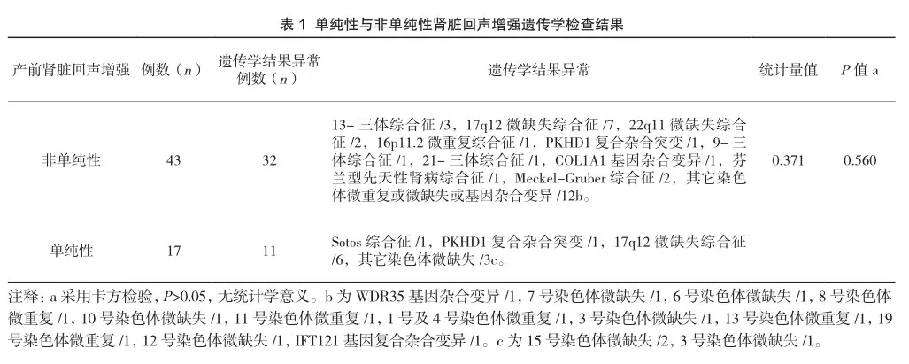
基因检测结果:60例胎儿中,孕妇年龄19~44岁,平均年龄(30.6±5.6)岁;超声诊断孕周17+5周~35周,平均孕周(26.8±4.4)周;遗传学结果异常43例(43/60,71.7%)。遗传学异常结果包括:17q12微缺失综合征13例(13/60,21.7%),13-三体综合征3例(3/60,5.0%),PKHD1复合杂合突变2例(2/60,3.3%),Meckel-Gruber综合征2例(2/60,3.3%),22q11微缺失综合征2例(2/60,3.3%),16p11.2微重复综合征1例(1/60,1.7%),芬兰型先天性肾病综合征1例(1/60,1.7%),COL1A1基因杂合变异1例(1/60,1.7%),21-三体综合征1例(1/60,1.7%),9-三体综合征1例(1/60,1.7%),Sotos综合征1例(1/60,1.7%),其它染色体微重复或微缺失或基因杂合变异15例(15/60,25.00%)。
单纯性肾脏回声增强17例(28.3%),非单纯性肾脏回声增强43例(71.7%);单纯性与非单纯性肾脏回声增强遗传学结果异常检出率差异性无统计学意义(P>0.05),见表1。
3 讨论
胎儿肾脏回声增强表现为肾脏回声高于肝脏及脾脏回声,可能与严重的肾脏疾病相关[5]。胎儿肾脏回声增强的病因有染色体疾病(9-三体综合征、13-三体综合征等)、基因组疾病(Wiedemann Beckwith综合征、17q12微缺失综合征)和单基因疾病(多囊肾病、肾小球囊性肾病、肾囊肿和糖尿病综合征、巴德特·比德尔综合征、代谢疾病等),也可为正常变异[2]。
本研究中,肾脏回声增强遗传学检查异常占比71.7%(43/60),单纯性及非单纯性肾脏回声增强遗传学检查异常的检出率比较差异无统计学意义。检出率最高的是17q12微缺失综合征,其次为13-三体综合征,其他有PKHD1复合杂合突变、Meckel-Gruber综合征、22q11微缺失综合征、16p11.2微重复综合征、芬兰型先天性肾病综合征、COL1A1基因杂合变异、21-三体综合征、9-三体综合征、Sotos综合征及其他染色体微缺失及微重复等。
产前肾脏回声增强与17q12微缺失之间存在显著高相关性[6],本研究中检出率占比最高的是17q12微缺失综合征。Sotos综合征胎儿期超声表现的报道主要集中于早孕期NT增厚、孕中期或孕晚期轻度侧脑室扩张及羊水多[7],而本研究中病例仅表现为双肾实质回声增强。PKHD1基因与成人型多囊肾相关,在胎儿期可表现为肾脏回声增强[8]。Meckel-Gruber综合征是一种致命的常染色体隐性先天性异常综合征,肾脏回声增强为主要特点。COL1A1基因杂合变异可引起成骨发育不全[9],目前并未找到成骨发育不全胎儿期肾脏回声增强的相关报道。
综上所述,胎儿肾脏回声增强基因检测异常结果占比较高,其中占比最高的为17q12微缺失综合征,COL1A1基因杂合变异及Sotos综合征与肾脏回声增强的相关性未找到相关报道,仍待进一步研究。胎儿肾脏回声增强的产前预后评估需要结合明确的基因检测结果、超声检查结果和胎儿家族史进行综合分析,因此产前诊断很重要。
参考文献
[ 1 ] H U A N G R , F U F , Z H O U H , e t a l . P r e n a t a l d i a g n o s i s i n t h e f e t a l h y p e r e c h o g e n i c k i d n e y s : a s s e s s m e n t u s i n g c h r o m o s o m a l m i c r o a r r a y a n a l y s i s a n d e x o m e s e q u e n c i n g [ J ] . H u m G e n e t , 2 0 2 3 , 1 4 2 ( 6 ) : 8 3 5 - 8 4 7 .
[2] DIGBY E L,LIAUW J,DIONNE J,et al.Etiologies a n d o u t c o m e s o f p r e n a t a l l y d i a g n o s e d hyperechogenic kidneys[J].Prenat Diagn,2021,41(4):465-477.
[3] SHUSTER S,KEUNEN J,SHANNON P,et al.Prenatal d e t e c t i o n o f i s o l a t e d b i l a t e r a l h y p e r e c h o g e n i c kidneys: etiologies and outcomes[J].Prenat Diagn,2019,39(9):693-700.
[4] 张素阁,陈传燕,王翔,等.产前超声诊断胎儿泌尿系统异常[J].中华实用诊断与治疗杂志,2015,29(4):399-400.
[5] 杨烨,陈桂兰,屈艳霞,等.超声检查对侵入性产前诊断价值[J].中华实用诊断与治疗杂志,2015,29(4):391-392,395.
[6] 张振辉,盘丽娟,谌奎芳,等.17q12微缺失综合征的产前超声特征及遗传学分析[J].中南大学学报(医学版),2021,46(12):1370-1374.
[7] CHEN C P,LIN S P,CHANG T Y,et al.Perinatal i m a g i n g f i n d i n g s o f i n h e r i t e d S o t o s syndrome[J].Prenat Diagn,2002,22(10):887-892.
[8] CAO J,CHEN A E,TIAN L,et al.Application of w h o l e e x o m e s e q u e n c i n g i n f e t a l c a s e s w i t h skeletal abnormalities[J].Heliyon,2022,8(7):9 8 1 9 .
[ 9 ] W E A V E R J S , R E V E L S J W , E L I F R I T Z J M , e t a l . C l i n i c a l m a n i f e s t a t i o n s a n d m e d i c a l i m a g i n g of osteogenesis imperfecta:fetal through adulthood[J].Acta Med Acad,2021,50(2):277-2 9 1 .
