Calcium-chelating peptides from rabbit bone collagen:characterization, identif ication and mechanism elucidation
2024-01-24FuhunYunYuFuLingHnkunZhuYongYuXinFengYiSunHongjieDiXinLiuZhengfngLiuYuhoZhng
Fuhun Yun, Yu Fu,b,, Ling M,b,c, Hnkun Zhu,b, Yong Yu,b,Xin Feng, Yi Sun, Hongjie Di,b, Xin Liu, Zhengfng Liu, Yuho Zhng,b,c,
a College of Food Science, Southwest University, Chongqing 400715, China
b Chongqing Key Laboratory of Speciality Food Co-Built by Sichuan and Chongqing, Chongqing 400715, China
c Key Laboratory of Luminescence Analysis and Molecular Sensing (Southwest University), Ministry of Education, Chongqing 400715, China
d Angel Yeast Co., Ltd., Yichang 443003, China
Keywords: Rabbit bone Collagen peptide Peptide-calcium chelate Chelation mechanism Liquid chromatography-tandem mass (LC-MS/MS)
ABSTRACT This study aimed to characterize and identify calcium-chelating peptides from rabbit bone collagen and explore the underlying chelating mechanism. Collagen peptides and calcium were extracted from rabbit bone by instant ejection steam explosion (ICSE) combined with enzymatic hydrolysis, followed by chelation reaction to prepare rabbit bone peptide-calcium chelate (RBCP-Ca). The chelating sites were further analyzed by liquid chromatography-tandem mass (LC-MS/MS) spectrometry while the chelating mechanism and binding modes were investigated. The structural characterization revealed that RBCP successfully chelated with calcium ions. Furthermore, LC-MS/MS analysis indicated that the binding sites included both acidic amino acids (Asp and Glu) and basic amino acids (Lys and Arg). Interestingly, three binding modes, namely Inter-Linking, Loop-Linking and Mono-Linking were for the first time found, while Inter-Linking mode accounted for the highest proportion (75.1%), suggesting that chelation of calcium ions frequently occurred between two peptides. Overall, this study provides a theoretical basis for the elucidation of chelation mechanism of calcium-chelating peptides.
1. Introduction
Calcium is an essential mineral for humans to maintain normal physiological activities and plays a signif icant role in bone growth,muscle contraction, and nerve transmission[1]. A number of health disorders, such as osteoporosis, rickets, and hypertension, can be induced by calcium deficiency[2]. At present, calcium supplements mainly include inorganic calcium, organic calcium and amino acid-calcium chelate[3]. In their practical application, several shortcomings, such as poor water solubility, low absorption rate and high cost, still exist[4]. By contrast, peptide-calcium chelate exhibits better solubility and thermal stability, which can prevent calcium precipitation during gastrointestinal digestion, thereby improving the absorption of calcium in the body[5]. Therefore, calcium-chelating peptides are promising to be used as an excellent calcium supplement with improved bioavailability.
With the improvement of living standards and changes in eating habits, rabbit meat become gradually a popular food worldwide.According to the Statistics Division of the Food and Agriculture Organization of the United Nations, the total global production of rabbits in 2020 was about 640 million[6]. During the processing of rabbit meat, a large amount of rabbit bone is annually generated,which is characterized by the high content of protein (41.47%) and minerals (58.98%)[4,7-8]. However, rabbit bones are normally discarded during processing, resulting in environmental pollution and waste of resources. Effective utilization of rabbit bones can bring economic benefits and reduce environmental pressure[7]. Due to its high stiffness and toughness, animal bone is extremely difficult to be degraded.Instant catapult steam explosion (ICSE) is an innovative technology based on the traditional steam explosion, swollen technology, steam spout and extrusion[9]. In previous studies, steam explosion was often used for the pretreatment of lignocellulose andAchyranthisbidentataeRadix[10-11]. ICSE is promising to be used for pretreatment of rabbit bone, which can contribute to the subsequent processing of rabbit bone and improved utilization rate of rabbit bone. Recent studies have indicated that a number of food-derived peptides possess a calciumchelating activity. It has been shown that various chelating sites in peptides vary greatly in their affinities for calcium ions depending on amino acid sequence of chelating sites[12]. According to several recent studies, the structural features that affect calcium-chelating activity have been mentioned, including certain amino acid composition and specific groups of amino acids[13]. For instance, in a recent study on porcine plasma-derived peptides, Asp, Glu, and Gly residues were identified as the main calcium-binding sites[14]. Furthermore, a number of amino acid residues can promote chelation reaction, such as carboxyl groups and sulfhydryl groups[13]. However, the chelating sites and chelating modes of mineral-chelating peptides have not been fully elucidated. Although spectroscopic technologies can infer the amino acid residues that are involved in the chelation reaction, they cannot accurately determine the chelating sites and calcium-binding modes. Therefore, it is of particular importance to further investigate and elucidate the chelation sites and their chelating mechanisms.
In recent years, peptide-calcium chelates from different animal bone sources have been reported, such as porcine bone, bovine bone and fish bone, etc.[4,15-16]. However, to the best of our knowledge,rabbit bone as an innovative source of calcium and bioactive peptides has received little attention. In this study, calcium and bioactive peptides were extracted from rabbit bone and applied for preparation of peptide-calcium chelates. Morphological analysis, structural characterization, and mechanistic elucidation of calcium-chelating peptides from rabbit collagen were performed by scanning electron microscopy (SEM), Zeta potential analyzer, Fourier transform infrared spectroscopy (FTIR), ultraviolet-visible absorption spectroscopy(UV-Vis), fluorescence spectroscopy, X-ray diffraction (XRD) and liquid chromatography-tandem mass spectrometry (LC-MS/MS).
2. Materials and methods
2.1 Materials and reagents
Rex rabbit bone (protein content, 31.57%) was obtained from Axingji Rabbit Breeding Base (Chongqing, China). Alcalase 2.4 L(activity 2.4 AU/g) was purchased from Novozymes Biotechnology Co., Ltd. (Beijing, China). Acetonitrile was purchased from Thermo Fisher Scientific (USA). All other chemical reagents used in this study were of analytical grade.
2.2 Pretreatment of rabbit bone
Instantaneous steam explosion treatment of rabbit bone was carried out through a steam explosion test rig (QBS-200B, Zhengdao Bio-energy Co., Ltd., Henan, China). The rabbit bones were washed with deionizing water to remove excessive meat and impurities,and then cut into chunks (4 cm in length). The treated rabbit bones were put into the chamber of the steam explosion equipment(250 g sample was added each time) and pressurized at 1.3 MPa.Samples were collected after the steam explosion. The upper layer of fat was removed by centrifugation at 5 000 ×gfor 20 min (Heraeus Multifuge × 3R, Thermo Fisher Scientific, USA). The remaining samples were further dried in an oven at 50 °C (101-4-S, Yuejin Medical Instrument Factory, Shanghai, China) until the moisture content was below 10%. According to different pressure holding times of steam explosion, the obtained samples were named as S1(8 min), S2 (5 min) and S3 (2 min). Rabbit bone subjected to ultrafine pulverizer treatment (WZJ-6, Bally Powder Engineering Technology Co., Ltd., Shandong, China) was employed as control. The treated rabbit bones (in the same way as the rabbit bones were treated before steam explosion) were immersed in boiling water for 30 min to remove fat and then placed in an oven for drying until the water content was < 10%. The dried rabbit bones were transferred to a superfine grinder for 15 min, and the final sample was recorded as Con.
2.3 Preparation and determination of rabbit collagen peptides (RBCP)
2.3.1 PreparationofRBCP
Enzymatic hydrolysis was conducted according to our previous study with slight modifications[17]. The solutions (10%) of rabbit bone samples (S1, S2, S3 and Con) were adjusted to pH 9.0 (PHS-25,Thunder Magnetic Analysis Instrument Factory, Hangzhou, China)and incubated in water bath at 55 °C for 10 min, then Alcalase(3%,m/m) was added to initiate enzymatic hydrolysis. Enzymatic hydrolysis lasted for 7 h, and samples (5 mL) were withdrawn at each hour. The hydrolysis reaction was terminated by heating at 95 °C in water for 15 min to inactivate enzymes. The solution was subsequently cooled to 25 °C. After centrifugation at 8 000 ×gfor 15 min, the supernatant was collected. The degree of hydrolysis was determined byo-phthalaldehyde (OPA) assay referring to our previous method[17]. OPA reagent (4 mL) was added to 200 μL of sample solution. The absorbance of solution was recorded at the wavelength of 340 nm after 2-min incubation at room temperature(25 °C) in the dark.
2.3.2Determinationofpeptidesizedistribution
The peptide size distribution of protein hydrolysates was determined using an HPLC system (Ultimate 3 000, Thermo Fisher Scientific, USA) equipped with a size exclusion chromatography column according to our previous studies[17-18]. The molecular weight ranges of the 5 fractions were > 5 000, 3 000−5 000, 1 000−3 000,500−1 000, < 500 Da. The percentage for each fraction was represented by its percentage area under the curve. Chromatographic conditions: mobile phase of 30% (V/V) acetonitrile (containing 0.1% trifluoroacetic acid); flow rate 0.5 mL/min; injection volume 20 μL; column temperature 25 °C; Chromatographic column:BioBasic SEC120 (7.8 mm × 300 mm, 5 μm). Detection wavelength: 214 nm. Cytochrome C (12 500 Da), Gly-Gly-Tyr-Arg(451.48 Da), bacitracin (6 511.51 Da), and human serum albumin(66 000 Da) were used as standards. The sample concentration was 1 mg/mL, and the sample or standard solution was passed through a 0.22 μm microporous membrane for determination. After measurement, the most suitable one was selected from four samples(S1, S2, S3 and Con) for the subsequent chelation reaction with bone calcium solution.
2.3.3Determinationofaminoacidsinrabbitbone
The amino acid composition was determined according to a previous study with slight modifications[19]using an automatic amino acid analyzer (L-8900, Hitachi, Tokyo, Japan).
2.4 Preparation of rabbit bone calcium solution (RBCS)
Bone calcium solution was prepared according to a previous method with slight modifications[20]. The remaining bone residue after enzymatic hydrolysis of rabbit bone was dried at 105 °C for 4 h. After drying, bone residue was ground and mixed with 0.4 mol/L HCl (bone to acid ratio of 1:20,m/m), and stirred in a water bath at 55 °C for 1 h.The supernatant solution was collected by centrifugation at 10 000 ×gand separated at 25 °C for 10 min. Finally, the bone calcium solution(5 mg/mL) was obtained and stored at 4 °C for future use.
2.5 Identification of calcium-chelating peptides
2.5.1 Preparationofrabbitcollagenpeptide-chelated calcium(RBCP-Ca)
RBCP-Ca was prepared according to a reported method[20]with minor modifications. In brief, the lyophilized RBCP were dissolved in de-ionized water to a concentration of 5 mg/mL, and then RBCS(5 mg/mL) with mass ratio (2:1,m/m)was added. The solutions were adjusted to pH 6.0 with 0.5% NaOH, and then stirred for 30 min at 40 °C. After the chelation reaction, absolute ethanol (5 times the volume)was added to remove free calcium to obtain calcium-peptide chelates.The mixture was centrifuged at 10 000 ×gat 4 °C for 10 min to collect the precipitate, freeze-dried and stored for further analysis.
2.5.2 Determinationofcalciumchelatingcapacity
The content of calcium was determined by flame atomic absorption spectrometer (TAS-986, Common to General Analysis,Beijing, China) in accordance with Sun et al.[21]. The absorbance value of the sample was measured at 422.7 nm, and the calcium content was quantified by comparing the calcium standard solution (0−6 μg/mL)[21].
2.6 Microstructure analysis
2.6.1 SEM
The microstructure of rabbit collagen peptide and rabbit collagen peptide-calcium chelate was analyzed by scanning electron microscope (Hitachi High Technologies Corp, Tokyo, Japan).Referring to a previous method[20], sample powder was poured onto the sample holder, fixed with double-sided tape, and then sputtered with gold to obtain a microscope scan image of sample at 2 000×magnification.
2.6.2 Zetapotentialandparticlesizedetermination
The zeta potential and size distribution were measured by the nanoparticle size and Zeta potential analyzer (Zetasizer Nano zsp,Malvern, UK) using the method of Zhang et al.[16]. The sample concentration was fixed at 0.1 mg/mL.
2.7 Spectroscopic analysis of RBCP and RBCP-Ca
2.7.1 FTIR
According to the previous method[22], each sample in powder form was loaded onto an FTIR (Nicolet 670, Thermo Fisher, Waltham,Massachusetts, USA) sample plate. Each sample was scanned 50 times with a resolution of 4 cm−1in the wavenumber range of 4 000−600 cm−1. Afterwards, the samples were compared in pairs by overlaying their FTIR spectra, and the infrared transmittance was analyzed.
2.7.2 UV-Visabsorptionspectrum
RBCP and RBCP-Ca were dissolved in de-ionized water at a concentration of 0.5 mg/mL. Then, the absorbance of the solution was measured in the range of 190−480 nm using UV-Vis spectrophotometer (UV-6100, Metash, Shanghai, China)[4].
2.7.3 Fluorescencespectrum
Fluorescence intensity was measured by referring to the method of Luo et al.[4]. RBCP and RBCP-Ca were dissolved in de-ionized water with the concentration of 0.5 mg/mL. Measurements were performed using a fluorescence spectrophotometer (F-2500, Hitachi,Tokyo, Japan). The fixed excitation wavelength was 275 nm, and the emission wavelength was 280−400 nm.
2.7.4 XRD
The X-ray diffractometer (X’Pert3 Powder10300, Panath,Netherlands) was used for XRD measurement. The measurement conditions were set as follows by the method of Malison et al. (Cu target X-ray tube, working voltage 40 kV, current 30 mA, scanning 2θrange 10°−70°, scanning speed 5°/min)[20].
2.8 LC-MS/MS analysis
The identification of RBCP and RBCP-Ca was implemented by LC-MS/MS according to our previous method[23]. HPLC (Easy nLC 1200, Thermo Fisher Scientific, Massachusetts, USA) system was used with C18, 3 µm, 100 Å, 75 µm × 15 cm column with 0.1% formic acid in phase A, and 0.1% formic acid and 80% acetonitrile/water in phase B. At the flow rate of 0.3 mL/min, the elution process was divided into 5 stages (95% A-phase from 0 min to 4 min, 90% A-phase from 4 min to 40 min, 72% A-phase from 40 min to 47 min, 62%A-phase from 47 min to 48 min, and 0% A-phase from 48 min to 60 min). Mass spectrometry (Orbitrap Fusion Lumos, Thermo Fisher Scientific, Massachusetts, USA) analytical conditions were capillary temperature (320 °C) and RF Lens (40), respectively, spray voltage 2.0 kV. The scanning range of parent ion wasm/z350−1 550. The sub ion scanning range starts fromm/z50. The primary resolution was set to 120 000@m/z200. The resolution was set to 120 000@m/z200,30 000@m/z200 for secondary. Ion screening window:m/z1.6. Fragmentation pattern: higher energy collision dissociation.Dynamic exclusion time: 60 s. Low-energy and high-energy collisionactivated dissociation of calcium-peptide chelate in the form of complexes can reflect calcium-binding site information. The chelating sites of calcium ions were identified based on the changed mass. The conventional modification was to deduct one hydrogen and add one calcium, at which the molecular weight was changed to 38.95 Da.The cross-linking modification was to deduct two hydrogens and add one calcium, and the changed molecular weight was 37.95 Da. Based on the obtained LC-MS/MS data results, PEAKS software was used to search against rabbit (Oryctolaguscuniculus) database to identify peptide sequences. Meanwhile, the binding modes of calcium ions and peptides were also analyzed.
2.9 Statistical analysis
All experiments were repeated in triplicate and the results were given as mean ± standard deviation (SD). SPSS statistical software was used to examine the differences for post-hoc Duncan test analysis, andP< 0.05 was regarded as statistically significant.
3. Results and discussion
3.1 Physicochemical properties of RBCP
Degree of hydrolysis (DH) of rabbit bone was measured after ICSE treatment, and the results are shown in Fig. 1A. With the extended hydrolysis time from 1 h to 7 h, DH of protein hydrolysate(S1, S2, S3 and Con) gradually increased. Compared with the other three groups, S1 showed the fastest growth tendency. The DH of S1 increased from 14.36% to 22.91%, DH of S2 increased from 13.42% to 19.93%, and DH of S3 increased from 8.47% to 16.90%.By contrast, the DH of Con increased from 9.29% to 14.37%. When the hydrolysis time reached 5 h, the increasing tendency became a plateau. At this time, the DH values of S1, S2, S3 and Con samples were 20.26%, 17.32%, 15.42% and 13.76%, respectively. This indicated that prolonged pressure maintenance time led to excessive degradation of rabbit bone protein structure during steam explosion treatment[9]. During enzymatic hydrolysis of collagen, bioactive peptides can be gradually released, and ICSE treatment was beneficial to enzymatic hydrolysis of rabbit bone. Furthermore, the results of peptide size distribution are displayed in Fig. 1B. With the enzymatic hydrolysis extending from 1 h to 7 h, the proportion of RBCP (S1, S2,S3 and Con) with molecular weight below 1 kDa gradually increased.After 1 h-hydrolysis, the proportion of RBCP (S1, S2, S3 and Con)with molecular weight below 1 kDa accounted for 51.2%, 48.3%,47.0% and 44.8%, respectively. According to previous studies, low molecular weight peptides have high calcium chelating activity, e.g.collagen peptides derived from fish skin-derived (molecular weight:180−2 000 Da)[24]. Thus, S1 was selected as the optimal sample for subsequent chelation reaction. Based on the abovementioned result,ICSE-treated rabbit bone sample (S1) was selected for chelation reaction after 5-h hydrolysis.

Fig. 1 Degree of hydrolysis of protein hydrolysates treated by Alcalase (S1, S2, S3 and Con) (A); Molecular weight distribution of S1 (B1), S2 (B2), S3 (B3) and Con (B4).
3.2 Calcium-binding ability of RBCP
In the present study, the effects of temperature (°C), time (min),pH and peptide/calcium (m/m) on the chelating capacity of RBCP-Ca were previous investigated by single-factor experiment. The results are shown in Fig. S1. It has been shown that the mass ratio and pH played a vital role, while the reaction time and reaction temperature exert no impact on the chelation reaction. The mass ratio of RBCP/RBCS exerted a significant impact on the chelation rate, showing an upward trend, followed by a significant decline (P< 0.05) (Fig. S1A). The calcium chelating activity was decreased with increasing mass ratio,which was due to excessive RBCP aggregation that impeded the chelation sites, resulting in the inability to chelate more calcium ions[25]. According to calcium-chelating rate, the optimal condition of mass ratio was fixed at 2:1, at which the binding rate of calcium reached 615.85 mg/g (P< 0.05). As can be seen in Fig. S1B, at pH 6.0,the chelating capacity of RBCP was 644.92 mg/g (P< 0.05). Since the initial pH of bone calcium solution was 3.0, its buffering effect made it difficult to adjust back to pH 7.0. Nevertheless, chelation was tested at pH 5.0−9.0, under which the results showed that the chelation rate increased slowly. This phenomenon might be due to the fact that when pH was low in the solution, the high content of H+in the solution could compete with calcium ions to provide electron groups, thus preventing calcium ions from chelating with peptides.However, when pH increased, the high content of OH−in the solution can form precipitates with calcium ions[15]. In order to avoid the influence of calcium hydroxide precipitation in chelates, pH 6.0 was finally selected for chelation reaction. Similarly, in the study of peptide-calcium chelate from chicken foot by-product, it has been shown that the chelating condition for peptides from chicken foot broth byproduct and bone calcium solution was optimal under weak acidic conditions[20].
According to previous reports, the calcium-binding capacity of peptides depends on the carboxy-calcium chelation of Asp and Glu[3].In addition, Arg and Lys also had a certain chelating ability in the study of Pacific cod peptide chelated calcium[26]. This indicated that the calcium-binding capacity of peptide has a certain relationship with the type and content of amino acids. The amino acid composition in rabbit bone was summarized in Table S1. The contents of Glu, Asp,Lys and Arg were 14.25%, 8.80%, 7.55% and 6.92%, respectively.This provided evidence that as main components of RBCP, Glu, Asp,Lys and Arg may exhibit the higher calcium binding capacity.
3.3 Structural characterization of RBCP-Ca
3.3.1 SEM and particle size distribution
SEM and particle size analysis could effectively characterize the microstructure of RBCP and RBCP-Ca. The SEM results are shown in Figs. 2A, B. Compared with RBCP, the surface of RBCP-Ca was rough with a granular structure. The results indicated that the coordinate bond between peptides and calcium ions caused the structural changes, while calcium ions promoted aggregation, thus forming the dense structure of RBCP-Ca[16,27]. The present results were similar to a previous study on the desalted duck egg white peptidecalcium chelate, in which calcium ions induced peptide folding and aggregation[28]. The particle size distribution results are shown in Fig. 2C, compared with the RBCP (220 nm), the main peak of particle size distribution of RBCP-Ca was at 1 480 nm. This demonstrated that the chelation reaction resulted in the increased average particle size of peptides. In the study of tilapia skin collagen peptide-iron chelation,the average particle size of tilapia skin collagen peptide-iron chelation was increased from 735.7 nm to 755.3 nm[29]. Furthermore, the zeta potential values of RBCP and RBCP-Ca were −22 and −4.95 mV,respectively. Zeta potential analysis further revealed that the chelation reaction between calcium ions and peptides resulted in the decreased negative charge on the surface of calcium chelate. Overall, all the changes observed in the microstructure of peptide-calcium chelate demonstrated that RBCP successfully chelated with calcium ions.

Fig. 2 SEM images of RBCP (A) and RBCP-Ca (B); Particle size distribution of RBCP and RBCP-Ca (C).
3.3.2 FTIR and XRD of RBCP and RBCP-Ca
Spectroscopy analysis (FTIR and XRD) was an effective means to observe the complex reaction process between mineral ions and organic ligands, which could provide information on the types of functional groups at the molecular level, and would effectively characterize the structural relationship between collagen peptides and mineral ions. Compared with RBCP, the FTIR spectra of RBCP-Ca showed obvious changes (Fig. 3A). After chelation reaction, three major shifts in the infrared absorption peaks (1 637.2 cm−1shifting to 1 645 cm−1, 1 539 cm−1shifting to 1 567 cm−1, and 1 394 cm−1shifting to 1 446 cm−1) were observed. In the FTIR, the peak at 1 637.2 cm−1representing the vibration of amide I (1 700−1 600 cm−1) shifted to 1 645 cm−1, indicating that -COOH was involved in the chelation reaction[16]. Similarly, the shift of absorption bands from 1 539 cm−1to 1 567.6 cm−1was mainly due to the stretching of C-N and N-H bonds,which suggested that Arg (1 538 cm−1) participated in the chelation reaction with calcium ions[30]. Moreover, the wavenumber shifted from 1 394 cm−1to 1 446 cm−1, implying that -COO- combined with calcium ions to form -COO-Ca[31]. The variation tendency of FTIR spectrum was similar to that of peptide-calcium chelate derived from cucumber seed peptide[31]. In addition, in the XRD pattern(Fig. 3B), the strongest diffraction peaks in the RBCP occurred between 2θ17°−21°, while there were no peaks at other diffraction angles. After chelation reaction between RBCP and calcium ions,a number of sharp diffraction peaks were observed, indicating the formation of new structure (peptide-calcium chelate). Diffraction peaks can be observed at 31.7° and 45.3°, which may be attributed to the presence of NaCl[32]. Some other diffraction peaks might be the interaction between peptides and calcium ions to form a new crystal structure RBCP-Ca[20]. Similarly, the chelation reaction between pentapeptide Phe-Val-Asp-Val-Thr from wheat germ protein hydrolysate and calcium ions led to a newly formed diffraction peak[33]. Our present study on RBCP-Ca was in good agreement with the abovementioned studies.

Fig. 3 FTIR spectra of RBCP and RBCP-Ca (A); XRD patterns of RBCP and RBCP-Ca (B); UV spectra of RBCP and RBCP-Ca (C); Fluorescence spectra of RBCP and RBCP-Ca (D).
3.3.3 UV-Vis absorption and fluorescence spectra of RBCP and RBCP-Ca
The UV-Vis absorption spectra are illustrated in Fig. 3C. RBCP exhibited multiple absorption peaks between 194−227 nm. After chelation reaction, the maximum absorption peak of RBCP was blueshifted. The UV spectra indicated that the interaction of calcium ions with RBCP resulted in the shift of absorption peak[4]. Generally,peptides can act as a ligand and interact with calcium ions, so the corresponding electron transitions and the spatial structure of chelate would alter, which suggested that peptides successfully chelated with mineral ions[34]. Moreover, compared with RBCP, the UV absorption spectra of RBCP-Ca were flatter than RBCP, and a weak peak was observed in the range of 265−285 nm, which might be due to the fact that RBCP contained several potential metal-chelating sites, such as -NH2and -COOH[35]. In the study of cucumber seed peptide-calcium chelate, the maximum absorption peak blue-shifted after chelation reaction, and the spectrum of CSP3-Ca was flatter than CSP3 in the range of 250−300 nm[31], which supported the present results. Similarly, the fluorescence intensity of RBCP-Ca was sharply decreased at the wavelength of 280−290 nm, and leveled off at the wavelength of 290−400 nm (Fig. 3D), suggesting that the chelation reaction between RBCP and calcium ions led to the structural changes in RBCP. Overall, the fluorescence intensity decreased after the addition of calcium ions. On the one hand, the decreased fluorescence intensity might be due to the calcium ions that caused the fluorescence quenching of calcium-binding peptides[4]. On the other hand, the new structure was formed by chromophores reacting with Ca2+, which resulted in the changed energy in the excited state,thereby decreasing the fluorescence intensity[31]. In the study of tilapia skin-derived collagen peptides, the fluorescence intensity was also decreased after peptides chelated with mineral ions, suggesting that peptides successfully chelated with metal ions[29].
Taken together, the microstructural and spectral analysis of RBCP and RBCP-Ca not only characterized the morphological features of peptide-chelated calcium, but also demonstrated that chelation reaction of RBCP and calcium ions occur mainly via carboxyl oxygen and amino nitrogen from peptides[36].
3.4 Identification and mechanism elucidation
In order to further elucidate the chelating sites and mechanism,liquid chromatography–high-resolution mass spectrometry, a robust method for analysis of chelating sites of peptide-calcium chelate, was employed. The peptide sequences of RBCP and parent proteins from rabbit protein databases were identified by LC-MS/MS. Based on the identification results, the most abundant peptides identified were derived from collagen type V α3 chain, collagen α-1(I) chain and collagen type IV α1 chain. Notably, some interesting peptides were derived from proteins with calcium-related functions, namely collagen type V α2 chain, collagen type III α1 chain, FAT atypical cadherin,calcium-transporting ATPase and Teneurin transmembrane protein.These proteins play a key role in promoting collagen development,regulating the skeletal system, promoting calcium-binding, catalyzing ATP hydrolysis and signaling transmission[37-38]. The abovementioned fact revealed that peptides in RBCP were involved in the chelation of calcium ions, regulating calcium transport and affecting signal transduction.
The total recognition times of peptides are shown in Fig. 4A,where the conventional spectrum was the peptide without Ca2+modification. A total of 6 382 peptides were identified in RBCP-Ca,including 6 366 calcium modifications. Inter-Linking type,Loop-Linking type and Mono-Linking type accounted for 75.1%, 3.2%and 21.4%, respectively. Furthermore, it has been shown that 99.7% of peptide-calcium chelates were identified in RBCP-Ca in comparison with RBCP (49%) (Fig. 4A). The present results suggested that RBCP indeed chelated calcium ions, which was consistent with the results of structural characterization (Figs. 2, 3). The chelating sites of calcium ions were also identified in RBCP, indicating that chelation reaction of RBCP simultaneously occurred during enzymatic hydrolysis. The protease can not only degrade rabbit bone collagen into collagen peptides, but also promote the release of bone calcium in rabbit bone. Protease can act on collagen fiber in bone, which can further degrade collagen fiber and cleave the precipitated calcium phosphate structure, resulting in the release of free Ca2+that in turn participated the chelation reaction. Although chelation reaction simultaneously occurred during enzymatic hydrolysis, the degree of chelation reaction was limited.

Fig. 4 Identification information on RBCP and RBCP-Ca by LC-MS/MS analysis (A); Summary of the identified chelating sites of RBCP-Ca (B); Schematic representation of three different chelating modes of calcium and RBCP (C).
In general, chelation sites and chelation modes are critical for elucidating the mechanism of chelation reaction. According to the previous reported binding sites of wheat germ protein hydrolysates to calcium, it was found that calcium-binding peptides were mainly composed of Glu, Arg, Asp and Gly[36]. In addition, the results of pig plasma-derived peptides and Pacific cod collagen peptides showed that Asp, Glu, Lys and Arg were the main calcium binding sites[14,26].Therefore, Glu, Arg, Asp and Lys were selected as the chelating sites for further identification in the present study. The identification results are shown in Fig. 4B. In terms of the chelating sites of RBCP-Ca and calcium ions, the contents of Lys and Arg were higher than those of Asp and Glu (2 071 for Asp, 2 496 for Glu, 3 359 for Lys, and 3 437 for Arg). This fact indicated that both acidic and basic amino acids could participate in the chelation reaction[3]. Carboxyl oxygen and amino nitrogen atoms in amino acids would form coordinate bonds with chelated calcium by donating electrons[3]. It was found that both amino and carbonyl groups were involved in the chelation reaction of Atlantic salmon ossein oligopeptides with calcium ions[39]. Similarly, the carboxyl oxygen and amino nitrogen atoms of whey protein-derived peptides could form coordinate bonds by donating electrons to peptide-calcium chelate[40]. Interestingly, the proportion of basic amino acids was higher than that of acidic amino acids at the chelation sites. This phenomenon might be related to the fact that the concentration of hydrogen ions was low in the weak acidic environment, while calcium ions were more easily to bind to amino nitrogen. The weak acid environment inhibited the ionization of carboxyl groups, resulting in the decreased extent of the chelation reaction with calcium ions. Nitrogen atoms as chelating atoms were reported to coordinate metal ions in a much easier fashion[41].Additionally, the schematic representation of chelating modes is illustrated in Fig. 4C. In general, three binding modes were identified, namely Mono-Linking, Inter-Linking and Loop-Linking. “Mono-Linking”mode: calcium ions were linked to only one peptide. “Inter-Linking”mode: two peptides were linked to each other by calcium ions.“Loop-Linking” mode: both ends of calcium ions were linked to the same peptide. Overall, the Inter-Linked type exhibited the highest proportion in the chelation reaction. According to LC-MS/MS spectra, in terms of Lys (4) and Arg (3) as chelating sites,Pro-Gly-Pro-Lys-Gly-Ala-Pro and Ser-Leu-Arg-Phe-Leu were linked to Ca2+(Inter-Linking mode) (Fig. 5A). By contrast, Ca2+was linked to Pro-Gly-Leu-Glu-Gly-Arg-Glu via Glu (4) and Arg (6) as chelating sites (Loop-Linking mode) (Fig. 5B). Val-Phe-Pro-Leu-Leu-Gln-Gly-Val-Pro-Gly-Asp-Leu-Gly-Ala-Pro was linked to Ca2+by Asp (11) as the binding site (Mono-Linking mode) (Fig. 5C). Inter-Linking type accounted for the highest proportion among three chelation modes,suggesting that this mode was easier to occur between calcium ions and rabbit bone peptides. During chelation reaction, metal ions can bind to multiple atoms to form a more stable coordination structure.Inter-Linking type contained two binding sites, while Mono-Linking type contained only one binding site. Therefore, Inter-Linking had a more stable and higher proportion. In contrast, the Loop-Linking type processed two chelating sites within the same peptide, which was more susceptible to steric hindrance due to the amino acid side chains within peptide chain[42]. As a result, it was frequently observed that calcium ions connect two peptides more easily during the chelation reaction.
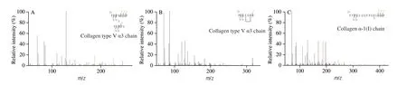
Fig. 5 Representative LC-MS/MS spectra of identified calcium-chelating peptides and modification sites. (A) Inter-Linking type; (B) Loop-Linking type;(C) Mono-Linking type.
4. Conclusions
In conclusion, the optimal conditions for preparation of RBCP-Ca were mass ratio of 2:1 (m/m) at pH 6.0 and 30 °C for 30 min. The chelating sites between RBCP and Ca2+mainly include Asp, Glu, Lys and Arg. Meanwhile, three major chelation modes of Inter-Linking,Loop-Linking and Mono-Linking were observed, among which Inter-Linking was predominant. In an acidic environment, calcium ions were more likely to chelate with amino nitrogen of basic amino acids within peptides, while the underlying mechanism remained to be further investigated. Overall, rabbit bone was a potential source of peptide-calcium chelates, which are promising to be used as functional food ingredients to promote the absorption of calcium in the body.
Conflicts of interest
The authors have no conflicts of interest to be declared.
Acknowledgments
This study was granted by the National Key R&D Program of China (2021YFD21001005), National Natural Science Foundation of China (31972102, 32101980), Special key project of Chongqing technology innovation and application development (cstc2021jscxcylhX0014), Chongqing Technology Innovation and Application Development Special Project (cstc2021jscx-tpyzxX0014).
Appendix A. Supplementary data
Supplementary data associated with this article can be found, in the online version, at http://doi.org/10.26599/FSHW.2022.9250125.
杂志排行
食品科学与人类健康(英文)的其它文章
- GUIDE FOR AUTHORS
- Call for Papers from Food Science of Animal Products
- Ascophyllum nodosum and Fucus vesiculosus ameliorate restenosis via improving inf lammation and regulating the PTEN/PI3K/AKT signaling pathway
- Alleviatory effect of isoquercetin on benign prostatic hyperplasia via IGF-1/PI3K/Akt/mTOR pathway
- Adsorption, in vitro digestion and human gut microbiota regulation characteristics of three Poria cocos polysaccharides
- Voluntary wheel running ameliorated the deleterious effects of high-fat diet on glucose metabolism, gut microbiota and microbial-associated metabolites