Structure elucidation,immunomodulatory activity,antitumor activity and its molecular mechanism of a novel polysaccharide from Boletus reticulatus Schaeff
2023-01-21SiyuanSuXiangDingYilingHouBininLiuZhouheDuJunfengLiu
Siyuan Su,Xiang Ding,Yiling Hou,Binin Liu,Zhouhe Du,Junfeng Liu
a Key Laboratory of Southwest China Wildlife Resources Conservation,College of Life Sciences,China West Normal University,Nanchong 637009,China
b Sericultural Research Institute,Sichuan Academy of Agricultural Sciences,Nanchong 637000,China
c College of Environmental Science and Engineering,China West Normal University,Nanchong 637009,China
Keywords:Boletus reticulatus Schaeff Polysaccharides Structure identif ication Immunomodulatory activity Antitumor activity
ABSTRACT A new water-soluble heteropolysaccharide with a molecular weight of 15 kDa was isolated from the fruiting bodies of Boletus reticulatus Schaeff.Structural characterization results revealed that B.reticulatus Schaeff polysaccharide (BRS-X) had a backbone of 1,6-linked α-D-galactose and 1,2,6-linked α-D-galactose which branches were mainly composed of a terminal 4-linked β-D-glucose and the ratio of D-galactose and D-glucose was 5:1.Bioactivity assays indicated that BRS-X displayed a strong proliferative activity in T cells and B cells and promoted the secretion of immunoglobulin G (IgG),IgE,IgD and IgM.In addition,BRS-X could facilitate the proliferation and phagocytosis of RAW264.7 cells and could significantly inhibit the growth of tumors in S180-bearing mice.The results of transcriptome sequencing analysis illustrated that total 46 genes enriched in MAPK and total 34 genes enriched in PI3K/Akt signaling pathways in BRS-X group.The protein VEGF and VEGFR expression were significantly reduced under the treatment with BRS-X.These findings provide a scientific basis for the edible and medicinal value of BRS-X.
1.Introduction
Cancer threatens human health because of its high mortality rate [1].Although therapeutic medicines and means are being updated and optimized,the side effects of these conventional therapies cause a series of adverse reactions [2,3].Therefore,anti-cancer medicines with good effects and low toxicity are urgently needed.In recent years,natural polysaccharides have received increasing attention in the biomedical field,because they not only have significant antitumor and immunomodulatory activities,but also have low toxic side effects [4-8].
Mushrooms have been used as food for centuries,not only because of its unique flavor,but also because of its important nutritional and medicinal value,owing to its rich bioactive compounds including proteins,polysaccharides,phenolic compounds,terpenes,etc.[9].Fungal immunomodulatory proteins (FIPs) obtained from edible and medicinal mushrooms are a family of enzymes,with a molecular weight approximately 13 kDa and highly similar amino acid sequences [10].FIPs induce the expression of cytokines (IFN-γ or IL-2,etc.) by binding to T cell receptors and activating PTK/PLC/PKCα/p38 signal pathways,which produces cytotoxic responses towards cancer cells [11].FIPs inhibit telomerase activity by regulating transcription and translation,induce release of intracellular calcium and up-regulation of apoptosis-related genes,activate the ER/calcium/CaN/Akt/ mTOR/p70S6K pathway to induce formation and accumulation of autophagosome,block the EGF/PI3K/Akt pathway and Stat3 pathway,and activate NF-κB/MMP-9 pathway,resulting in the destruction of genomic integrity of cancer cells,apoptosis of cancer cells and inhibition of tumor invasion and migration [12].The potent antitumor activity of phenols isolated from mushrooms has also been confirmed.Phenolic compounds suppress NF-κB activity by blocking the activation of IκBαto regulate expression of NFκB-dependent anti-apoptotic proteins,proliferative proteins and metastatic proteins,and restrain focal adhesion kinase (FAK)-mediated ERK1/2 and PI3K/Akt signal transduction,thereby inhibiting MMP-2,MMP-9 and uPA,leading to the growth,migration and invasion of tumor cells to be hindered [13].Terpenes from various mushrooms are cytotoxic to several cancer cell lines (A549 cells,HeLa cells,HT29 cells,HepG2 cells,4T1 cells,MCF-7 cells) [13].They stimulate tumor cells to elicit the expression of proteins that play an important role in preventing the cell cycle from being in the G0/G1 phase,cell apoptosis and oxidative stress [14],and they also possess immunomodulatory activities,including stimulating nuclear factor NF-κB and MAPK signaling pathway [15].According to reports,mushroom polysaccharides exhibit a variety of biological functions,including anti-cancer,anti-diabetic,anti-inflammatory,antibacterial,anti-oxidant,hypoglycemic and immunomodulatory activities [16-19].The anti-tumor activity of mushroom polysaccharides is realized by activating the immune system and directly inhibiting the growth of cancer cells.They bind to different membrane receptors (TLR,CR3,Dectin-1,NLR,RLR) triggering a series of signal transduction responses,which can enhance the immune response of B cell secreting antibodies and generate a systemic T cell immune response against tumor antigens,encouraging tumor regression and animal survival [20,21].By means of regulating the Bcl-2 genome encoded a large number of proteins related to apoptosis,inhibiting the production of NO and MAPK and Akt signal transduction involved angiogenesis,and restricting the ERK1/2 or ERK5 pathway to arrest the cell cycle in the G1 phase,mushroom polysaccharides promote the apoptosis of cancer cells and inhibit their migration [22].Theβ-glucan fromLentinus edodes(Berk.) Sing had significant anti-tumor activity,and was successfully developed as an anti-tumor medicine through clinical trials [23].
Mushrooms of the genusBoletusare widely distributed in the world,and are one of the most delicious and consumed edible fungi [24].A number of studies have been reported about the bioactivities of their polysaccharides (Table 1),such as antiglycation,antidiabetic,antioxidant,immunoregulatory and antitumor activities in various tumors.BoletusreticulatusSchaeff belonging to the family fungi,Basidiomycota,Agaricomycetes,Boletales,Boletaceae andBoletusis widely found under the mixed forest in the summer and autumn.Due to its delicious and delicate textures,rich protein content,low fat content and relatively high content of polyunsaturated fatty acid and tocopherols,B.reticulatusSchaeff has good antioxidant properties and can be used in the food,cosmetics and pharmaceutical industries [25,26].However,little is known about functional ingredients of it.In the light of the strong biological activities of the genusBoletuspolysaccharides and the absent researches of immunoregulatory and antitumor activities potential ofB.reticulatusSchaeff,this study focused on the polysaccharide fromB.reticulatusSchaeff.
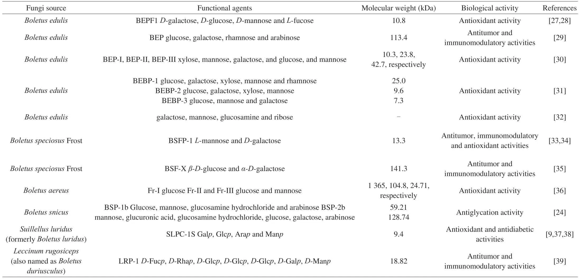
Table 1 Source,composition and bioactivity of some polysaccharides from the genus Boletus mushrooms.
In the present study,a novel water-soluble polysaccharide(BRS-X) was extracted,isolated and purified from the fruiting bodies ofB.reticulatusSchaeff from Xiaojin County,China.Its chemical structure was first elucidated.In addition,the immunoregulatory and anti-tumor activities of BRS-X were determined using the cell modelin vitroand S180-bearing mice modelin vivo.This study also aimed to identify signaling pathways in tumor cells to help determine the molecular mechanism underlying the anti-tumor activity of BRS-X.
2.Materials and methods
2.1 Materials and chemicals
Fresh fruiting bodies ofB.reticulatusSchaeff were collected fromXiaojin County,China.A voucher specimen was stored in the Key Laboratory of Southwest China Wildlife Resources Conservation,College of Life Sciences,China West Normal University.The ethanol was purchased from Swancor Shanghai Fine Chemical Co.,Ltd.(Shanghai,China).Trifluoroacetic acid (TFA),standard monosaccharide and dextran were purchased from Sigma-Aldrich(Shanghai) Trading Co.,Ltd.(Shanghai,China).DEAE-cellulose(DE-52) was purchased from Beijing Solarbio Science&Technology Co.,Ltd.(Beijing,China).Cell counting kit (CCK-8 cell counting kit)was purchased from Dojindo Molecular Technologies,Inc.(Shanghai,China).Lipopolysaccharide (LPS) fromEscherichia coli055:B5,PBS buffer,0.5% Trypsin-EDTA,neutral red and dimethyl sulfoxide(DMSO) were purchased from Sigma-Aldrich Inc.(Missouri,USA).RPMI1640 medium (phenol red free) and fetal bovine serum (FBS)were purchased from Gibco Inc.(New York,USA).Mouse ELISA(enzyme linked immunosorbent assay) Kit (VEGF,VEGFR) were purchased from Boster Biological Technology Co.Ltd.(Wuhan,China).Mouse ELISA (enzyme linked immunosorbent assay) Kit(IgG,IgD,IgE,IgM) were purchased from KALANG Biological Technology Co.Ltd.(Shanghai,China).All analytical reagents were of analytical grade.
2.2 Extraction and purification of polysaccharides from B.reticulatus Schaeff
Fresh fruiting bodies ofB.reticulatusSchaeff were dried in an oven,and the dried fruiting bodies were put into a pulverizer and crushed into powder.DryB.reticulatusSchaeff powder (400 g) was boiled in water for 3 times (6 h for each) [40].The supernatant was concentrated after centrifugation.Three times volume of 95% ethanol were added in the supernatant to precipitate crude polysaccharides.The crude polysaccharides were purified by DEAE cellulose (DE-52)column and dialysis (7 000 Da,Biosharp),drying in vacuum freezedrying machine (ALPHA2-4LD plus,Christ).The finalB.reticulatusSchaeff polysaccharide was named BRS-X.
2.3 Determination of the molecular weight
The molecular weight of BRS-X was determined via highperformance gel permeation chromatography (HPGPC) [17,41].A total of 10 mg BRS-X was weighted,dissolved in 1 mL distilled water and filtered (0.22 μm pore).The measured data was analyzed via Empower Pro GPC software (version B.01.02;Agilent Technologies Inc.,USA) and the dextran standards dextran standards (mw: 20 000,45 000,65 000,125 000,195 000) were used as references.
2.4 Monosaccharide composition analysis
The monosaccharide composition of BRS-X was analyzed by high performance liquid chromatography (HPLC) (Agilent 1100,USA).A total of 20 mg BRS-X was dissolved in 5 mL trifluoroacetic acid (TFA)solution (2 mol/L) and hydrolyzed for 6 h (100 °C).The supernatant was dried and washed with distilled water to remove residual TFA(repeated 3 times).Operating conditions: RID detector;injection volume: 10 μL;column temperature: 35 °C [3].The monosaccharide samples were used as standards.
2.5 Methylation analysis and GC-MS
Methyl iodide reagent was used to obtain methylated BRS-X [42].The dry methylated product was dissolved in 2 mol/L TFA,then hydrolyzed for 6 h (100 °C).Silane reagent was used to prepare derivatized product detected by GC-MS (Agilent 7890A,USA).The initial temperature set at 80 °C and maintained for 3 min,with a linear increase to 200 °C at a rate of 10 °C/min,then maintaining at 200 °C for 10 min [43].
2.6 Nuclear magnetic resonance (NMR) assay
A total of 50 mg BRS-X was dissolved in 500 μL D2O [35].The1H NMR spectra,13C NMR spectra,1H-1H COSY spectra (the1H-1H COSY spectrum reflects the coupling relationship between adjacent hydrogen),HMQC spectra (the information given by the HMQC spectrum is the relationship between directly connected carbon and hydrogen) and HMBC spectra (the HMBC spectrum provides the correlation between carbon and hydrogen separated by two or three bonds) were analyzed by the Varian Unity INOVA 400/45 (Varian Medical Systems,USA) and internal standard was tetramethylsilane [34].
2.7 Cell lines and reagents
Macrophages RAW264.7 cell line,B cell line,T cell line and S180 cell line (sarcoma cell) were purchased from the cell bank of the Typical Culture Preservation Committee of the Chinese Academy of Science (Shanghai,China).All cells were cultivated in RPMI 1640 medium with 10% FBS,1% penicillin (100 IU/mL) and streptomycin (100 mg/L) at 37 °C with 5% CO2.
2.8 T cells,B cells and RAW264.7 cells proliferation assay
Pharmacological evaluation of BRS-X on T cells,B cells and RAW264.7 cells proliferation was examined via CCK-8 method [44].Cells (1 × 10⁵cells/mL) were added to a 96-well plate (100 μL/well)and incubated for 24 h (5% CO2,37 °C) [3].Different concentrations of BRS-X (final concentration 5,10,20 μg/mL) were added to the 96-well plate (100 μL/well),incubated for 24 h.LPS (final concentration 5 μg/mL) and the cell culture medium were used as positive control and negative control,respectively [35].Proliferation assay was analyzed via instructions of CCK-8.The value of optical density (OD)was detected at 450 nm.The way of calculating cell viability was:cell proliferation rate (%)=[(As-Ac)/(Ac-Ab)]× 100,whereAcwas the absorbance of negative control group,Abwas the absorbance of blank group,Aswas the absorbance of experimental groups.The Leica Microsystems inverted fluorescence microscope (DMI4000B,Germany) was used to observe the cell morphology and cell numbers alterations.
2.9 Immunoglobulin analysis in vitro
In order to understand alterations of IgG,IgE,IgD and IgM secreted by B cells,ELISA kits were applied to detecte these immune factors.
2.10 Pharmacological evaluation for macrophage phagocytic activity
RAW264.7 cells (1 × 10⁵cells/mL) were pre-cultured in the 96-well plate (100 μL/well) for 24 h.RPMI 1640 medium,LPS(final concentration 5 μg/mL) and different concentrations of BRS-X(final concentration 5,10,20 μg/mL) were added to the 96-well plate(100 μL/well),respectively.After incubated for 24 h,neutral red reagent (0.075 g/L) or fluorescent microspheres reagent (1 × 10⁷fluorescent microspheres/mL) was added to the 96-well plate.After 30 min,the neutral red reagent or fluorescent microspheres reagent was discarded and washed three times with PBS,following by adding lysis buffer (ethanol : glacial acetic acid,1:1) (200 μL/well).After incubated for 2 h,the OD at 540 nm (neutral red reagent) or 505 nm(fluorescent microspheres reagent) was measured.
2.11 The inhibitory effect of BRS-X on S180 cells in vitro
The procedure was the same as 2.8.It should be noted that the final concentration of mannanide (positive control) was 5 μg/mL,and the final concentration of BRS-X was 2.5,5,10 μg/mL.The inhibition activity of BRS-X on tumor cellsin vitrowas calculated according to the following formula: Inhibition activity (%)=[(Ac-As)/(Ac-Ab)]×100,whereAcwas the absorbance of negative control group,Abwas the absorbance of blank group,Aswas the absorbance of experimental groups.
2.12 Animals
Twenty Kunming female mice (6 weeks,(18 ± 2) g) were purchased from North Sichuan Medical College,Nanchong,China.In order to avoid casualties caused by aggressive behavior among male mice,docile female mice were selected as the experimental subjects.Before the experiment,all mice which housed in pairs were placed in suitable laboratory conditions and allowed to adapt to the facilities for one week.All animal experimental procedures were approved by the Animal Care and Use Committee of North Sichuan Medical College.
2.13 Ascites tumor-bearing mice model
S180 tumor cells were inoculated into the left axilla of all health mice.Mice were randomly divided into three groups (n=5).After 24 h,BRS-X (20 mg/kg) and positive control mannatide (20 mg/kg)were given by intragastric administration.The model control group was given with the equal volume of saline.The treatments fed to mice were daily for 8 continuous days.After 8 days,the mice were sacrificed,then bodies and tumor tissues were weighed accurately.The tumor inhibition rate was calculated according to the following formula: Inhibition rate (%)=(the mean tumor tissues weight of model control group -the mean tumor tissues weight of treated group)/the mean tumor tissues weight of model control group × 100.
2.14 Transcriptome analysis of tumor tissues
The collected tumors of control group,mannatide group and BRS-X group were used to carry out transcriptome assay.The collected tumors of control group,mannatide group and BRS-X group were used to carry out transcriptome assay.The tumors,quickly freezed by liquid nitrogen,were sent to Novogene Bioinformatics Technology Co.Ltd.,Beijing,China.The remaining tumor tissues were stored at -80 °C.The total RNA of the sample was extracted by the Trizol method.Agarose gel electrophoresis was used to detect whether the RNA was contaminated.The Agilent 2100 Bioanalyzer detected the integrity of RNA.After the samples were tested to be qualified,the cDNA library was constructed,which was sequenced by Next-Generation Sequencing (NGS) technology based on Illumina hiseq sequencing platform.By removing contaminated reads,reads with unknown base N content >5% and low-quality reads,highquality clean reads can be obtained.
2.15 KEGG enrichment analysis
The counted genes need to meet these conditions: FPKM(fragments per kilobase of transcript sequence per millions reads mapped) value was more than 0.3 and relative transcriptional inhibition rate or relative transcriptional promotion rate was more than 5% .The relative transcriptional inhibition rate was calculated according to the following formula: Relative transcriptional inhibition rate (%)=(model group gene relative transcription amount -treatment group gene relative transcription amount)/model group gene relative transcription amount × 100.Specially,the gene relative transcription amount (%)=gene transcription amount/atcbgene transcription amount × 100,and theatcbgene as an example for the reference gene.These genes were introduced into KEGG(Kyoto Encyclopedia of Genes and Genomes) to analyze cell signaling pathways drawn by Pathway Builder Tool 2.0 software.The biological function and expression mechanism of protein in tumor cells were analyzed by National Center for Biotechnology Information (NCBI).
2.16 Cytokines determination of tumor tissues
Protein of tumor tissue was extracted on ice by tissue lysate containing proteinase inhibitor.The purity and concentration of extracted protein was detected by Ultramicro ultraviolet spectrophotometer (Thermo NanoDrop 2000,USA).In order to understand alterations of protein vascular endothelial cell growth factor receptor (VEGFR) and vascular endothelial cell growth factor(VEGF) in BRS-X group compared with model control group,ELISA kits were used to detect these factors.
2.17 Statistical methods
Data were showed as the mean ± standard deviation (SD).All statistical comparisons were analyzed using a one-way analysis of variance (ANOVA) test followed by Student Newman Keuls test with SPSS 17.0 software.Values ofP<0.05 were considered a statistically significant difference.
3.Results and discussion
3.1 Chemical characterization of BRS-X
3.1.1 Monosaccharide composition and molecular weight
The results of HPLC could directly reflect the composition and ratio of monosaccharides of the polysaccharide (not shown).The retention time of the standards was 14.214 min forD-glucose,16.704 min forD-galactose and 18.614 min forD-mannose,respectively.Compared with the retention time of the monosaccharide standard,the two peaks in the results of monosaccharide analysis of polysaccharides were glucose (the retention time was 14.208 min) and galactose (the retention time was 16.692 min),respectively.The ratio of galactose to glucose in BRS-X was determined to be around 5:1 based on the area ratio of the two peaks.Therefore,the HPLC results indicated that BRS-X was composed of two kind of monosaccharides,D-galactose andD-glucose,and their ratio was about 5:1.
The results were in line with those of reported studies,which indicated that glucose and galactose were monosaccharides commonly found in fungus [9].The highest monosaccharide content of polysaccharide LRP-1,BEP,BSFP-1,BSF-X was glucose (39.13%) [39],galactose (35.56%) [29],mannose (69.78%) [34],and glucose (about 2/3) [35],respectively,revealing that polysaccharides of different species ofBoletushad various monosaccharide composition and content.The polysaccharides inBoletusfungus existed different galactose contents as well.The BRS-X had very high galactose contents,which was strongly different from glucose as the main monosaccharide in numerous edible fungi.It was interesting to note that polysaccharides with galactose as the main chain have attracted the attention of researchers and have been reported to have a variety of biological activities [38].For instance,Zheng et al.[45]found that the mushroom polysaccharide LCP-2 (with the highest galactose content) compared with LCP-1 and LCP-3 from the Boletaceae family had the strongest ability to promote the proliferation activity of RAW264.7 cells.The polysaccharide BEP with galactose as the backbone could inhibit the metastasis of renal cancerin vivoand enhanced the body’s immunity [29].On the other hand,the content of galactose in BRS-X (approximately 5/6) was higher than that in LCP-2 (47.21%),BEP (35.56%) and BSF-X (about 1/3),suggesting that BRS-X possessed a higher degree of solubility in water [46].In general,the higher the solubility of polysaccharides,the stronger the biological activities and the good solubility is better for industrial applications [47].
The molecular weight and purity of BRS-X were determined via a HPGPC method.The HPGPC elution curve of BRS-X had a single peak,which indicated that BRS-X had high homogeneity (Fig.1A).The weight-average molecular weight (mw) was 150 935 Da,peak molecular weight (mp) was 14 138 Da,number-average molecular weight (mn) was 4 093 Da and the polydispersity was 36.88.
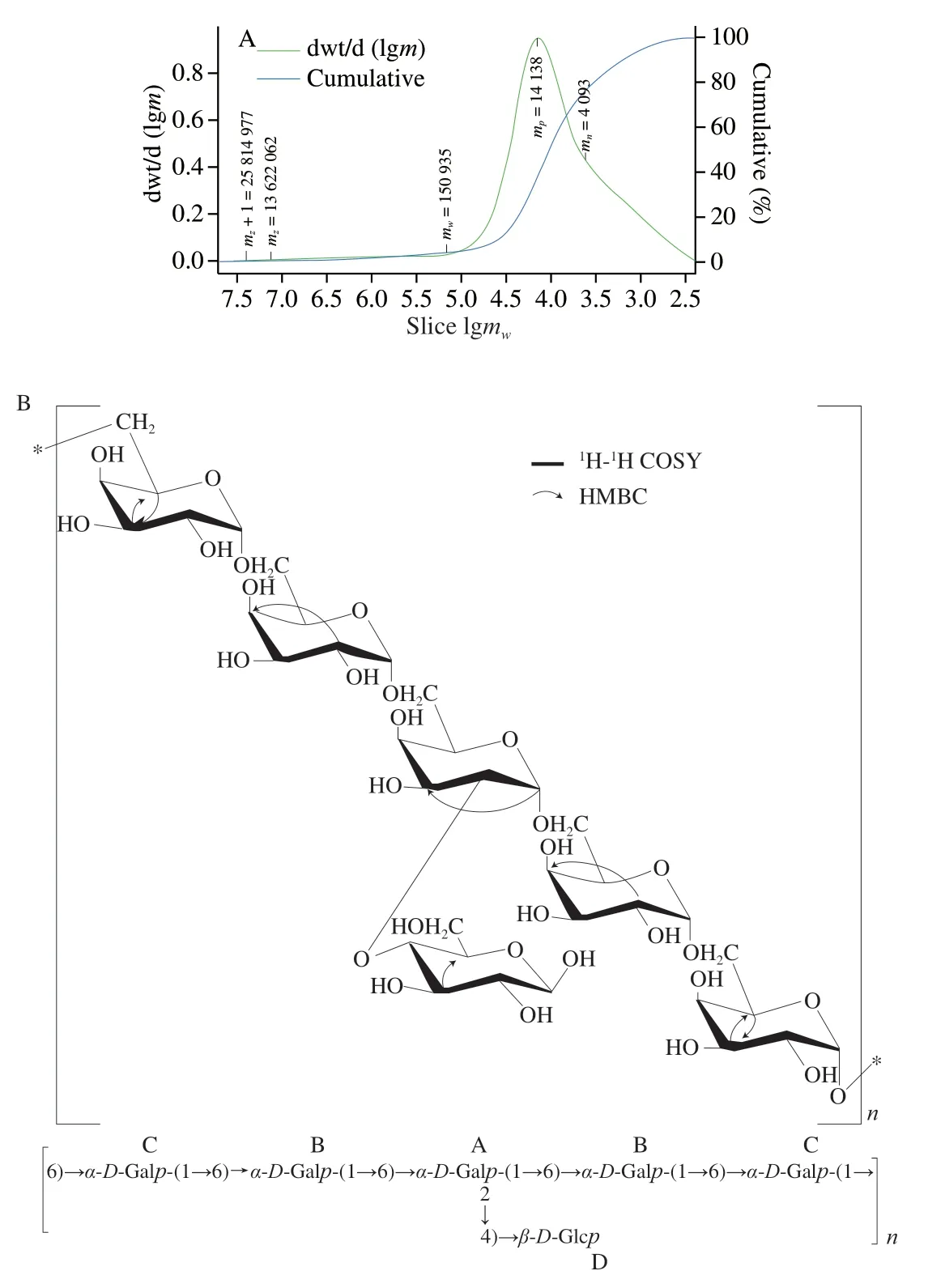
Fig.1 The molecular weight of BRS-X (A);Predicted chemical structure and key 2D-NMR correlations of BRS-X (B).
On account of different structures,different polysaccharides are in possession of different molecular weights,and the molecular weight is considered to be a critical factor affecting the biological activity of polysaccharides [48].In general,polysaccharides with large molecular weight have better pharmacological activities [22,49].Polysaccharides BSP-2b with highermwobtained fromBoletus snicusexhibit stronger antiglycation effects than BSP-1b [24].The(1→3)-β-glucan fraction extracted and purified fromGrifola frondosawith the highest molecular weight (800 kDa) displays the most potent antitumor and immunomodulatory activities [50].However,few low molecular weight polysaccharides have comparable activities [51],indicating that different polysaccharides have different ranges of optimal molecular weight for bioactivities.
3.1.2 Methylation analysis
The glycosidic linkage patterns and molar ratios of monosaccharide residues were determined by methylation and GC-MS analysis (Table 2).As summarized in Table 2,the derivatives of BRS-X were identified as 3,4-Me2-Galpand 1,2,3,6-Me4-Glcpwith the molar ratio of 5:1 approximately,which was in accordance with the results of HPLC.Considering that OH on C2 is not easy to be methylated due to steric hindrance,which results in incomplete methylation in the experiment,it is concluded that BRS-X is composed of 1,6-Galp,1,2,6-Galpand 4-Glcp.On the basis of monosaccharide composition and methylation results,it was inferred that the main chain of BRS-X was 1,6-α-D-Galpand the side chain was a 4-β-D-Glcplinked to the O-2 of Galp(Fig.1B).

Table 2 Linkage patterns of sugar residues from the polysaccharide (BRS-X).
It was reported that polysaccharides mainly composed of glucans linked byβ-(1→3) bonds usually possessed strong biological activity,because the helical conformation formed was a very important factor for intracellular biological recognition,andβ-(1→6) bondlinked glucans exhibited lower activity due to its flexibility in conformation [51,52].However,researches showed BSF-X consisting of glycosidic linkagesβ-(1→4) andα-(1→6) [35],BSFP-1 being withα-(1→6),andα-(1→4) [34],BEP retainingα-(1→6),andα-(1→3) [29],they all had significant immunomodulatory and anti-tumor activities,indicating that glycosidic linkage was only one factor affecting the activity of polysaccharide.
3.1.3 NMR spectrum analysis
To further comprehend the full structural characterization of BRS-X,the polysaccharide was analyzed by way of NMR spectroscopy (1H NMR,13C NMR,1H-1H COSY,HMQC and HMBC).In the1H NMR (400 Hz) spectrum (Fig.2A),the hydrogen signals atδ4.93,4.93 and 4.85 were assigned to anomeric protons ofα-pyranose unit,while the signal atδ4.64 was attributed to anomeric protons ofβ-pyranose forms [17,47].The peaks atδ4.9 were assigned to anomeric protons signals of 1,2,6-linkedα-D-Galp(A) and 1,6-linkedα-D-Galp(B),whereas the resonances atδ4.85 and 4.64 were attributed to anomeric protons signals of 1,6-linkedα-D-Galp(C) and 4-linkedβ-D-Glcp(D),respectively.The peaks atδ3.32-4.07 were composed of a large number of overlapping hydrogen signals,which were attributed to protons from C-2 to C-6 in each monosaccharide [53,54].The signal atδ1.1 was attributed to the methyl signal of ethanol because the ethanol precipitation method was used to extract polysaccharides.The hydrogen signal for deuteroxide wasδ4.70 [35,47].

Fig.2 The 1H NMR spectra of BRS-X (A);The 13C NMR spectra of BRS-X (B);1H-1H COSY spectrum of BRS-X (C).
In the13C NMR spectra (Fig.2B),the inexistence of chemical shift withinδ106-109 indicated no furan rings in BRS-X [3].The signals atδ101.41,97.79,97.78 and 97.72 were anomeric carbon peaks.The chemical shifts atδ101.41 and 97.79 were assigned to anomeric carbon signals of 1,6-linkedα-D-Galp(B) and 1,2,6-linkedα-D-Galp(A),and the resonances atδ97.78 andδ97.72 were attributed to anomeric carbon signals of 1,6-linkedα-D-Galp(C)and 4-linkedβ-D-Glcp(D),respectively.The strong chemical signals withinδ80-55 could be attributed to carbon signals of C2-C6 of glucose and galactose.The chemical signals of each monosaccharide residue in BRS-X were determined by comparing with the literature[3,35,55,56].
Each proton signal of monosaccharides in BRS-X could be obtained by1H-1H COSY spectrum.In the1H-1H COSY spectrum(Fig.2C),the signal A (δ4.93/3.53) represented the correlation between H-1 and H-2 of the 1,2,6-linkedα-D-Galp(A),the signal B(δ4.93/3.65) represented the correlation between H-1 and H-2 of the 1,6-linkedα-D-Galp(B),the signal C (δ4.85/3.68) represented the correlation between H-1 and H-2 of the 1,6-linkedα-D-Galp(C) and the signal D (δ4.64/3.36) represented the correlation between H-1 and H-2 of the 4-linkedβ-D-Glcp(D),respectively [54,57].Other signals in the1H NMR spectra of BRS-X were assigned in the light of the aforementioned results (not shown).
In addition,the direct correlation of carbon and hydrogen signals could be obtained by HMQC spectra.In the HMQC spectrum (Fig.3A),the signal A (δ4.93/97.79) represented the correlation between H-1 and C-1 of the 1,2,6-linkedα-D-Galp(A),the chemical signal B(δ4.93/101.41) represented the correlation between H-1 and C-1 of the 1,6-linkedα-D-Galp(B),the signal C (δ4.85/97.78) represented the correlation between H-1 and C-1 of the 1,6-linkedα-D-Galp(C)and the signal D (δ4.64/97.72) represented the correlation between H-1 and C-1 of the 4-linkedβ-D-Glcp(D),respectively.Other signals in the13C NMR spectra of BRS-X were assigned in accordance with the aforementioned results (not shown).
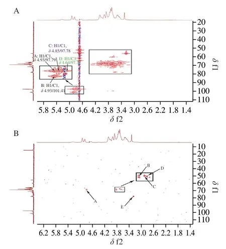
Fig.3 HMQC spectrum of BRS-X (A);HMBC spectrum of BRS-X (B).
HMBC provided a correlation spectrum between carbon and hydrogen separated by two or three bonds.In the HMBC spectrum of BRS-X (Fig.3B),the signal A (δ4.93/70.11) conformed to the correlation between H-1 and C-3 of the 1,2,6-linkedα-D-Galp(A),the signal B (δ3.84/70.14) was in accord with the correlation between H-3 and C-5 of the 1,6-linkedα-D-Galp(C),the signal C (δ3.74/68.00)tallied with the correlation between H-5 and C-3 of the 1,6-linkedα-D-Galp(C),the signal D (δ3.65/68.30) indicated the correlation between H-2 and C-4 of the 1,6-linkedα-D-Galp(B) and the signal E(δ3.28/78.03) represented the correlation between H-3 and C-5 of the 4-linkedβ-D-Glcp(D),respectively.The analysis results of the NMR spectra were consistent with the GC-MS analysis.
3.2 Biological activities of BRS-X
T lymphocytes and B lymphocytes are the main cells that mediate the body’s specific immunity.Macrophages have many immune functions,such as immune defense,immune surveillance,immune regulation and antigen presentation.By presenting antigens to T lymphocytes and B lymphocytes,macrophages can phagocytize and remove foreign bodies,aging and dead cells,and thus participate in the non-specific and specific immune response of the body [58].
3.2.1 Effects of BRS-X on the proliferation of T cells in vitro
The analysis of results showed that BRS-X group (final concentration 5,10,20 μg/mL) could significantly promote the proliferation of T cells compared with the negative control group(Fig.4A).When the concentration of BRS-X was 20 μg/mL,the proliferation efficiency of T cells reached the maximum and increased by 38.88% .At the same concentration (5 μg/mL),the proliferation rate of T cells stimulated by BRS-X (14.77%) was higher than that stimulated by LPS (13.00%).Cell morphology observation demonstrated that compared with cells in negative control group,cells stimulated by BRS-X formed large clusters and increased in number(Fig.4B).
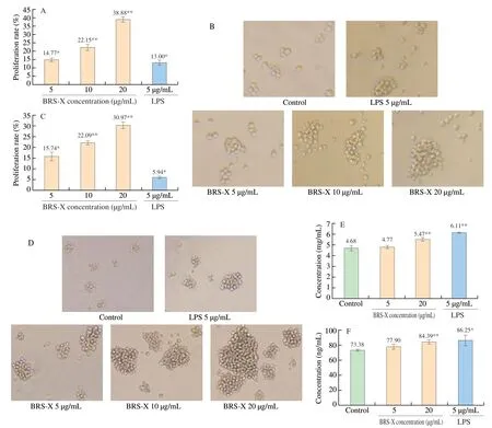
Fig.4 Effect of BRS-X on T cells and B cells in vitro.(A) Proliferation rate of T cells stimulated by BRS-X.(B) The effect of BRS-X on T cell morphology (10× 10).(C) Proliferation rate of B cells stimulated by BRS-X.(D) The effect of BRS-X on B cell morphology (10 × 10).(E) The secretion of IgG in B cells stimulated by BRS-X.(F) The secretion of IgE in B cells stimulated by BRS-X.(G) The secretion of IgD in B cells stimulated by BRS-X.(H) The secretion of IgM in B cells stimulated by BRS-X.* P <0.05 and ** P <0.01 in comparison with negative control group.

Fig.4 (Continued)
3.2.2 Effects of BRS-X on the proliferation of B cells in vitro
The results indicated that compared with the negative control group,the BRS-X group (final concentration 5,10,20 μg/mL)was capable of significantly promoting the proliferation activity of B cells in a certain concentration range (Fig.4C and 4D).When the concentration of BRS-X was 20 μg/mL,the proliferation effect was maximized,and the proliferation rate was 30.97% .Moreover,the proliferation rate of 5 μg/mL BRS-X was higher than that of 5 μg/mL LPS.
Studies have reported that polysaccharides can promote the proliferation and differentiation of T and B lymphocytes by accelerating the progression of the cell cycle from G0/G1 phase to S phase and G2/M phase [59].In our previous research,Lactarius deliciosusGray polysaccharide (LDG-M),B.speciosusFrost polysaccharide (BSF-X) andTricholoma matsutakepolysaccharide(TMP-B) could promote the proliferation of immune cells and the percentage of cells in S phase and G2/M phase increased significantly [35,60,61].Polysaccharides can also promote lymphocytes proliferation by activating immune cell surface pattern recognition receptors (PRRs) to trigger intracellular signal transduction such as MAPK and NF-κB [62,63].However,due to the wide variety of polysaccharides,different pathways of animal immune regulation and the interaction between signal pathways make immune regulation of polysaccharide very complicated.Therefore,the mechanism of polysaccharide BRS-X on animal immune regulation still needs to be further studied.
3.2.3 Effects of BRS-X on B cells secreting immunoglobulin in vitro
Compared with the negative control group,BRS-X was able to promote B cells secreting IgG,IgE,IgD and IgM (Figs.4E-H).When the concentration of BRS-X was 20 μg/mL,the secretion of IgG,IgE and IgD reached the maximum,which increased 16.99%,15.01%,25.17%,respectively.The results of IgM demonstrated that BRS-X could also slightly promote the release of IgM from B cells,which increased 5.86% .
3.2.4 Effects of BRS-X on the proliferation of RAW264.7 cells in vitro
The results revealed that BRS-X (final concentration 5,10,20 μg/mL) could significantly promote the proliferative activity of RAW264.7 cells (Fig.5A).When the concentration of BRS-X was 10 μg/mL,the proliferation efficiency of RAW264.7 cells reached the maximum and increased by 122.49%,which was higher than that of the positive control group (LPS).The results of cell morphology showed that the macrophages generally adhered to the wall and showed an elliptical shape and after being activated,they often extended pseudopodia and showed irregular shapes under the stimulation of BRS-X (Fig.5B).
3.2.5 Effects of BRS-X on the phagocytic function of RAW264.7 cells in vitro
Neutral red,a macromolecular substance,entered macrophages via endocytosis,which had a strong absorption peak at 540 nm [64].Compared with the negative control group,BRS-X significantly facilitated the phagocytosis of RAW264.7 cells (Fig.5C).When BRS-X reached a concentration of 10 μg/mL,the phagocytosis activity reached the maximum,which was 67.74% .Furthermore,the cell phagocytosis activity at a concentration of 5 μg/mL BRS-X was even greater than 5 μg/mL LPS.
When the fluorescent microspheres were phagocytized by RAW264.7 cells,an inverted fluorescence microscope could observe their fluorescence signals [65].Compared with the negative control group,BRS-X significantly accelerated the phagocytosis of RAW264.7 cells (Fig.5D and 5E).When the concentration of BRS-X was 10 μg/mL,the phagocytosis activity increased 10.45% higher than that of LPS group.
Phagocytosis is an important early step to effectively eliminate pathogens and an important way for macrophages to exert immune function.The above results indicated that BRS-X not only promoted the proliferation of RAW264.7,but also enhanced the phagocytic function of RAW264.7,which may play an important role in resisting pathogen invasion,removing abnormal cells and cell debris,and maintaining body homeostasis.
3.2.6 The inhibitory effect of BRS-X on S180 cells in vitro
The results showed that BRS-X (final concentration 2.5,5,10 μg/mL) significantly inhibited the growth of S180 cells compared with the negative control groupin vitro.When the concentration of BRS-X was 10,5,2.5 μg/mL,the inhibition rates were 39.74%,35.71%,and 22.12%,respectively (Fig.5F).
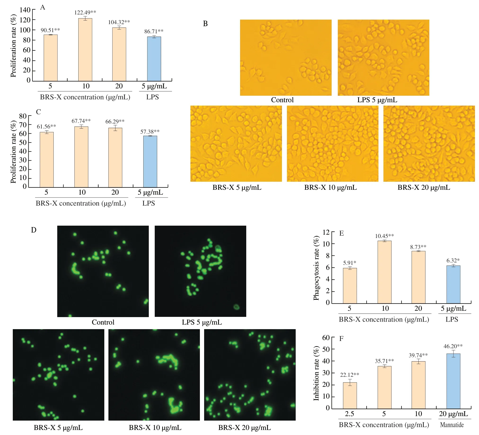
Fig.5 Effect of BRS-X on RAW264.7 cells and S180 cells in vitro.(A) Proliferation rate of RAW264.7 cells stimulated by BRS-X.(B) The effect of BRS-X on RAW264.7 cell morphology (10 × 10).(C) Phagocytosis rate of RAW264.7 cells stimulated by BRS-X (neutral red).(D) The map of RAW264.7 cells phagocytosing fluorescent microspheres.(E) Phagocytosis rate of RAW264.7 cells stimulated by BRS-X (fluorescent microspheres).(F) Inhibition rate of BRS-X on the growth of S180 cells.* P <0.05 and ** P <0.01 in comparison with negative control group.
3.2.7 The inhibitory effect of BRS-X on S180 tumor in vivo
Each group of mice and tumor tissues were collected.The results showed that there was no significant difference in weight of mice bodies between BRS-X group and model control group,indicating that BRS-X had no influence on the growth of mice.However,the growth of tumor was significantly inhibited by BRS-X compared with model control group and its inhibition rate was 37.86% (Table 3 and Fig.6A).
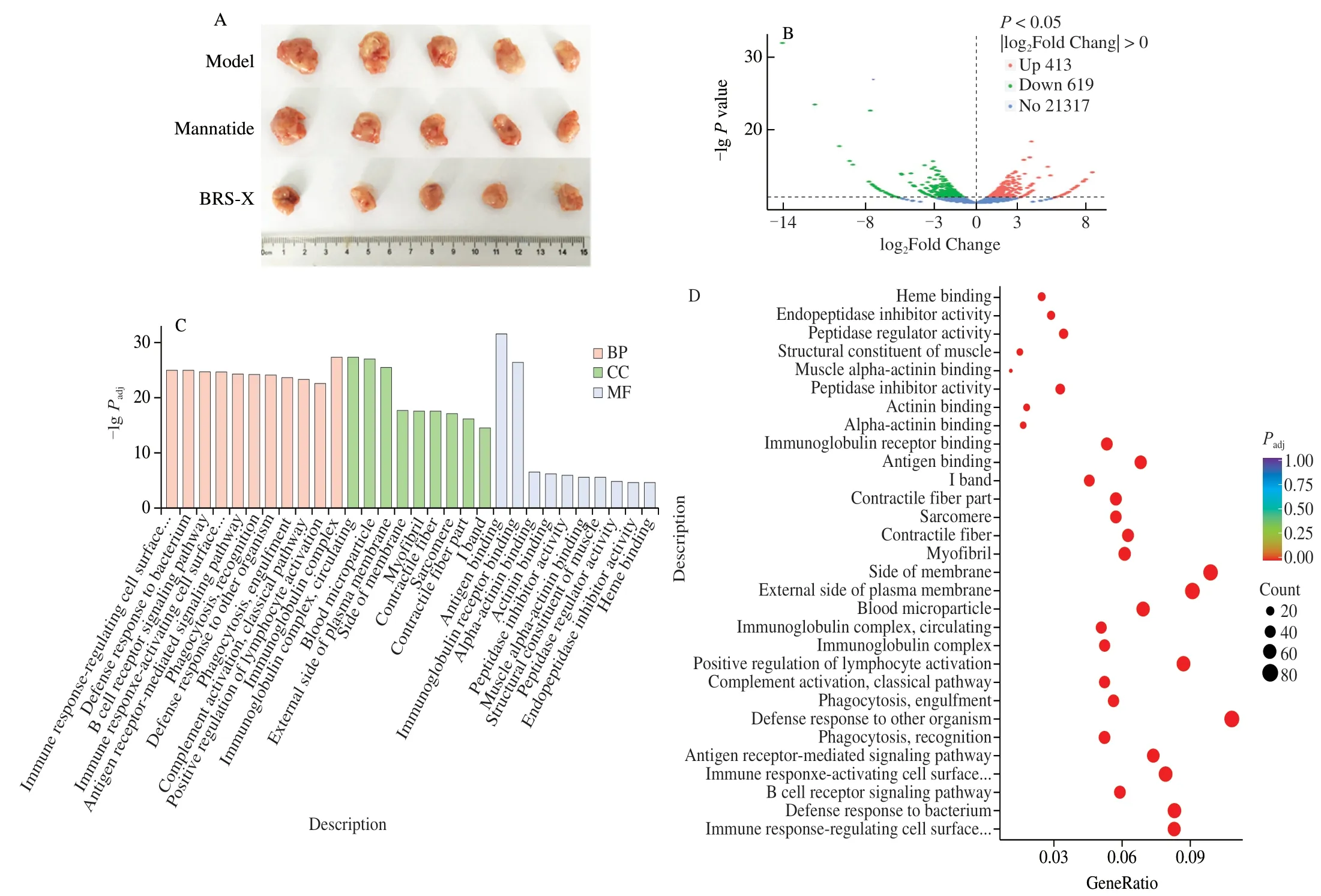
Fig.6 Transcriptome analysis of BRS-X on S180-bearing mice.(A) Tumors size of S180-bearing mice.(B) Volcano map analysis of genes in BRS-X group compared with model group.(C) The GO functional classification of differential genes in BRS-X group compared with model group.(D) The GO enrichment analysis of the differential genes of in BRS-X group compared with model group.

Table 3 Inhibitory effect of the polysaccharide (BRS-X) on S180-bearing mice.
The S180-bearing mice model has been widely used to explore the anti-tumor activity of polysaccharidesin vivo.The polysaccharide BSF-X ofB.speciosusForst with a moleculer weight of 141.3 kDa was composed of glucose and galactose,which was the same as the monosaccharide composition of BRS-X,and its main chain was connected byβ-(1→4) glycosidic linkages [35].BSF-X exhibited effective inhibition of the growth of S180 tumors,and the inhibition rates were 33.33% (20 mg/kg) (lower than BRS-X) and 61.35% (40 mg/kg),respectively [35].The polysaccharide BSF-A with a moleculer weight of 13.3 kDa was made up of galactose and mannose with the main chain linked byα-(1→4) glycosidic bonds,of which the inhibition rate of S180 tumor growth was 53.12% (20 mg/kg) and 62.45% (40 mg/kg),respectively [33,34].Interestingly,the tumor suppressive effects of BRS-X and BSF-X were much lower than that of BSF-A when the dose was 20 mg/kg,but the inhibitory effects of BSF-X and BSF-A were very similar when the dose was 40 mg/kg,indicating that not only the structure of polysaccharides could affect the anti-tumor activity,but also the concentration of polysaccharides.Within a certain concentration range,the anti-tumor effect of some polysaccharides changes greatly,and some polysaccharides change little.
3.2.8 Transcriptome analysis
The transcriptome analysis of tumor tissues showed that there were 413 up-regulated genes,619 down-regulated genes and 21317 unchanged genes in total (Fig.6B).According to the GO (Gene Ontology) functional classification of tumor transcriptome unigenes,31255 unigenes in BRS-X group were annotated and classified into three categories: biological process (BP),cellular component (CC),and molecular function (MF) (Fig.6C).Statistics of the number of genes in each category found that genes of defense response to other organism and positive regulation of lymphocyte activation accounted for a large proportion in the category of biological process,genes of side of membrane and external side of plasma membrane accounted for a large proportion in the category of cellular component,and genes of antigen binding and immunoglobulin receptor binding accounted for a large proportion in the category of molecular function,respectively (Fig.6D).Via analyzing the genes in KEGG pathways in cancer,there were 136 down-regulated genes and 21 up-regulated genes in BRS-X group compared with model group.
3.2.8.1 Genes with relative transcriptional inhibition rate more than 25%
There were 23 down-regulated genes with relative transcriptional inhibition rate more than 25% (Fig.7A).Thewnt10bgene encodes the WNT10B protein (Wingless-type MMTV integration site family,member 10B) [66].Thewnt10bgene was down-regulated in BRS-X group,suggesting that BRS-X could inhibit the abnormal activation of WNT10B protein and regulate the conduction of the Wnt/β-catenin signaling pathway dependent on WNT10B protein.The geneatf4of the activating transcription factor/cyclic AMP response element binding protein (ATF/CREB) family encodes an endoplasmic reticulum stress-related protein,which can induce the expression of certain genes,such as vascular endothelial growth factor (VEGF) and genes related to amino acid,glucose and lipid metabolism [67,68].Theatf4gene was down-regulated in BRS-X group,indicating that BRS-X could inhibit angiogenesis around tumors,protein synthesis and energy production in tumor cells.Themapk9gene (mitogenactivated protein kinase 9) encodes c-Jun N-terminal kinase 2 (JNK2)belonged to the mitogen-activated protein kinase family [69].Themapk9gene was down-regulated in BRS-X group,indicating that BRS-X could inhibit the abnormal activation of the proto-oncogenec-junand inhibit the proliferation of vascular endothelial cells by regulating the conduction of the MAPK signaling pathway and blocking the source of tumor nutrition.Thepik3cdgene encodes the catalytic subunitδof phosphatidylinositide 3-kinase (PIK3CD),which catalyzes the phosphorylation of phosphatidylinositol 4,5-bisphosphate (PIP2) to phosphatidylinositol 3,4,5-triphosphate(PIP3) [70].Thepik3cdgene was down-regulated in BRS-X group,suggesting that BRS-X could inhibit the conduction of PI3K/Akt signaling pathway by reducing the concentration of the second messenger,thereby inhibiting the growth of tumor cells.The downregulatedpdgfdgene encodes platelet derived growth factor-D(PDGF-D),suggesting that BRS-X could inhibit angiogenesis and metabolism of tumor cells by inhibiting vascular smooth muscle cells and endothelial cells from the G0/G1 phase to the division cycle.The genefgfr3,a member of the fibroblast growth factor receptor family,encodes a transmembrane tyrosine kinase receptor protein [71].Thefgfr3gene was down-regulated in BRS-X group,revealing that BRS-X could inhibit the generation of proliferation signals in tumor cells by reducing the production of cell surface receptor proteins.
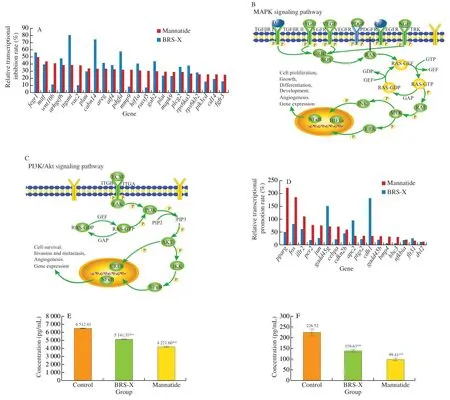
Fig.7 KEGG analysis of BRS-X on S180-bearing mice.(A) Genes with a relative transcriptional inhibition rate more than 25% in the BRS-X group.(B) Genes with a relative transcriptional promotion rate more than 10% in the BRS-X group.(C) MAPK signaling pathway in tumor tissues stimulated by BRS-X.(D) PI3KAkt signaling pathway in tumor tissues stimulated by BRS-X.(E) The effect of BRS-X on VEGFR expression in tumor tissues.(F) The effect of BRS-X on VEGF expression in tumor tissues.* P <0.05 and ** P <0.01 in comparison with negative control group.
3.2.8.2 Genes with relative transcriptional inhibition rate below25%
In addition,there were 113 down-regulated genes with relative transcriptional inhibition rate below 25% .Interestingly,31 genes enriched in the mitogen-activated protein kinases (MAPK) signaling pathway (Fig.7B),includingntrk1(23.62%),casp3(21.88%),ikbkg(21.69%),braf(17.11%),sos2(16.25%),cdc42(14.62%),grb2(14.47%),araf(11.78%),map2k1(10.87%),kras(10.02%),mapk1(8.53%),vegfd(8.12%),nras(7.87%),egfr(6.52%),vegfr(6.47%) etc.MAPK signaling pathway could transmit extracellular stimuli (such as cytokines IL-1β,IL-6,TNF-α and nerve growth factors) to cells [72],which could cause a series of cascade reactions,and eventually regulated the activity of transcription factors and the expression of some genes,which played an important role in regulating many physiological functions,cell growth,differentiation and proliferation [73,74].
In the MAPK signaling pathway,the expression of epidermal growth factor receptor family geneegfrwas down-regulated,suggesting that BRS-X played an important role in regulating the tumor microenvironment and the connection between cells.The genegrb2(growth factor receptor bound protein 2) encodes an adaptor protein [75].Thegrb2gene was down-regulated in BRS-X group,revealing that BRS-X could regulate the adhesion between tumor cells or between tumor cells and extracellular matrix,the stability of extracellular matrix structure,the flexibility of cytoskeleton and the migration ability of tumor cells.The down regulation of the genesos2(son of sevenless homolog 2) indicated that BRS-X could regulate GTP/GDP exchange and related protein activities.The generasencodes a guanosine triphosphate (GTP) binding protein that can hydrolyze GTP into GDP [76].Therasgene was down-regulated in BRS-X group,suggesting that BRS-X could inhibit the signal transduction of tumor cell growth and differentiation by regulating the concentration of GTP/GDP.The down regulation of RAF (rapidly accelerated fibrosarcoma) kinase family genesraf1,arafandbrafindicated that BRS-X could regulate activities of apoptosis-related protein and cell movement.The mitogen-activated extracellular signal-regulated kinase (MEK) and extracellular signal-regulated kinase (ERK) of the MAPK family are encoded by the genesmap2k1andmapk1,respectively [77,78].Themap2k1andmapk1genes were down-regulated in BRS-X group,suggesting that signaling pathways related to proliferation,differentiation and survival in tumor cells were weakened under the stimulation of BRS-X.The protein encoded by nuclear factor kappa B (NFκB) family genesnfkb1andrelaformed p50/p65 dimer protein [79].Thenfkb1andrelagenes were downregulated in BRS-X group,indicating that BRS-X was able to reduce the transcription of target genes related to proliferation,invasion and metastasis in tumor cells.Hypoxia inducible factor-1 (HIF-1) is a heterodimeric transcription factor encoded by the genehif1aand the gene aryl hydrocarbon receptor nuclear translocator (arnt) [80].The geneshif1aandarntwere down-regulated in BRS-X group,indicating that BRS-X could block the energy metabolism of nucleosides,amino acids,and sugars in tumor cells,tumor cell survival and angiogenesis by regulating the transcription of related genes.
And there were 29 genes enriched in the phosphatidylinositol 3-kinase/protein kinase B (PI3K/Akt) signaling pathway(Fig.7C),includingmdm2(21.72%),ptk2(17.51%),jak1(16.13%),fn1(15.66%),bcl2l11(14.31%),akt3(13.93%),itgb1(12.77%),kras(10.02%),bcl2(9.51%),nras(7.87%),itgav(7.55%),foxo3(7.36%),cdk4(6.44%) etc.PI3K/Akt signaling pathway is mediated by enzyme-linked receptors,which can regulate cell life activities.This pathway can be activated with the participation of a variety of growth factors,cytokines,extracellular matrix and also plays an important role in cell proliferation,apoptosis,tissue inflammation,tumor growth,and invasion [81].
In the PI3K/Akt signaling pathway,the genesitgavanditgb1encode theαsubunit andβsubunit of integrin,respectively [82].The genesitgavanditgb1were down-regulated in BRS-X group,suggesting that BRS-X could inhibit the adhesion,movement and signal transduction of tumor cells by reducing the combination of tumor cells and extracellular matrix.The focal adhesion kinase(FAK) encoded by the geneptk2belongs to the family of nonreceptor tyrosine kinases (NRPTK),which can regulate the cell cycle and tumor microenvironment [83].The geneptk2was downregulated in BRS-X group,indicating that BRS-X could inhibit the proliferation of endothelial cells and tumor cells by increasing the ratio of cells in the G2/M phase,leading to vascular formation disorder and tumor cell apoptosis.The geneakt3belongs to serine/threonine kinase family,which is a key molecule in the PI3K/Akt signaling pathway,suggesting that BRS-X could inhibit the activation of downstream proteins by reducing the concentration of AKT protein.The geneikbkgencodes the regulatory subunit γ of inhibitor of nuclear factor kappa-B kinase (IKK),which is also known as the essential regulatory factor of NFkB [84].The geneikbkgwas down-regulated in BRS-X group,indicating that BRS-X could retain NFkB in the cytoplasm of tumor cells and inhibit oncogene expression.The genecreb3l4,a member of the cyclic adenosine monophosphate responsive element binding protein(CREB) family,encodes a nuclear transcription factor [85].The genecreb3l4was down-regulated in BRS-X group,indicating that BRS-X could inhibit the synthesis of metastasis-related proteins in tumor cells by reducing gene transcription.The B-lycytoma-2(BCL2) protein encoded by the genebcl-2is an important factor that regulates cell apoptosis located in the outer membrane of mitochondria [86].The genebcl-2was down-regulated in BRS-X group,suggesting that BRS-X could promote tumor cell apoptosis by destroying mitochondrial permeability.
3.2.8.3 Genes with relative transcriptional promotion rate more than 10%
There were 17 up-regulated genes with relative transcriptional promotion rate more than 10% (Fig.7D).For instance,the growth arrest and DNA damage inducible 45 gene (gadd45) is a stress response gene with the function of tumor suppression [87].The genesgadd45βandgadd45γwere up-regulated in BRS-X group,suggesting that BRS-X could maintain the stability of the genome in tumor cells and inhibit tumor growth by promoting DNA damage repair and inducing G2/M block in the cell cycle.The protein NFKBIA encoded by the NF-κB inhibitor alpha gene (nfkbia) is an inhibitor that regulates the entry of the transcription factor NF-κB into the nucleus [88].The genenfkbiawas up-regulated in BRS-X group,indicating that BRS-X could prevent the activation of the NF-κB signaling pathway in tumor cells and inhibit the continuous expression of oncogenes.The fms-like tyrosine kinase 3 ligand protein (FLT3L) encoded by theflt3lgene is an early hematopoietic growth factor that can significantly amplify the number of dendritic cells (DC),natural killer cells (NK)and cytotoxic T lymphocytes (CTL)in vivoandin vitro[89].The geneflt3lwas up-regulated in BRS-X group,indicating that BRS-X may strengthen the body’s anti-tumor immune response by activating cellular immunity.
3.2.9 Cytokines determination
The amount of protein expression will reveal the level of transcription and translation of genes in tumor tissues of mice.To further verify the signaling pathways,the expression of VEGFR(Fig.7E) and VEGF (Fig.7F) protein was analyzed by ELISA method.Compared with the negative control group,the expression level of VEGFR in the BRS-X group prominently reduced by 21.05%,and VEGF reduced by 38.36%,both with a statistical significance(P<0.01).The development of tumors is closely related to angiogenesis,and the signaling pathway composed of VEGF and VEGFR is one of the strongest positive regulatory pathways in regulating angiogenesis [90].The expression of VEGF and VEGFR protein was down-regulated under the stimulation of BRS-X,suggesting that BRS-X could inhibit endothelial cell proliferation and luminal formation,and regulate capillary angiogenesis,thereby inhibiting the proliferation,invasion and migration of tumor cells.
4.Conclusion
Biological activities of polysaccharides were influenced by its structure characteristics,including the monosaccharide composition,the molecular weight,the water solubility and glycosidic bonds,etc.[91].In this study,a new polysaccharide (BRS-X) was isolated from the fruiting bodies ofB.reticulatusSchaeff.BRS-X had a molecular weight of 15 kDa and the composition was primarily galactose and glucose with the ratio of 5:1.Structural identification results indicated that the main chain of BRS-X was 1,6-α-D-Galpand the side chain was a 4-β-D-Glcplinked to the O-2 of Galp.Cells(T cells,B cells and RAW264.7 cells) cultured with BRS-X showed high proliferation activity.Furthermore,BRS-X could enhance phagocytosis capacity of RAW264.7 cells and stimulate B cells to secrete more IgG,IgE,IgD and IgM.Results of S180-bearing mice model assay indicated that BRS-X played an anti-tumor role.Then,we performed a transcriptome analysis of the tumor tissues of the model control group,BRS-X group and positive control group,which allowed us to know the expression and differences of genes and to screen out key genes involved in the molecular mechanism of the anti-tumor activity of BRS-X.Especially,the anti-tumor activity of BRS-Xin vivowas manifested through MAPK and PI3K/Akt signaling pathways and the protein VEGF and VEGFR expression were prominently reduced under the treatment with BRS-X.This study provided a scientific basis for the diversified utilization ofB.reticulatusSchaeff resources and introduced a novel polysaccharide (BRS-X) as a valuable source which revealed unique antitumor and immunoregulatory properties.
Declaration of competing interest
The authors have declared that no competing interest exists.
Acknowledgements
This project was supported by the Open Project Program of Irradiation Preservation Technology Key Laboratory of Sichuan Province,Sichuan Institute of Atomic Energy (FZBC2020009),the Open Research Fund Program of Departmental and Municipal Co-construction of Crops Genetic Improvement of Hill Land Key Laboratory of Sichuan Province (2021CGIHL02),Science and Technology Support Project of Nanchong Science and Technology Bureau of Sichuan Province (20YFZJ0053 and 20YFZJ0054),and the Sericulture Innovation Team of Sichuan Province(SCCXTD-2021-17).
Appendix A.Supplementary data
Supplementary data associated with this article can be found,in the online version,at http://doi.org/10.1016/j.fshw.2022.07.067.
杂志排行
食品科学与人类健康(英文)的其它文章
- Colloidal nanoparticles prepared from zein and casein:interactions,characterizations and emerging food applications
- Biological factors controlling starch digestibility in human digestive system
- Preparation methods,biological activities,and potential applications of marine algae oligosaccharides: a review
- Development of hyaluronic acid-based edible film for alleviating dry mouth
- Mushroom β-glucan and polyphenol formulations as natural immunity boosters and balancers: nature of the application
- Preparation of multicore millimeter-sized spherical alginate capsules to specifically and sustainedly release fish oil
