Application of extracellular vesicles from mesenchymal stem cells promotes hair growth by regulating human dermal cells and follicles
2022-08-01RamyaLakshmiRajendranPrakashGangadaranMiHeeKwackJiMinOhChaeMoonHongYoungKwanSungJaetaeLeeByeongCheolAhn
Ramya Lakshmi Rajendran, Prakash Gangadaran, Mi Hee Kwack, Ji Min Oh, Chae Moon Hong, Young Kwan Sung, Jaetae Lee, Byeong-Cheol Ahn
Ramya Lakshmi Rajendran, Prakash Gangadaran, Ji Min Oh, Chae Moon Hong, Jaetae Lee,Βyeong-Cheol Ahn, Department of Nuclear Medicine, School of Medicine, Kyungpook National University, Daegu 41944, South Korea
Prakash Gangadaran, Mi Hee Kwack, Young Kwan Sung, Βyeong-Cheol Ahn, BK21 FOUR KNU Convergence Educational Program of Biomedical Sciences for Creative Future Talents,Department of Biomedical Sciences, School of Medicine, Kyungpook National University,Daegu 41944, South Korea
Mi Hee Kwack, Young Kwan Sung, Department of Immunology, School of Medicine, Kyungpook National University, Daegu 41944, South Korea
Chae Moon Hong, Jaetae Lee, Βyeong-Cheol Ahn, Department of Nuclear Medicine, Kyungpook National University Hospital, Daegu 41944, South Korea
Abstract BACKGROUND Dermal papillae (DP) and outer root sheath (ORS) cells play important roles in hair growth and regeneration by regulating the activity of hair follicle (HF) cells.AIM To investigate the effects of human mesenchymal stem cell-derived extracellular vesicles (hMSC-EVs) on DP and ORS cells as well as HFs. EVs are known to regulate various cellular functions. However, the effects of hMSC-EVs on hair growth, particularly on human-derived HF cells (DP and ORS cells), and the possible mechanisms underlying these effects are unknown.METHODS hMSC-EVs were isolated and characterized using transmission electron microscopy, nanoparticle tracking analysis, western blotting, and flow cytometry. The activation of DP and ORS cells was analyzed using cellular proliferation,migration, western blotting, and real-time polymerase chain reaction. HF growth was evaluated ex vivo using human HFs.RESULTS Wnt3a is present in a class of hMSC-EVs and associated with the EV membrane. hMSC-EVs promote the proliferation of DP and ORS cells. Moreover, they translocate β-catenin into the nucleus of DP cells by increasing the expression of β-catenin target transcription factors (Axin2,EP2 and LEF1) in DP cells. Treatment with hMSC-EVs also promoted the migration of ORS cells and enhanced the expression of keratin (K) differentiation markers (K6, K16, K17, and K75) in ORS cells. Furthermore, treatment with hMSC-EVs increases hair shaft elongation in cultured human HFs.CONCLUSION These findings suggest that hMSC-EVs are potential candidates for further preclinical and clinical studies on hair loss treatment.
Key Words: Mesenchymal stem cells; Extracellular vesicles; Hair growth; Dermal papillae; Outer root sheath cells
lNTRODUCTlON
Hair loss is a common and progressive condition affecting both men and women. Within hair follicles(HFs), cells and their secretory factors undergo complex and intricate interactions for the progression of the HF cycle from telogen to anagen[1,2]. Hair loss can be stopped and hair regrowth can be improved to a certain extent by minoxidil or finasteride treatment, but complete recovery is not possible. Hair transplant surgery is another option to avoid baldness. It is not a cure for male pattern baldness and is associated with complications such as edema, and rarely bleeding, folliculitis, numbness of the scalp,telogen effluvium, and infection[3,4]. The dermal papilla (DP) and outer root sheath (ORS) cells support the regulation of the hair cycle. However, they gradually lose their key hair-inducing properties under pathological conditions[5]. The restoration of DP and ORS cell functions is required to promote hair regrowth.
Extracellular vesicles (EVs) are spherical vesicles that are released by nearly all cells into the extracellular milieu, and are found in body fluids and culture media. EVs comprise functional lipids,proteins, and nucleic acids, and act as mediators of intercellular communication. EVs are classified as exosomes, small EVs, microvesicles, and apoptotic bodies. Exosomes are released by cellular multivesicular bodies, whereas microvesicles are formed by the outward budding of the plasma membrane; both are secreted under normal cellular conditions. In contrast, apoptotic bodies form during cell death[6,7].
In recent years, EVs have emerged as potential therapeutic candidates for various diseases, including ischemic diseases, wound healing, and hair regrowth, by delivering their cargo to target cells[8-12]. EVs or nanovesicles from DP cells[13-17], fibroblasts[18,19], stem cells[11,20], macrophages[21,22] and neural progenitor cells[23] have been shown to have potential therapeutic effects on hair growth in recent studies. Nearly half of these studies have reported enhanced hair regrowth using DP cells as the source cells, which showed potential as therapeutic candidates for hair regrowth. However, clinical translation of EVs derived from DP cells is limited because HFs are not readily available for isolating DP cells, and they gradually lose key hair-inducing properties uponin vitroculture[13,24]. Stem cells, which can be easily isolated from bone marrow (BM), adipose tissue, and the umbilical cord and generated using induced pluripotent stem cells, have been used for regenerative therapies in the last few decades,including hair regeneration[25-28]. In our previous report, we studied the efficacy of mesenchymal stem cell (MSC)-derived EVs on hair regrowth in addition to the efficacy of mouse BM-MSC-EVs on human DP cells using a mouse model[11]. In another study, human MSC-EVs (hMSC-EVs) were used in a mouse model[29].
In this study, we investigated the effects of human BM-MSCs-EVs (hBM-MSCs-EVs) on hair growth.Additionally, we examined the possible molecular mechanisms responsible for hair regrowth. Finally,human DP cells, human ORS cells, and human HFs were treated with hMSC-EV and then examined for the activation of DP and ORS cells and their effects on hair shaft elongation in human HFs.
MATERlALS AND METHODS
Cell culture
BM-MSCs (normal, human; PCS-500-012™) were purchased from the American Type Culture Collection(Manassas, VA, United States). Cells were cultured in Dulbecco′s Modified Eagle′s (DMEM)-F12 medium (HyClone, Logan, UT, United States) supplemented with 10% EV-depleted fetal bovine serum(FBS; Hyclone; ultracentrifuged at 120000 × g for 18 h at 4 °C) and antibiotics (1% penicillin-streptomycin) (Gibco, Carlsbad, CA, United States) and maintained at 37 °C and 5% CO2.
Isolation and culture of human DP and ORS cells
During hair transplantation of male patients with androgenic alopecia, biopsy specimens from the occipital scalps were obtained after receiving consent. The Medical Ethics committee of Kyungpook National University Hospital (Daegu, Korea) approved all the described studies (IRB No. KNU 2018-0155). The HFs were dissected to isolate DP cells from the bulbs, and the cells were transferred to tissue culture dishes coated with bovine type I collagen and cultured in low-glucose DMEM (HyClone, Logan,UT, United States) supplemented with 1% antibiotic-antimycotic and 20% heat-inactivated FBS at 37 °C.The cells were cultured for seven days with medium replacement every three days. The cells were then cultured in low-glucose DMEM supplemented with 10% heat-inactivated FBS in 100-mm culture dishes.Once the cells reached subconfluence, they were harvested using 0.25% trypsin and 10 mmol/L ethylenediaminetetraacetic acid (EDTA) in phosphate-buffered saline (PBS) (split at a 1:5 ratio). Cells from passage 2 were used for further experiments[30].
The same hair specimens were used to isolate ORS cells. The hair shaft and bulb regions of the HFs were removed (to avoid contamination by other cells). HFs were trimmed and immersed in DMEM supplemented with 20% FBS in tissue culture dishes coated with rat collagen type I (Corning,Kennebunk, ME, United States). Cells were cultured for three days, and the medium was changed to keratinocyte growth medium, EpiLife medium (Gibco BRL) with 1% antibiotic-antimycotic solution,and 1% EpiLife defined growth supplement medium. After reaching subconfluence, the cells were harvested using 0.25% trypsin and 10 mmol/L EDTA in PBS (split at a 1:5 ratio) and maintained in EpiLife medium. Cells from passage 2 were used for further experiments[18].
Isolation of hMSC-EVs
hMSC-EVs were isolated from the culture medium of human BM-MSCs (from passage 3 to 6) by ultracentrifugation as previously described[10]. The culture medium was centrifuged at 1500 × g for 10 min to remove the cells. Next, it was centrifuged at 4000 × g for 20 min to remove the cell debris. The collected culture media was filtered through a 0.45-μm syringe filter and ultracentrifuged at 100000 × g for 60 min. The collected hMSC-EV pellets were resuspended in PBS and ultracentrifuged at 100000 × g for 60 min. The hMSC-EVs were then reconstituted in 50-100 μL PBS and stored at -80 °C until use. All ultracentrifugation procedures were performed at 4 °C using an SW28 rotor (Beckman Coulter). A Pierce bicinchoninic acid (BCA) protein assay kit (Thermo Fisher Scientific, MA, United States) was used to measure the amount of EVs.
Transmission electron microscopy
hMSC-EV pellets were resuspended in 100 μL of 2% paraformaldehyde. The samples were then added to Formvar or carbon transmission electron microscope (TEM) grids, and the membranes were air-dried for 20 min in a clean environment. The grids were washed with PBS (100 μL) and incubated in 50 μL of 1% glutaraldehyde for five minutes. The grids were then washed with distilled water for 7 × 2 min cycles and observed under an HT 7700 TEM (Hitachi, Tokyo, Japan) to view the morphology of the hMSC-EVs[9].
Nanoparticle tracking analysis
The measurement of hMSC-EVs was performed by nanoparticle tracking analysis (NTA) using NanoSight LM10 (Malvern). hMSC-EVs were diluted 1000-fold with Milli-Q water, and then a sterile syringe was used to inject the sample into the chamber while ensuring that no bubbles were present.Measurements (n= 5) were performed and evaluated using the NanoSight NTA software. The NanoSight software found that the measured values were the same as the measured particle sizes.
Western blot analysis
Western blotting was performed as previously described[31]. To extract proteins, whole cells and EVs were treated with radio immunoprecipitation assay buffer (Thermo Fisher Scientific) containing a cocktail of protease inhibitors. Total protein concentration was measured using a Pierce BCA Protein Assay Kit (Thermo Fisher Scientific). Equal quantities of proteins (10 μg) were separated using 10%sodium dodecyl sulfate-polyacrylamide gel electrophoresis and transferred to polyvinylidene fluoride membranes (Millipore, Burlington, MA, United States). Blots were probed with primary antibodies against Alix (dilution 1:4000; Abcam, Cambridge, MA, United States), cytochrome C (dilution 1:2500;Abcam), GM130 (dilution 1:5000; Abcam), Wnt3a (dilution 1:2500; Abcam), PCNA (dilution 1:5000; Cell Signaling Technology, Danvers, MA, United States), and anti-rabbit secondary antibodies (dilution 1:8000; Cell Signaling Technology, Danvers, MA, United States) conjugated to horseradish peroxidase.Signals were detected using enhanced chemiluminescence (GE Healthcare, Waukesha, WI, United States) according to the manufacturer’s protocol. Blot images were cropped and prepared using MS PowerPoint (Microsoft, CA, United States).
Flow cytometry
Flow cytometry was performed as previously described[21]. hMSC-EVs were attached to 4 μm aldehyde or sulfate latex beads (Invitrogen, Carlsbad, CA, United States) by mixing 5 μg of the sample with 10 μL of beads for 15 min. The final volume was made up to 1 mL using PBS and mixed for 2 h in a rotary shaker. The sample reaction was stopped by adding 100 mmol/L glycine (1 mL) and 2% bovine serum albumin in PBS for 30 min in a rotary shaker. EVs were bound to beads and incubated overnight at 4 °C with Wnt3a. The beads were then incubated for 60 min at 37 °C with a fluorescein isothiocyanate(FITC)-labeled anti-rabbit antibody. They were resuspended in 1 mL PBS for flow cytometric analysis using a BD FACS Aria III instrument, asperthe manufacturer’s instructions (BD Biosciences, Franklin Lakes, NJ, United States).
The EV internalization assay
hMSC-EVs were labeled with DiD dye (hMSC-EVs/DiD) as described previously[11]. DP or ORS cells (1× 10⁴) were cultured on eight-well chamber slides and incubated overnight. The DP was then incubated with unlabeled hMSC-EVs (10 μg/mL) and hMSC-EVs/DiD (5 and 10 μg/mL) for 2 h at 37 °C in 5%CO2. The ORS cells were then incubated with unlabeled hMSC-EVs (5 μg/mL) and hMSC-EVs/DiD (2.5,5 μg/mL) for 2 h at 37 °C in 5% CO2. The cells were subsequently fixed in paraformaldehyde and mounted using mounting medium with 4′, 6-diamidino-2-phenylindole (DAPI) (Vector Laboratories,Burlingame, CA, United States). A confocal laser scanning microscope (LSM 800 with AiryScan, Zeiss,Oberkochen, Germany) was used to observe and record the cellular internalization of hMSC-EVs into DP or ORS cells.
In vitro cell proliferation assay
DP or ORS cells were seeded (0.5 × 10⁴/well) in 96-well plates and maintained overnight at 37 °C and 5% CO2. Cells treated with hMSC-EVs (DP cells: 2, 4, 6, 8, and 10 μg/mL) and ORS cells (1-5 μg/mL)were maintained for 24 h at 37 °C and 5% CO2. CCK8 (10 μL) (CCK8 assay kit, Dojindo Molecular Technologies, Kyushu, Japan) solution was added to each well. Two hours later, according to the manufacturer's instructions, a spectrophotometer was used to measure the optical density at 450 nm to observe the cell proliferation rate.
β-catenin localization in DP cells by immunofluorescence assay
DP cells (1 × 10⁴) were seeded on an eight-well chamber slide and incubated overnight. hMSC-EVs (10 μg/mL) were added and incubated for an additional 24 h. The cells in the chamber were then fixed with 4% paraformaldehyde, probed with a primary anti-β-catenin antibody (dilution 1:200; Cell Signaling Technology) overnight and washed with PBS. The fixed cells were then incubated with Alexa Fluor FITC-conjugated anti-rabbit antibody for 60 min at room temperature for 45 min. Slides were washed three times with PBS and mounted using mounting medium with DAPI (Vector Laboratories). Images were analyzed using a confocal microscope (LSM 5 exciter, Zeiss, Oberkochen, Germany).
β-catenin trans-localization in DP cells by western blotting
DP cells (1 × 10⁶) were seeded on a 6-well plate and incubated overnight. Next, hMSC-EVs (5 and 10 μg/mL) were added and incubated for an additional 24 h. The nuclear fraction was isolated using an NE-PER™ Nuclear and Cytoplasmic Extraction Reagents kit (Thermo Fisher Scientific) according to the manufacturer’s instructions.
Real-time polymerase chain reaction
Cells were lysed using TRIzol solution (Invitrogen) and total RNA was extracted according to the manufacturer’s instructions. A real-time polymerase chain reaction (RT-PCR) was performed as described previously[21] using the SsoAdvancedTM Universal SYBR Green Supermix (Bio-Rad,Hercules, CA, United States) in a CFX96 touch-RT-PCR system (Bio-Rad). The PCR primer sequences used in this study are listed in the Supplementary Table 1.
Cell migration assay
Migration assays were performed in 24-well cell culture inserts containing trans-parent PET membranes with 8.0-mm pores (BD Biosciences). Human ORS cells were seeded on the upper chamber insert at 5 ×10³/well in 0.5 mL serum-free medium containing 0, 2.5, or 5 μg/mL hMSC-EVs and cultured for 24 h.The medium was supplemented with 10% FBS in the lower chamber as a chemoattractant. After 24 h,the cells on the lower surface were fixed with 2% paraformaldehyde, stained with crystal violet, viewed under phase-contrast microscopy, and enumerated.
Hair shaft elongation of human HFs
Human HFs were isolated and cultured as described previously[32]. HFs were treated with varying concentrations of hMSC-EVs (0, 0.1, 0.5, and 1 μg/mL) and hair shaft elongation was measured on day 6.
Statistical analysis
The mean ± SD is used to express all data. Two-group comparisons were performed using Student’s ttest in Microsoft Excel (Microsoft, Redmond, WA, United States) or GraphPad Prism 9 software version 9.0.0 (121) (GraphPad Software, San Diego, Inc., CA, United States). Statistical significance was set atP<0.05.
RESULTS
Characterization of hMSC-EVs and detection of Wnt3a associated with EV-membrane
The morphology of the isolated hMSC-EVs was analyzed using TEM. TEM imaging of hMSC-EVs showed that most hMSC-EVs were spherical, which is the classical morphology of EVs. Moreover,hMSC-EVs were intact and undamaged after the isolation procedure (Figure 1A). The results of NTA of hMSC-EVs showed that their average diameter was 168.4 ± 78.4 nm (Mode: 144.3 nm) (Figure 1B).Western blotting analysis of EV biomarkers revealed that Alix was present in hMSC-EVs. Cytochrome C(a mitochondrial protein) and GM130 (a Golgi apparatus protein), which are negative EV markers, were absent in hMSC-EVs, confirming that hMSC-EVs were not contaminated with other cells or organelles.Moreover, the presence and enrichment of Wnt3a were greater in hMSC-EVs than in hMSCs(Figure 1C). Flow cytometry was used to confirm the location of Wnt3a in hMSC-EVs, which showed that 34.22% of hMSC-EVs had Wnt3a on their membranes (Figure 1D and E).
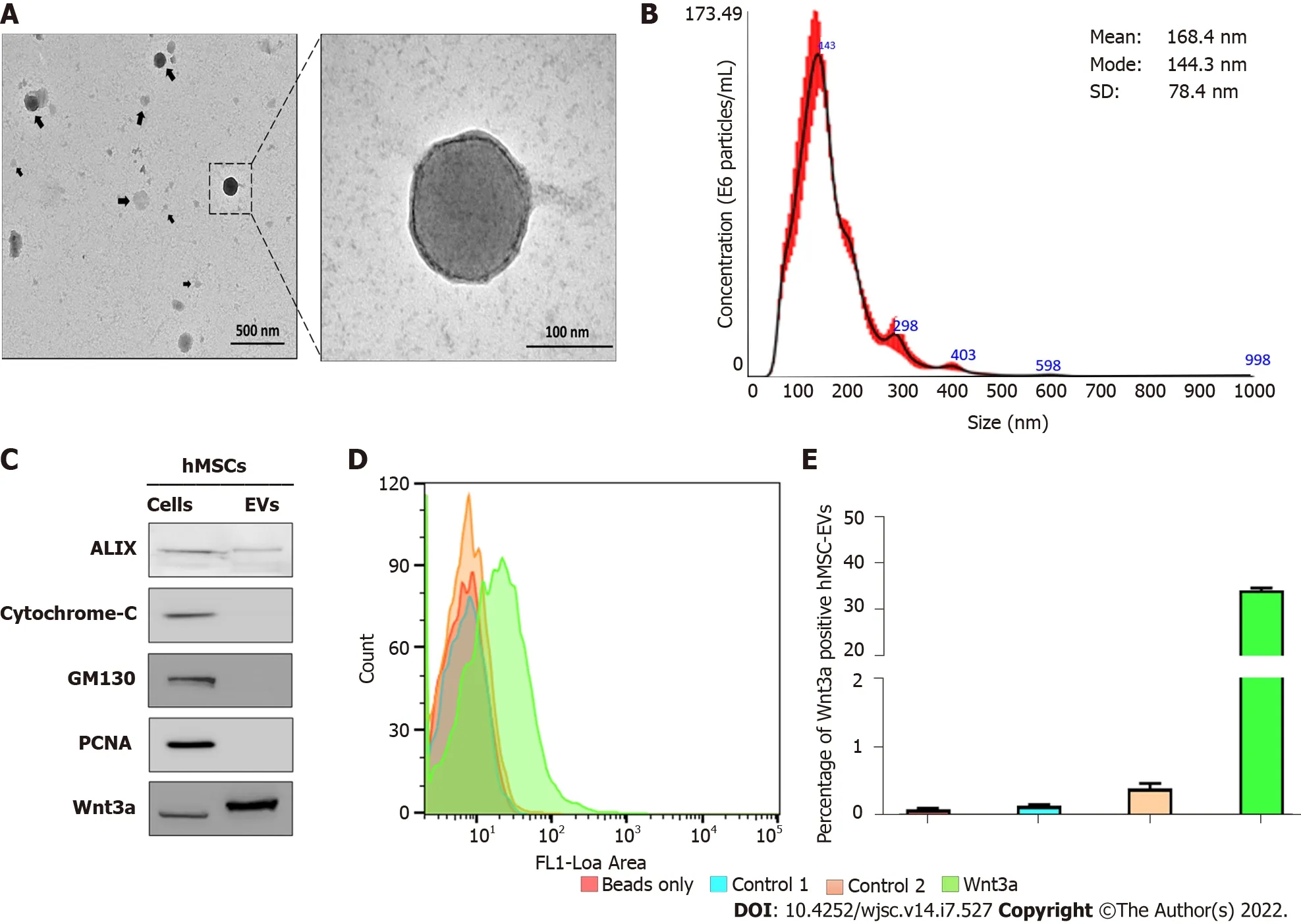
Figure 1 lsolation and characterization of human mesenchymal stem cell-derived extracellular vesicles. A: The morphology of human mesenchymal stem cell-derived extracellular vesicles (hMSC-EVs) was confirmed using transmission electron microscopy (scale bars: 500 and 100 nm); B: hMSCEV size was determined using nanoparticle tracking analysis (n = 5); C: Western blot analysis using Alix, cytochrome C, GM130, PCNA, and Wnt3a antibodies on hMSCs and hMSC-EVs; D and E: Flow cytometry count graphs of only beads, control 1 (beads + hMSC-EVs), control 2 [beads + hMSC-EVs + Secondary fluorescein isothiocyanate (FITC) antibody], and Wnt3a (beads + hMSC-EVs + Wnt3a antibody + Secondary FITC antibody) (n = 3). The values obtained from experiments are shown mean ± SD. FITC: Fluorescein isothiocyanate; hMSC-EVs: Human mesenchymal stem cell-derived extracellular vesicles, NTA: Nanoparticle tracking analysis.
hMSC-EVs promote the proliferation and activation of DP cells
To examine the interaction and integration of hMSC-EVs with recipient DP cells, hMSC-EVs were labeled with DiD dye, and the labeled hMSC-EVs/DiD cells were incubated with DP cells for 4 h.Confocal microscopy showed that hMSC-EVs interacted and integrated inside the cells (Figure 2A). The effects of hMSC-EVs on the proliferation of DP cells were examined, and the results showed that hMSCEV treatment significantly increased the proliferation of DP cells (P< 0.001) with 2-6 μg/mL of hMSCEVs and (P< 0.01) with 8-10 μg/mL of hMSC-EVs (Figure 2B). Since hMSC-EVs showed the presence of Wnt3a, we examined the translocation of β-catenin into the nucleus of DP cells after treatment with hMSC-EVs (10 μg/mL), which revealed a strong signal in the nucleus of DP cells (Figure 2C). In addition, we observed a dose-dependent increase in β-catenin levels in the nuclear fraction of hMSC-EVtreated cells compared with that of control-treated cells (Figure 2D). Furthermore, we examined the expression of Wnt/β-catenin target transcription factors (Axin2, EP2 and LEF1). RT-PCR results showed that there was a significant (P< 0.001 orP< 0.01) upregulation of Axin2, EP2 and LEF1 expression in DP cells in a dose-dependent manner compared to the control (Figure 2E).
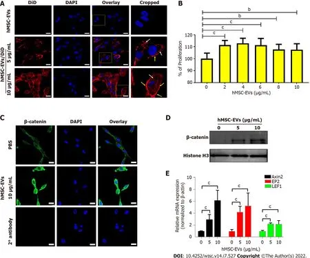
Figure 2 lnteraction of human mesenchymal stem cell-derived extracellular vesicles with dermal papillae cells leads to cell proliferation and activation of Wnt/β-catenin signaling. A: Dermal papillae (DP) cells incubated for 2 h with non-labeled human mesenchymal stem cell-derived extracellular vesicles (hMSC-EVs) (10 μg/mL) and DiD-labeled hMSC-EVs (5 and 10 μg/mL; hMSC-EVs/DiD) (scale bar: 20 μm); B: DP cell proliferation was determined using a CCK8 assay 24 h after treatment with 0-10 μg hMSC-EVs (n = 5); C: β-catenin immunofluorescence assay in DP cells after 24 h of treatment with hMSC-EVs (10 μg/mL) (scale bar: 20 μm); D: The levels of β-catenin in the nuclear fraction of DP cells treated with hMSC-EVs (5 and 10 μg/mL) with histone H3 used as a loading control for nuclear fraction; E: Quantitative real-time polymerase chain reaction results of mRNA expression of Axin2, EP2 and LEF1 in DP cells treated with hMSC-EVs (5 and 10 μg/mL) for 24 h (n = 3). The values obtained from experiments are shown mean ± SD (bP < 0.01; cP < 0.001. Student’s t-test was used for comparison). hMSC-EVs: Human mesenchymal stem cell-derived extracellular vesicles; DP: Dermal papillae.
hMSC-EVs promote the proliferation and migration of human ORS cells
Confocal microscopy revealed the interaction and integration of hMSC-EVs into ORS cells (Figure 3A).The effect of hMSC-EVs on the proliferation of ORS cells was investigated. The results showed that hMSC-EV treatment significantly increased the proliferation of ORS cells (P< 0.001) at 1-5 μg/mL(Figure 3B). As the migration of ORS cells is a hallmark of hair elongation, we examined the migration of ORS cells using hMSC-EVs. After treatment with hMSC-EVs (2.5 and 5 μg/mL), ORS cells showed significantly increased migration in a dose-dependent manner at both concentrations (P< 0.01 at 2.5 μg/mL andP< 0.001 at 5 μg/mL) (Figure 3C and D). Furthermore, we examined the expression of keratin (K) differentiation markers (K6, K16, K17, and K75) in ORS cells after treatment with hMSC-EVs(2.5 and 5 μg/mL). RT-PCR results showed a significant upregulation of all K mRNAs in a dosedependent manner compared to the control. K75 showed the highest expression (P< 0.001), followed by K16 (P< 0.001) and K6 (P< 0.001) at both concentrations; K17 showed significant upregulation at 2.5 μg/mL (P< 0.05); and hMSC-EV treatment at 5 μg/mL showed no significant difference (P> 0.05)compared to the control (Figure 3E).
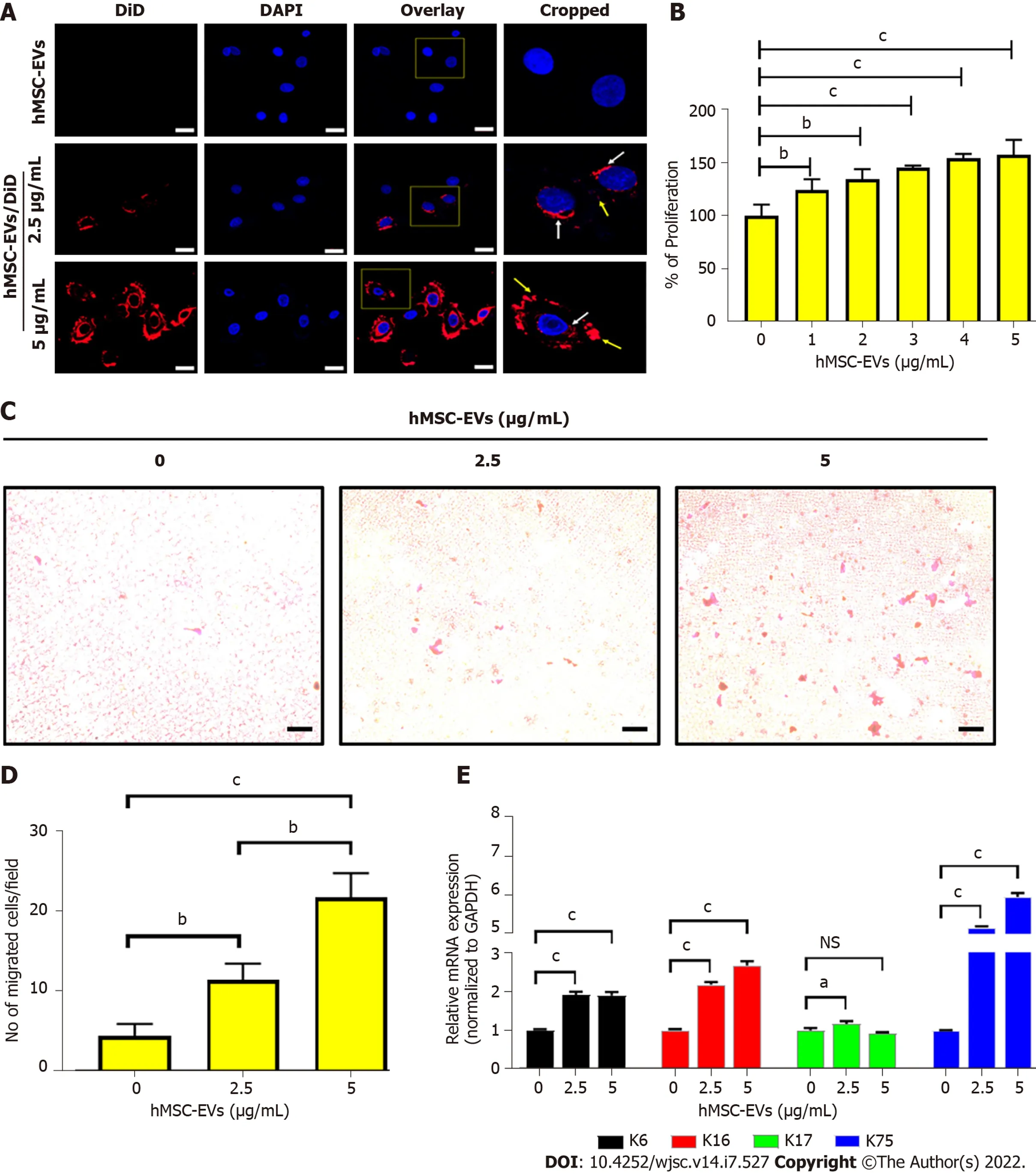
Figure 3 lnteraction of human mesenchymal stem cell-derived extracellular vesicles with outer root sheath cells leads to cell proliferation, migration, and differentiation. A: Outer root sheath (ORS) cells incubated for 2 h with non-labeled human mesenchymal stem cell-derived extracellular vesicles (hMSC-EVs) (5 μg/mL) and DiD-labeled hMSC-EVs (2.5 and 5 μg/mL; hMSC-EVs/DiD) (scale bar: 20 μm); B: ORS cell proliferation was determined using a CCK8 assay 24 h after treatment with 0-5 μg/mL hMSC-EVs (n = 4); C and D: Phase-contrast microscopy images of migrated ORS cells 24 h after treatment with hMSC-EVs (2.5 and 5 μg/mL; scale bar: 50 μm); the quantified data of migrated cells are shown in (A) (n = 3); E: Quantitative real-time polymerase chain reaction results of mRNA expressions of keratin (K) 6, K16, K17, and K75 in ORS cells treated with hMSC-EVs (5 and 10 μg/mL) for 24 h (n = 3).The values obtained from experiments are shown mean ± SD (aP < 0.05; bP < 0.01; cP < 0.001. Student’s t-test was used for comparison). NS: Not significant; hMSCEVs: Human mesenchymal stem cell-derived extracellular vesicles; ORS: Outer root sheath.
hMSC-EVs elongate human HFs
To examine the elongation of hair shafts, mini-organ cultures were performed using human scalp HFs.The HFs were treated with hMSC-EV (0, 0.05, and 0.01 μg/mL) and Wnt inhibitor-XAV939 (5 μM)treatments for six days; the results showed that hMSC-EVs increased hair shaft length significantly (P<0.01) at 0.05 μg/mL and (P< 0.001) at 0.1 μg/mL compared to control (vehicle). The XAV939 treatment significantly (P< 0.001) reduced the hair shaft elongation compared to control (vehicle). Combination treatment with hMSC-EV (0.05 and 0.01 μg/mL) and Wnt inhibitor-XAV939 (5 μM) significantly (P<0.001) abolished hMSC-EVs-induced hair shaft elongation (Figure 4).
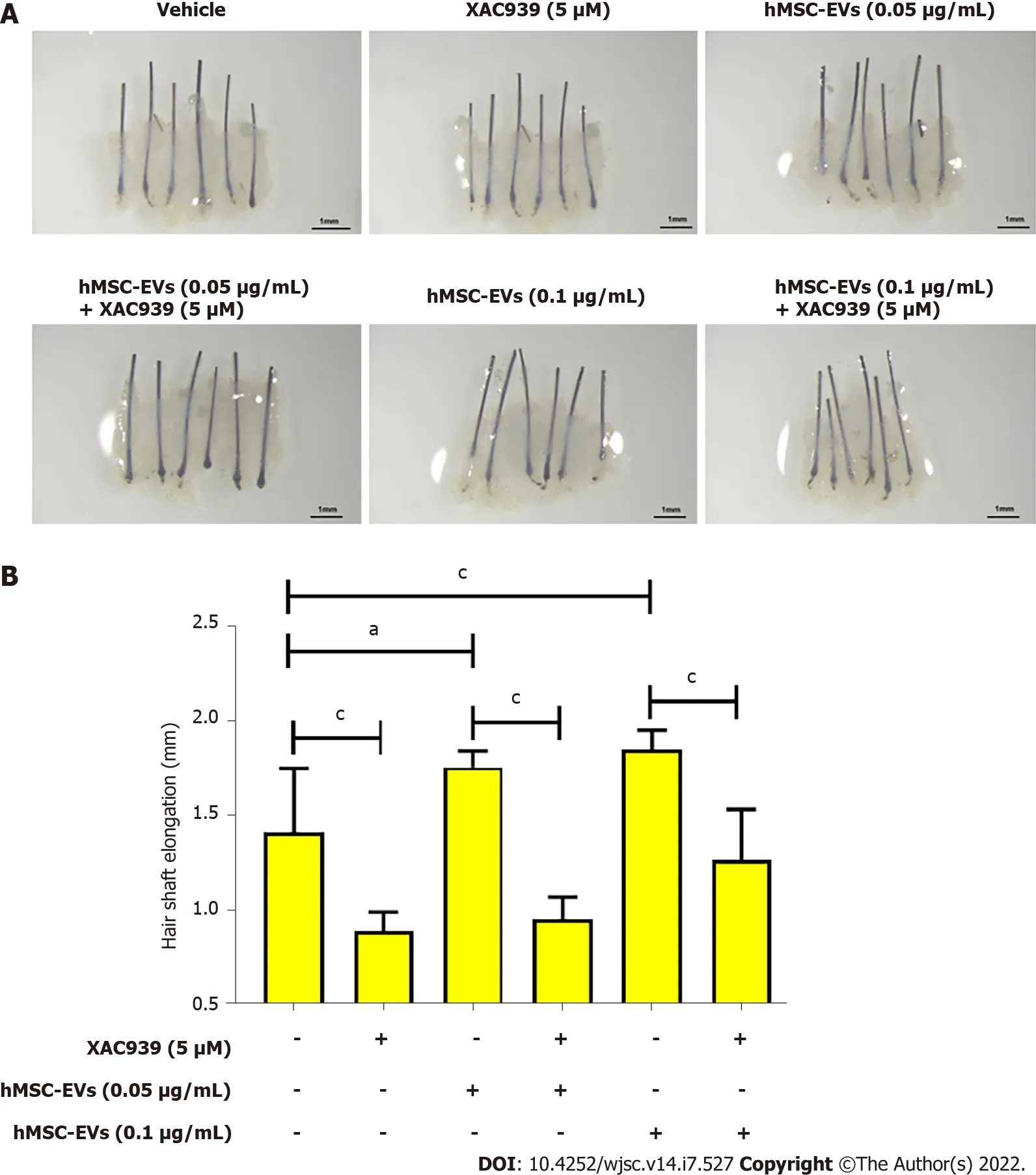
Figure 4 Human mesenchymal stem cell-derived extracellular vesicles treatment promoted human hair follicle shaft elongation. A:Representative images of human hair follicles of an individual after human mesenchymal stem cell-derived extracellular vesicles (0, 0.05, and 0.01 μg/mL) and XAV939 (5 μM) treatments; B: Quantified data of hair shaft elongation on day 6 (n = 6). (aP < 0.05; cP < 0.001. Student’s t-test was used for comparison). hMSC-EVs:Human mesenchymal stem cell-derived extracellular vesicles.
DlSCUSSlON
EVs were isolated from the hMSC culture medium by serial centrifugation, filtration, and ultracentrifugation. The isolated hMSC-EVs displayed intact EV morphology (round) and size distribution.Moreover, hMSC-EVs were enriched in Alix (a typical biomarker of EVs) and lacked cytochrome C (a mitochondrial marker), GM130 (a golgi marker), and PCNA (a nuclear marker), which confirmed that our hMSC-EVs were not contaminated with cell organelles, consistent with previous reports[9-11]. The Wnt/β-catenin signaling cascade is crucial for the development and maintenance of HFs[13,33]. The presence of Wnt3a in hMSC-EVs was confirmed, and Wnt3a was more enriched in EVs than in cells.Several previous studies[34-37] have well documented the enrichment of Wnt proteins on EVs.Furthermore, a significant proportion of Wnt3a (34.22%) was associated with the EV membranes. Our previous study with macrophage-and fibroblast-derived EVs also showed that they have > 90%(macrophage-derived EVs) or > 70% (fibroblast-derived EVs) associated with the EV membrane[18,21]and A recent study showed that Wnt3a, Wnt5a, and Wnt7a were present on the surface of small EVs isolated from a mouse hippocampal cell line (HT-22), which is in agreement with our current study[37].
To exert the therapeutic effects of any EV, an interaction with target/recipient cells or internalization into target/recipient cells is needed[6,7,13]. Our results revealed that hMSC-EVs actively interacted and integrated into DP cells. In the hair growth process, activation and maintenance of the Wnt/β-catenin signaling cascade in DP cells are crucial[1,11]. In this study, we observed increased proliferation of DP cellsin vitroon treatment with hMSC-EVs. Most studies on various EVs have shown an increase in DP cell proliferation upon treatment[11,13,15,17]. Furthermore, our results revealed that treatment of DP cells with hMSC-EV translocated β-catenin into the nucleus, which is a requirement for the activation of hair-inducing transcription factors[38,39]. Additionally, hMSC-EVs increased the expression of hairinducing transcription factors in DP cells (Axin2, EP2 and LEF1). Similar results were observed in other studies that used EVs for treatment[15,18,21].
ORS cells are a putative source of stem cells with therapeutic capacity. Survival, migration, and differentiation are important for HF maintenance[40,41]. Our results show that the interaction and integration of hMSC-EVs into ORS cells increased cellular proliferation and migration, which are necessary for hair growth. Furthermore, hMSC-EV treatment increased the expression of differentiation markers (K6, K16,K17, and K75), indicating the differentiation of cultured ORS cells into follicular lineages[42]. Finally, we investigated the hair-inducing properties of hMSC-EVs on human HFs. We observed that hMSC-EVs increased hair shaft length, which was abolished by the Wnt inhibitor. These findings suggest a potential therapeutic effect of hMSC-EVs in human HFs through Wnt/β-catenin signaling. Several other studies using EVs in HFs have reported an increase in hair shaft elongation[15,16,18,21].
In the present study, we showed the enrichment of Wnt3a in hMSC-EVs and some association of Wnt3a with the EV membrane. However, compared to macrophage-and fibroblast-derived EVs, hMSCEVs showed a lower association between Wnt3a and the membrane[18,21,37]. We have not ruled out that other proteins and miRNAs may play a role in hair regrowth because a few recent studies have shown that miRNA-100, miR-NA-140-5p, and miRNA-218-5p play certain roles in hair regrowth[13,16,23]. Further, a complete proteomic and miRNA analysis is needed to reveal a more complete understanding of hair growth promoted by hMSC-EV treatment.
CONCLUSlON
The present study demonstrated that hMSC-EVs enhance hair growth by activating HF cells and HFs.Thus, hMSC-EVs could be therapeutic candidates for hair loss treatment.
ARTlCLE HlGHLlGHTS
Research background
Hair loss is one of the most common disorders in both sexes. Despite the availability of several treatment options, no definitive treatment method is currently available. Application of extracellular vesicles (EVs) has been suggested as a possible new treatment modality for hair loss.
Research motivation
Although cell-derived EV treatments have shown reasonable efficacy in hair loss studies, the molecular mechanisms and therapeutic effects are still relatively unknown.
Research objectives
We examined the effects of human mesenchymal stem cell-derived EVs (hMSC-EVs) on human dermal papillae (DP), outer root sheath (ORS) cells, and hair follicles (HF).
Research methods
Human DP cells, ORS cells, and HFs were treated with various amounts of hMSC-EVs to investigate the effect of hMSC-EVs on human cellsin vitroandex vivo.
Research results
The Wnt3a-containing hMSC-EVs treatment increased the proliferation of DP cells and the Wnt/βcatenin signaling cascade and activated transcription related to hair growth. Similarly, hMSC-EV treatment increased the proliferation, migration, and keratin differentiation of ORS cells. Theex vivotreatment with hMSC-EVs increased human HF shaft elongation.
Research conclusions
Application of hMSC-EVs may be a new potential strategy for hair loss treatment.
Research perspectives
Our findings demonstrate the effects of hMSC-EVs on hair cells and HFs.
FOOTNOTES
Author contributions:Rajendran RL and Gangadaran P contributed equally to this study; Rajendran RL, Gangadaran P and Ahn BC contributed to the conception and design of the study, data interpretation, and funding acquisition;Rajendran RL, Gangadaran P, Kwack MH, Oh JM and Hong CM contributed to the methodology, data acquisition,and analysis; Rajendran RL and Gangadaran P wrote the original draft of the article; Kwack MH, Oh JM, Hong CM,Sung YK and Lee J drafted, reviewed and edited the manuscript, and contributed to project administration; The study was led by Ahn BC, and all of the authors read and approved the final version of the manuscript.
Supported byBasic Science Research Program through the National Research Foundation of Korea (NRF), Funded by the Ministry of Education, No. NRF-2019R1I1A1A01061296 and No. NRF-2021R1I1A1A01040732; Korea Health Technology R & D Project through the Korea Health Industry Development Institute, Funded By the Ministry of Health & Welfare, Republic of Korea, No. HI15C0001.
lnstitutional review board statement:This study was approved by the Institutional Review Board (IRB) of Kyungpook National University Hospital, No. KNU-2018-0161 and conducted in accordance with the principles of the Declaration of Helsinki.
lnformed consent statement:All study participants or their legal guardian provided informed written consent about personal and medical data collection prior to study enrolment.
Conflict-of-interest statement:All the authors report no relevant conflicts of interest for this article.
Data sharing statement:No additional data are available.
ARRlVE guidelines statement:The authors have read the ARRIVE guidelines, and the manuscript was prepared and revised according to the ARRIVE guidelines.
Open-Access:This article is an open-access article that was selected by an in-house editor and fully peer-reviewed by external reviewers. It is distributed in accordance with the Creative Commons Attribution NonCommercial (CC BYNC 4.0) license, which permits others to distribute, remix, adapt, build upon this work non-commercially, and license their derivative works on different terms, provided the original work is properly cited and the use is noncommercial. See: https://creativecommons.org/Licenses/by-nc/4.0/
Country/Territory of origin:South Korea
ORClD number:Ramya Lakshmi Rajendran 0000-0001-6987-0854; Prakash Gangadaran 0000-0002-0658-4604; Mi Hee Kwack 0000-0002-7381-6532; Ji Min Oh 0000-0001-8474-7221; Chae Moon Hong 0000-0002-5519-6982; Young Kwan Sung 0000-0002-5122-4616; Jaetae Lee 0000-0001-5676-4059; Byeong-Cheol Ahn 0000-0001-7700-3929.
S-Editor:Fan JR
L-Editor:A
P-Editor:Yu HG
杂志排行
World Journal of Stem Cells的其它文章
- Therapeutic potential of dental pulp stem cells and their derivatives:lnsights from basic research toward clinical applications
- Role of hypoxia preconditioning in therapeutic potential of mesenchymal stem-cell-derived extracellular vesicles
- Application of exosome-derived noncoding RNAs in bone regeneration: Opportunities and challenges
- Metabolic-epigenetic nexus in regulation of stem cell fate
- Stem cell therapy for insulin-dependent diabetes: Are we still on the road?
- Prodigious therapeutic effects of combining mesenchymal stem cells with magnetic nanoparticles
