Investigating the influence of moxibustion on colonic mucosal barrier in rats with dextran sulfate sodium-induced ulcerative colitis
2022-02-24SHENYa沈亚CUIYunhua崔云华SHIZheng施征WUHuangan吴焕淦WANGZhaoqin王照钦WULuyi吴璐一LUYuan陆嫄HUANGYan黄艳LIUYanan刘雅楠LONGJunyi龙俊燚LINYaying林亚莹MAZhe马喆
SHEN Ya (沈亚), CUI Yunhua (崔云华), SHI Zheng (施征),, WU Huangan (吴焕淦),, WANG Zhaoqin (王照钦),WU Luyi (吴璐一), LU Yuan (陆嫄),, HUANG Yan (黄艳),, LIU Yanan (刘雅楠),, LONG Junyi (龙俊燚),LIN Yaying (林亚莹), MA Zhe (马喆),
1 Yueyang Hospital of Integrated Traditional Chinese and Western Medicine, Shanghai University of Traditional Chinese Medicine,Shanghai 200437, China
2 Shanghai Research Institute of Acupuncture and Meridian, Shanghai 200030, China
3 Department of Aeronautics and Astronautics, Shanghai Key Laboratory of Acupuncture Mechanism and Acupoint Function, Fudan University, Shanghai 200433, China
4 Key Laboratory of Acupuncture and Immunological Effects, Shanghai University of Traditional Chinese Medicine, Shanghai 200030,China
Abstract
Keywords: Moxibustion Therapy; Medicinal Cake-partitioned Moxibustion; Moxa Stick Moxibustion; Point, Tianshu (ST25);Colitis, Ulcerative; Transforming Growth Factor beta1; Rats
Ulcerative colitis (UC) is a chronic non-specific inflammatory bowel disease. The lesions often involve the mucosa and submucosa of rectum or colon. The main clinical symptoms are abdominal pain, diarrhea,and bloody pus stool. Epidemiological data show that the prevalence of UC is higher in Western developed countries[1]. The incidence of UC in China had increased to 24.5 per 100 000 people in 2014[2]. UC is a complex condition with irregular and recurrent acute attacks, and classified as a refractory disease by the World Health Organization[3]. At present, the pathogenesis of UC is not yet clear, but the current consensus considers that UC is caused by the interaction of many factors, such as susceptibility genes, immune regulation disorders,intestinal mucosal barrier defects, and environment[4-5].The destruction of intestinal mucosal barrier and continuous inflammation are the reasons making it difficult to treat and last for a long duration[6]. Intestinal mucosal barrier damage causes an increased permeability to increase the harmful bacteria and foreign antigens entering the intestinal lumen, and further induce immune regulation disorders and inflammatory reactions in the body, leading to UC[7].Therefore, protecting the integrity of the intestinal mucosal barrier is one of the strategies to inhibit the occurrence and development of UC.
Common clinical treatment drugs for UC, including glucocorticoids, monoclonal antibodies, and 5-aminosalicylic acid, may show some problems, such as adverse reactions, drug resistance, and dependence[8-9].Moxibustion is a therapy of traditional Chinese medicine(TCM). It can unblock the meridians, Qi, and blood through warm stimulation, regulate the Zang-Fu organs,and strengthen the healthy Qi to eliminate the pathogenic factors, thus effectively alleviating the clinical symptoms of UC patients[10]. Studies have shown that moxibustion has a regulatory effect on the intestinal mucosal barrier function and can inhibit the inflammatory response of the colonic mucosa of UC rats[11]. Our previous study found that moxibustion pretreatment can protect the intestinal mucosal barrier of UC rats[12]. Here, we observed the effects of herbal cake-partitioned moxibustion and mild moxibustion on the intestinal mucosal barrier of rats under the physiological and UC pathological conditions to reveal the mechanism of moxibustion in UC treatment.
1 Materials and Methods
1.1 Experimental animals and grouping
Forty inbred, specific-pathogen-free grade, male Sprague-Dawley rats, weighing 160-200 g, were purchased from Shanghai Slack Laboratory Animal Co.,Ltd., China [License No. SCXK (Shanghai) 2012-0002].Rats were kept in the Experimental Animal Center of Shanghai University of Traditional Chinese Medicine.Feeding environment: 12 h/12 h light/dark alternate,average room temperature at (20±2) ℃, and indoor relative humidity of 50%-70%. After 7 d of adaptive feeding, the rats were randomly divided into a normal group and a modeling group, with 20 rats in each group.The experimental UC model was established by drinking 4% dextran sulfate sodium (DSS) for 7 d. Two modeled rats and two normal rats were randomly selected for model identification. After the UC model was confirmed successfully prepared, the remaining 18 modeled rats were randomly divided into a model group, a model +herbal cake-partitioned moxibustion group, and a model + mild moxibustion group, with six rats in each group; the remaining normal rats were randomly divided into a normal group, a normal + herbal cake-partitioned moxibustion group, and a normal + mild moxibustion group, with six rats in each group. All experimental operations were conducted under the guidance of the Animal Ethics Committee of Shanghai University of Traditional Chinese Medicine, and the experimental animals were properly handled in accordance with theGuiding Opinions on the Treatment of Experimental Animalspromulgated by the Ministry of Science and Technology of the People’s Republic of China.
1.2 Drugs and reagents
Moxa stick, moxa wool (Nanyang Hanyi Moxa Co., Ltd.,China); herbal powder [Rou Gui(Cortex Cinnamomi),Fu Zi(Radix Aconiti Lateralis Praeparata),Hong Hua(Flos Carthami), etc.], and edible rice wine (Zhejiang Jiashan Rice Wine Co., Ltd., China); DSS (molecular weight:36-50 kDa, Cat. No. 9011-18-1, specification: 500 GM,MP Biomedicals, USA); primary Claudin antibody(concentration: 1:500, Cat. No. ab1509, Abcam, UK);Occludin (concentration: 1:200, Cat. No. ab167161,Abcam, UK); transforming growth factor beta1 (TGF-β1)(concentration: 1:200, Cat. No. ab92486, Abcam, UK);mucin 2 (MUC2, ready to use, Cat. No. ab76775, Abcam,UK); junction adhesion molecular 1 (JAM1,concentration: 1:100, Cat. No. ab180821, Abcam, UK);mice and rabbit general immunohistochemistry kits,blocking solution, primary antibody diluent and secondary antibody diluent (Shanghai Weiao Biotechnology Co., Ltd., China); hematoxylin and eosin(Nanjing Jiancheng Technology Co., Ltd., China).
1.3 Modeling and intervention methods
1.3.1 Animal UC model preparation[13]
The 4% DSS aqueous solution was prepared by mixing the drinking water with DSS powder. An experimental UC rat model was established by drinking the 4% DSS aqueous solution for 7 d. The 18 UC rats received a maintenance dose of 1% DSS aqueous solution during the treatment period.
1.3.2 Intervention methods
Herbal cake-partitioned moxibustion: A herbal cake(8 mm in diameter and 4 mm in thickness) was placed on Tianshu (ST25) with a moxa cone (1 cm in diameter of the base and 90 mg in mass) on the top for moxibustion,once a day, two moxa cones per acupoint each time, for seven consecutive days. The location of Tianshu (ST25)refers to theExperimental Acupuncture Science[14].
Mild moxibustion: A piece of moxa stick of 7 mm in diameter and 20 mm in length (2 g in mass) specifically for rats was used. Moxibustion was performed about 2 cm over Tianshu (ST25) for 10 min each time and once a day, for seven consecutive days in total.
Rats in the normal group and model group only received the same grasping and fixing as those in the intervention groups.
1.4 Specimen collection and processing
After the intervention, rats were anesthetized by intraperitoneal injection of 2% sodium pentobarbital solution [40-50 mg/(kg·bw)]. After successful anesthesia,rats were fixed, and the colon from the pubic symphysis to the cecum was collected to observe the general condition by opening the colon along the mesentery.Approximately 8 cm of the colon was selected from the bottom to the top. About 1 cm distal colon was fixed in 4% paraformaldehyde solution and the remaining section was cut, mixed, and equally divided into two parts to be frozen in liquid nitrogen for 1 h, and then placed in a -80 ℃ refrigerator for later use.
1.5 Hematoxylin-eosin (HE) staining
The 4% paraformaldehyde-fixed colon tissue was dehydrated, embedded in paraffin, and then sliced (4 μm in thickness). The slices were routinely dewaxed to water by using xylene Ⅰ and Ⅱ for 20 min, respectively.Then 100%, 90%, 80%, and 70% ethanol was used in sequence for 5 min each to make the tissue samples washable with water and combined with water-soluble staining. After being washed with double distilled water for 5 min × 2 times followed by hematoxylin dip staining for 2-3 min, the slices were further subjected to running water for 10 min, 1% hydrochloric acid ethanol for 1-2 s,running water for 5 min, and eosin dip staining for 2-3 min. The 70%, 80%, 90%, and 100% ethanol was respectively applied for 1-2 s to remove the residual water in the tissue. Transparency was performed with xylene Ⅰ and Ⅱ for 15 min, respectively. Mounted the slides and observed them under the microscope. The histopathology was scored according to the scoring standard established by OBERMEIER F,et al[15].
1.6 Western blotting (WB) method
Rat colon tissue was homogenized, fully lysed by adding an appropriate amount of RIPA lysate, and centrifuged at high speed to collect the supernatant. The protein concentration of each sample was determined by the BCA protein quantitative method, and the sample amount was adjusted according to the concentration during denaturing and loading. The proteins were electrophoresed, transferred onto the membrane,blocked in a blocking solution (5% skimmed milk powder)for 1 h at room temperature, and incubated with the primary antibody on the shaker at 4 ℃ overnight. After washing, the membrane was incubated at room temperature for 1 h with the secondary antibody. Again,after washing, the membrane was developed and imaged to calculate the gray value with the BIO-RAD system.
1.7 Immunohistochemistry
After deparaffinization, hydration, antigen retrieval,and blocking, the paraffinized sections were incubated with the primary antibody at 4 ℃ overnight, and then the second antibody. After washing with PBS, color rendering was achieved with DAB color-developing solution. The sections were dehydrated and mounted after the nuclei were stained with hematoxylin for 1 min.Three to five fields of view on each slice were randomly selected and imaged after observation under an optical microscope with identical brightness. Image-Pro Plus(IPP) software was used to analyze and calculate the positive area (Area) and the integral optical density (IOD)of the positive target in each photo, to calculate the average optical density (AOD) of the positive target (AOD= IOD ÷ Area), thus to determine the average value of each field of view on the slice, indicating the positive target AOD of the slice.
1.8 Statistical processing
Statistical analysis was performed using SPSS version 21.0 statistical software. The measurement data were first tested for normality. The multiple groups were compared by one-way analysis of variance (ANOVA)method if the variances of the normal distribution data were uniform, and Dunnett’s T3 method was used for pairwise comparisons for data with uneven variance.Nonnormal distribution data were compared by the nonparametric test between groups, and the pairwise comparisons were achieved by the rank-sum test(Kruskal-WallisH). Normal distribution data were shown as mean ± standard deviation (±s). Nonnormal distribution data were shown as median (lower quartile,upper quartile) [M (QL, QU)]. All analyses were performed by two-tailed tests, withα=0.05 as the test level, andP<0.05 indicating the statistically significant difference.
2 Results
2.1 Histopathological observation of colon tissue in UC rats
The colon mucosal epithelium of rats in the normal group, the normal + herbal cake-partitioned moxibustion group, and the normal + mild moxibustion group were intact, with neatly arranged glands, evenly distributed interstitium, and no congestion or edema. The colonic mucosal epithelium in the model group was incomplete with obvious ulcers, disordered gland arrangement, and many inflammatory cell infiltrations in the mucosa and submucosa, accompanied by hemorrhage and edema. In the model + herbal cake-partitioned moxibustion group and the model + mild moxibustion group, the colonic mucosal epithelium was partially repaired, showing healing ulcers, irregularly arranged glands, and a small amount of inflammatory cell infiltration in the mucosa and submucosa, which was significantly reduced compared with the model group (Figure 1).

Figure 1. Histopathological observation of rat colon tissue in each group (HE staining, ×200)
Compared with the normal group, the colonic histopathological score of the model group was significantly higher (P<0.01), but the colonic histopathological scores in the normal + herbal cakepartitioned moxibustion group and the normal + mild moxibustion group were not statistically different(P>0.05). Compared with the model group, the colonic histopathological scores of rats in the model + herbal cake-partitioned moxibustion group and the model +mild moxibustion group were statistically significantly lower (P<0.05). There was no statistical difference in the score between the model + herbal cake-partitioned moxibustion group and the model + mild moxibustion group (P>0.05), (Figure 2).

Figure 2. Histopathological score of rat colon tissue
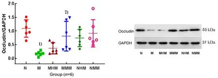
Figure 3. Occludin protein expression in rat colon tissue in each group
2.2 Intestinal mucosal barrier-related protein expression in UC rats
2.2.1 Occludin protein expression in rat colon tissue
Compared with the normal group, the Occludin protein expression in rat colon tissue in the model group was significantly reduced (P<0.01). However, the Occludin protein expression in the colon tissue of rats in the normal + herbal cake-partitioned moxibustion group and the normal + mild moxibustion group was not significantly different from that in the normal group(P>0.05). Compared with the model group, the Occludin protein expression in the colon tissue of the model +herbal cake-partitioned moxibustion group was increased, but the difference was not statistically significant (P>0.05); the Occludin protein expression of rat colon tissue in the model + mild moxibustion group was significantly increased (P<0.01). There was no significant difference between the model + herbal cakepartitioned moxibustion group and the model + mild moxibustion group (P>0.05), (Figure 3).
2.2.2 Claudin protein expression in rat colon tissue
Compared with the normal group, the Claudin protein expression in the colon tissue in the model group was significantly decreased (P<0.01). There were no significant differences in the Claudin protein expression in the colon tissue in the normal + herbal cakepartitioned moxibustion group and the normal + mild moxibustion group compared with the normal group(P>0.05). Compared with the model group, the Claudin protein expression in the model + herbal cakepartitioned moxibustion group and the model + mild moxibustion group had an upward trend, but the differences were not statistically significant (P>0.05).There was no significant difference between the model +herbal cake-partitioned moxibustion group and the model + mild moxibustion group (P>0.05), (Figure 4).
2.2.3 JAM1 protein expression in rat colon tissue
The positive JAM1 protein expression was brown and mainly distributed in the cytoplasm of intestinal epithelial cells and glandular epithelial cells. In the normal group, the normal + herbal cake-partitioned moxibustion group, and the normal + mild moxibustion group, densely distributed tan positive expression was seen in the cytoplasm, suggesting a large amount of JAM1 protein expression. Only a small amount of JAM1 protein was positively expressed in the model group(Figure 5).

Figure 4. Claudin protein expression in rat colon tissue in each group

Figure 5. JAM1 protein expression in rat colon tissue in each group (immunohistochemistry, ×200)
Compared with the normal group, the JAM1 protein expression in the model group was significantly decreased (P<0.01); there was no significant difference in the JAM1 protein expression in rat colon tissue in the normal + herbal cake-partitioned moxibustion group or the normal + mild moxibustion group (P>0.05).Compared with the model group, the JAM1 protein expression in the colon tissue in the model + herbal cakepartitioned moxibustion group and the model + mild moxibustion group was significantly increased (P<0.01).There was no significant difference in the JAM1 protein expression between the model + herbal cake-partitioned moxibustion group and the model + mild moxibustion group (P>0.05), (Figure 6).
2.2.4 MUC2 protein expression in rat colon tissue
The positive MUC2 protein expression was brown,concentrated in the colon mucosal goblet cells and a small number of epithelial cells. A large amount of positive MUC2 protein expression can be seen in the normal group, the normal + herbal cake-partitioned moxibustion group, and the normal + mild moxibustion group; the MUC2 protein expression intensity in rat colon tissue in the model group, the model + herbal cakepartitioned moxibustion group, and the model + mild moxibustion group was weaker than that of the normal group (Figure 7).
Compared with the normal group, the MUC2 protein expression in the colon tissue in the model group was significantly reduced (P<0.01). The MUC2 protein expression in rat colon tissue in the normal + herbal cakepartitioned moxibustion group and the normal + mild moxibustion group was not statistically different from that in the normal group (P>0.05). Compared with the model group, the MUC2 protein expression in the model+ herbal cake-partitioned moxibustion group and the model + mild moxibustion group was significantly increased (P<0.01). There was no significant difference in the MUC2 protein expression between the model +herbal cake-partitioned moxibustion group and the model + mild moxibustion group (P>0.05), (Figure 8).
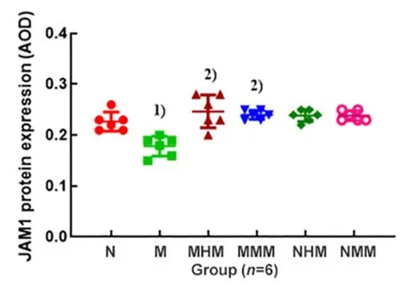
Figure 6. Comparison of the JAM1 protein expression in rat colon tissue
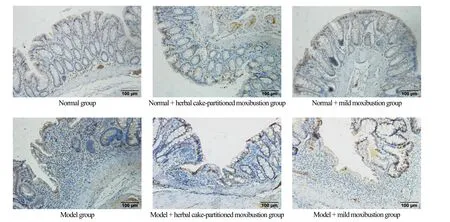
Figure 7. MUC2 protein expression of rat colon tissue in each group (immunohistochemistry, ×200)
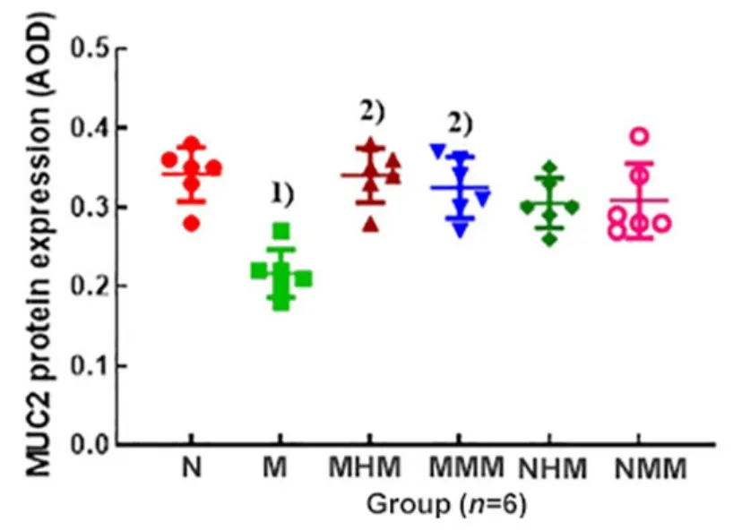
Figure 8. Comparison of the MUC2 protein expression in rat colon tissue in each group
2.3 Effect of moxibustion on the TGF-β1 protein expression in the colon tissue of UC rats
The positive TGF-β1 protein expression was yellow,mainly located in the intestinal epithelial cells. The normal group, the normal + herbal cake-partitioned moxibustion group, and the normal + mild moxibustion group all showed a large amount of positive TGF-β1 protein expression; the model group, the model + herbal cake-partitioned moxibustion group, and the model +mild moxibustion group showed different degrees of positive TGF-β1 protein expression in the colon tissue(Figure 9).
Quantitative analysis showed that the TGF-β1 protein expression in the colon tissue in the model group was significantly down-regulated compared with the normal group (P<0.01). The TGF-β1 protein expression in rat colon tissue in the normal + herbal cake-partitioned moxibustion group and the normal + mild moxibustion group was not significantly different from that in the normal group (P>0.05). Compared with the model group,the TGF-β1 protein expression in the colon tissue in the model + herbal cake-partitioned moxibustion group and the model + mild moxibustion group was significantly upregulated (P<0.01). There was no significant difference in the TGF-β1 protein expression between the model +herbal cake-partitioned moxibustion group and the model + mild moxibustion group (P>0.05), (Figure 10).
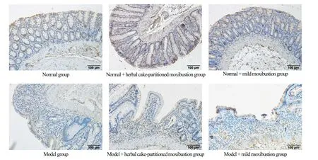
Figure 9. TGF-β1 protein expression in rat colon tissue in each group (immunohistochemistry, ×200)
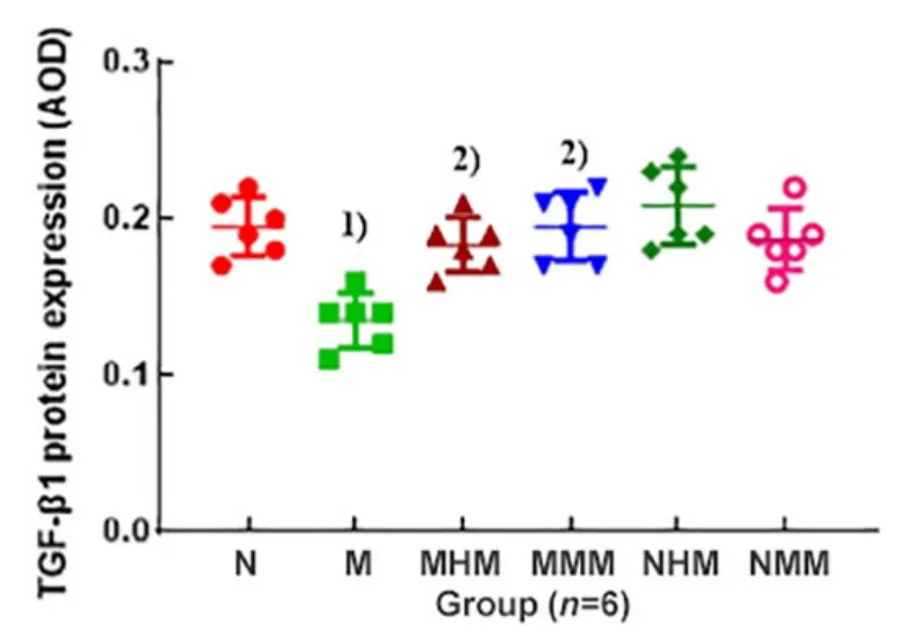
Figure 10. Comparison of the TGF-β1 protein expression in rat colon tissue in each group
3 Discussion
According to symptoms, UC can be classified into the categories of “diarrhea”, “dysentery”, or “bloody stool” in TCM[16]. The pathogenesis is mainly the deficiency in the root cause and excess in the symptoms. UC can be caused by physical weakness, exogenous infection,improper diet, emotional disturbance, etc. Tianshu (ST25)is the Front-Mu point of the large intestine. It can recruit Qi and blood to the intestines and stomach; meanwhile,it can regulate the Qi movement to promote ascending the clear and descending the turbid. The harmony of Yin and Yang, and the smooth Qi movement are very important for the treatment of intestinal diseases,especially UC. Moxibustion at Tianshu (ST25) can alleviate the symptoms and play an overall regulatory role in UC treatment[17].
The structure of the intestinal mucosal barrier is mainly the mucosal basement membrane, the intestinal epithelial cell layer, and the mucus layer on its surface. Its integrity is essential for water and nutrient absorption in the intestine and preventing the invasion of pathogenic microorganisms[18]. The tight junction (TJ) between intestinal epithelial cells mainly constitutes the mechanical barrier of the intestinal mucosa. Occludin,Claudin, and junction adhesion molecules are the main TJ proteins. They can maintain the stability of TJ and the selective permeability of the intestinal epithelium,playing a decisive role in the intestinal mucosal barrier[19].Experimental studies have found that, compared with the normal group, the TJ protein expression level in UC rats is significantly reduced and related to the severity of UC[20].
Our previous study also showed that pretreatment with herbal cake-partitioned moxibustion or mild moxibustion at Tianshu (ST25) can regulate the intestinal mucosal barrier-related proteins such as Occludin, MUC2,and ZO-1 in UC rats. In this study, rats were subjected to moxibustion intervention after UC model preparation,and it was found that the TJ protein expression in the intestinal mucosa of UC rats was increased. Both herbal cake-partitioned moxibustion and mild moxibustion significantly up-regulated the JAM1 protein expression in the colon tissue, and mild moxibustion also significantly up-regulated the Occludin protein expression in the colon tissue. Network structure change of the MUC2 affects the permeability of the intestinal mucosal barrier and induces intestinal mucosal inflammation[21].Compared with the normal individuals, the colon mucous layer of UC patients becomes thinner, and the MUC2 expression is significantly reduced, which makes the condition of UC worse[22]. MUC2-deficient mice have clinical manifestations similar to UC, including weight loss, abdominal pain, proctoptosis, hematochezia, etc.[23].In this study, we observed that the MUC2 protein expression in UC model rats was significantly lower than that in the normal group. After the intervention of herbal cake-partitioned moxibustion or mild moxibustion, the MUC2 protein expression in the colon tissue of UC rats was significantly increased, suggesting that the herbal cake-partitioned moxibustion and the mild moxibustion both can improve the permeability and the integrity of the intestinal mucosa of UC rats by promoting the MUC2 protein expression, playing important roles in protecting the intestinal mucosal barrier.
Reduction of intestinal mucosal barrier-related proteins can increase the permeability of intestinal mucosa, which then encourages harmful bacteria and foreign antigens to enter the intestinal lumen, and subsequently produces many inflammatory factors to induce the intestinal mucosal inflammation[24]. TGF-β1 is an anti-inflammatory cytokine closely related to UC, and also plays an important role in the pathological process of UC[25]. LIU X,et al[26]found that activation of the TGF-β1 signaling pathway could regulate the T cell differentiation and inhibit the Th17 and interleukin-17 expression, suggesting that TGF-β1 can suppress inflammation in the UC colon. This experiment found that compared with the normal rats, the TGF-β1 protein expression in the colon tissue of the model rats was significantly reduced. Both the herbal cake-partitioned moxibustion and the mild moxibustion promoted the TGF-β1 protein expression, suggesting that moxibustion can up-regulate the TGF-β1 expression, and thus reduce UC inflammation.
Here, two moxibustion methods were used to treat UC,namely, herbal cake-partitioned moxibustion and mild moxibustion. Mild moxibustion applies moxibustion by keeping the lighted moxa sticks away from the skin of the body sites. Herbal cake-partitioned moxibustion applies moxibustion by placing the herbal cake made of specific medicines between the moxa cone and the acupoints,simultaneously exerting the mild heat of moxa fire and the medicinal effect of the Chinese medicines. This study showed no statistical difference in regulating the intestinal mucosal barrier-associated proteins or antiinflammatory cytokines expression between the herbal cake-partitioned moxibustion and the mild moxibustion,suggesting that the two moxibustion methods have similar effects in improving UC inflammation. In addition,this study also found that neither herbal cakepartitioned moxibustion nor mild moxibustion influenced the colonic histopathology and intestinal mucosal barrier-related proteins expression in normal rats. Consequently, moxibustion has no adverse effects on normal rats and can be applied for health care and disease prevention in healthy people. In summary, both herbal cake-partitioned moxibustion and mild moxibustion can protect the integrity of the intestinal mucosal barrier by increasing the expression of intestinal mucosal barrier-related proteins and TGF-β1 protein in UC rats, and improve the pathological status of the colons, thereby reducing the colon mucosal inflammation. However, shortcomings remain in this study, such as the mechanism of moxibustion in regulating mechanical barrier-related molecules is still unclear, and the relationship between the mechanical barrier and biological barrier needs to be further explored. It is necessary to combine the research results of intestinal flora to deeply understand the protective mechanism of moxibustion regarding the intestinal mucosal barrier in the future.
Conflict of Interest
Author WU Huangan is a member of the Editorial Board of Journal of Acupuncture and Tuina Science. The paper was handled by other editors and has undergone a rigorous peer-review process. Author WU Huangan was not involved in the journal’s review or decisions related to this manuscript.
Acknowledgments
This work was supported by National Natural Science Foundation of China (国家自然科学基金项目, No.81774405, No. 81873372, No. 81973955, No. 82004475);Training Program of the Excellent Talents of Shanghai Municipal Health System (上海市卫生系统优秀人才培养计划, No. 2018YQ11); Scientific Research Project of Shanghai Municipal Commission of Health and Family Planning (上海市卫生和计划生育委员会科研课题, No.20184Y0038); Special Clinical Research Project in the Health Industry of Shanghai Municipal Commission of Health and Family Planning (上海市卫生和计划生育委员会卫生行业临床研究专项, No. 201840096).
Statement of Human and Animal Rights
The treatment of animals in this experiment conformed to the ethical criteria.
Received: 7 April 2021/Accepted: 17 June 2021
猜你喜欢
杂志排行

Journal of Acupuncture and Tuina Science的其它文章
- Effects of electroacupuncture with different frequencies on hippocampal neuronal apoptosis and JNK signaling pathway in rats with vascular dementia
- Effects of electroacupuncture pretreatment on M1 polarization of alveolar macrophages in rats with acute lung injury
- Clinical observation on row needling at the Governor Vessel on the head for poststroke insomnia
- Effects of Tuina combined with graded motor imagery on the upper-limb motor function and quality of life of patients with hemiplegia after stroke
- Effects of acupuncture combined with Brunnstrom staging on upper-limb motor function, cerebral arterial blood flow velocity, and brain function remodeling after stroke
- Observation of the efficacy and safety of the Song-Relaxing and Zhen-Vibrating abdomen manipulation for patients with prediabetes