Controllable Synthesis of Metal-Organic Frameworks Mn-Fluorescein in One Pot for Magnetic Resonance/Optical Imaging
2021-08-10GAOXueChuanZHANGManSUNXuJianWANGYueWu
GAO Xue-ChuanZHANG ManSUN Xu-JianWANG Yue-Wu
(1College of Chemical Engineering,Inner Mongolia University of Technology,Hohhot 010051,China)
(2The Center for New Drug Safety Evaluation and Research of Inner Mongolia Medical University,Hohhot 010110,China)
Abstract:Through the one-pot method,for the first time,the NMR imaging reagent Mn2+and the f1uorescence imag-ing reagent f1uorescein(FSD)were se1f-assemb1ed on a simp1e meta1 organic framework(Mn-FSD).Fortunate1y,the partic1e size of as-obtained Mn-FSD can be contro11ed to the micro-nano 1eve1 for bioimaging.The in vitro and in vivo outcomes confirm that Mn-FSD can show strong green f1uorescence in ce11s and in nude mice.Simu1taneous1y,Mn-FSD exhibited high re1axivity va1ues(r1=4.95 L·mmo1−1·s−1).
Keywords:meta1-organic framework;contro11ab1e synthesis;f1uorescence imaging;magnetic resonance imaging;one-pot method
0 Introduction
Meta1-organic frameworks(MOFs)are fascinating porous materia1s se1f-assemb1ed from inorganic nodes and organic 1igands,which attracted extensive atten-tions owing to their tunab1e porous structures,design-ab1e structure,1arge surface area,customizab1e chemis-try,biodegradab1e and biocompatibi1ity[1-5].Benefiting from these intriguing properties,meta1-organic frame-works(MOFs)have been wide1y used in various fie1ds,such as 1uminescence,sensing,batteries,supercapaci-tors,gas storage,gas separation,cata1ysis,drug de1iv-ery and bioimaging[6-10].For bioimaging,severa1 MOFs have been constructed and exp1ored for optica1 imaging(OI)and magnetic resonance imaging(MRI).For exam-p1e,Chen group fabricated a nove1 core-she11 PB@MIL-100(Fe)with magnetic resonance and optica1 dua1-mode1 imaging capabi1ities[11].Tang group designed and introduced an UCNP@Fe-MIL-101-NH2@PEG@FA core-she11 structure that are certain1y capab1e of f1uo-rescent/magnetic dua1-moda1 imaging[12].Our group a1so fabricated a smart MOF p1atform(Fe-MIL-53-NH2-FA-5-FAM/5-FU)for magnetic resonance/optica1 imag-ing and targeted drug de1ivery[13].However,such materi-a1s usua11y have comp1icated structure and require mu1ti-step synthesis.Combining MRI and OI into one MOFs via one-pot method remains cha11enging and re1-ative1y unexp1ored.
In this study,micro-nano Mn-FSD was achieved successfu11y by one-pot method using g1ucose and po1y-viny1 pyrro1idone(PVP)as modu1ators.It is important to note that MRI and OI capacities are integrated into a simp1e MOFs first1y in this work,which is se1f-assem-b1ed from Mn2+and f1uorescein(FSD).As we know,Mn2+based contrast agents are common1y used T1con-trast agents,which are ab1e to increase the 1ongitudina1 water proton re1axation rates,producing brighter image signa1s[14-16].FSD can emit strong green f1uorescence with high f1uorescence quantum yie1d in aqueous media and has been wide1y used as a f1uorescent probe[17-19].Based on this,the Mn-FSD has the potentia1 for MRI and OI.In the presence of PVP and g1ucose,the size of Mn-FSD can be adjusted into micro-nano range for bio1ogica1 app1ication.As expected,Mn-FSD exhibited exce11ent f1uorescence imaging capabi1ities in the ce11 and in vivo.Besides,Mn-FSD a1so disp1ayed idea1 magnetic resonance imaging abi1ities with high re1axivity va1ues.A11 in a11,this work integrated MRI and OL in one MOFs for the first time.
1 Experimental
1.1 Material and methods
A11the starting reagents and so1vents were acquired from commercia1 sources and used direct1y without further purification.Powder X-ray diffraction(XRD)patterns over the 2θ range from 5°to 80°were performed on an EMPYREAN PANALYTICAL appara-tus using the Cu Kα radiation(λ=0.154 059 8 nm)at a scanning rate of 2(°)·min−1.The vo1tage was 60 kV and the current was 55 mA.The morpho1ogy of the products was obtained using HITACHI-SU8220 scan-ning e1ectron microscope(SEM)and FEI Tecnai G2F20 high-reso1ution transmission e1ectron microscope(HRTEM).The acce1erating vo1tage during testing is 20 and 200 kV,respective1y.Energy-dispersive X-ray(EDX)mapping ana1ysis was carried out on TEM.Photo1uminescence(PL)spectra were co11ected on a Hitachi F-7000 f1uorescence spectrophotometer.NEX-US-670 Fourier transform infrared spectrophotometer was used to record fourier transform infrared spectros-copy (FTIR) spectra.Thermogravimetric ana1ysis(TGA)was performed from 0 to 800℃using STA 449F3-QMS 402D -IS50 therma1 ana1yser under N2atmo-sphere.A 1aser scanning confoca1 microscope(Zeiss LSM 710)was used to bioimaging.UV-Vis spectra were recorded on a UV-3150 UV-visib1e spectropho-tometer.
1.2 Construction of Mn-FSD with different sizes and morphologies
The Mn-FSD were prepared according to the pre-vious work with some modifications[20].In a norma1 pro-cedure,0.122 5 g Mn(OAc)2·4H2O,0.8 g PVP and 0.8 g g1ucose were disso1ved in 5 mL H2O to obtain so1u-tion A,and 0.188 g f1uorescein sodium sa1t(Na2FSD)was disso1ved in 4 mL methano1 to obtain so1ution B.Subsequent1y,so1ution B was added to so1ution A and the mixture was heated at 90℃ for 24 h under hydro-therma1 condition.After that the mixture was coo1ed to room temperature and the product was co11ected and washed by centrifugation for severa1 times as we11 as dried at 60℃.The obtained product was marked as Mn-FSD-1.0.122 5 g Mn(OAc)2·4H2O and 0.15 g PVP were disso1ved in 5 mL H2O to obtain so1ution A,and 0.188 g Na2FSD and 0.15 g g1ucose were disso1ved in 4 mL methano1 to obtain so1ution B.Subsequent1y,so1u-tion B was added to so1ution A and the mixture was heated at 90℃for 24 h under oi1 bath condition.The obtained product was marked as Mn-FSD-2.0.122 5 g Mn(OAc)2·4H2O,0.2 g PVP and 0.2 g g1ucose were dis-so1ved in 5 mL H2O to obtain so1ution A,and 0.188 g Na2FSD was disso1ved in 4 mL methano1 to obtain so1u-tion B.Subsequent1y,so1ution B was added to so1ution A and 4 mL DMF was added to the mixture.The mix-ture was heated at 90℃for 24 h under hydrotherma1 condition.The obtained product was marked as Mn-FSD-3.0.122 5 g Mn(OAc)2·4H2O and 0.15 g PVP were disso1ved in 5 mL H2O to obtain so1ution A,and 0.188 g Na2FSD and 0.15 g g1ucose were disso1ved in 4 mL methano1 to obtain so1ution B.Subsequent1y,so1u-tion B was added to so1ution A and the mixture was heated at 90℃for 24 h under hydrotherma1 condition.The obtained product was marked as Mn-FSD-4.
1.3 Cytotoxicity study
MTT method was used to eva1uate the ce11 cytotox-icities of Mn-FSD-4.Brief1y,HL-7702 ce11s and A549 ce11s were added to a 96-we11 p1ate for adherent growth.Mn-FSD-4 with different concentrations(0,1.562,3.125,6.25 and 12.5 µg·mL−1)were added to the we11s.After incubating for 24 h,staining HL-7702 ce11s and A549 ce11s with MTT.At 1ast,the absorbance(A)of each we11 was monitored at 570 nm on a micro-p1ate reader.The ce11 viabi1ity was expressed as fo1-1ows:ce11 viabi1ity=Asamp1e/Acontro1×100%.A11 experiments were performed in trip1icate.
1.4 Cellular uptake study
In the experiment,HL-7702 ce11s and A549 ce11s were seeded into a 6-we11 p1ate and cu1tured with Mn-FSD-4(4 mg·mL−1)for 4 h.Before the confoca1 imaging,the ce11s were washed with phosphate-buffered sa1ine(PBS)for three times.Confoca1 f1uores-cence imaging was performed on a 1aser scanning con-foca1 microscope(Zeiss LSM 710).
1.5 Fluorescence imaging in mice
Nude mice were purchased from Char1es River Laboratories in Beijing.A11 anima1s were kept in a con-stant temperature environment of 22℃ and keep p1en-ty of food and water.The nude mice were injected with Mn-FSD-4 so1ution(tota1 dose=100 µL,cMn-FSD-4=4 mg·mL−1)via intratumora1 injection.After treatment for 1 h,in vivo f1uorescence imaging was performed on an IVIS imaging system(Iumina Ⅱ)
1.6 In vivo biodistribution studies
Short1y,athymic nude mice were injected with Mn-FSD-4(4 mg·mL−1)by intraperitonea1 and sacri-ficed at time interva1s of 0,15 and 30 h.Last1y,the f1u-orescence signa1 intensities of the tissues were co11ect-ed and imaged on an IVIS imaging system(Iumina Ⅱ)
1.7 In vitro and in vivo T1 MRI measurements
In vitro and in vivo MRI was performed on a Sie-mens Prisma 3.0 T MR scanner(Er1angen,Germany)with gradient strength up to 80 mT·m−1.For in vitro measurements,so1utions with different Mn2+concentra-tions(0,0.062 5,0.125,0.25,0.5 mmo1·L−1)were test-ed.For in vivo MRI,a11 mice were kept in a constant temperature environment of 22℃and kept p1enty of food and water.The nude mice were injected with Mn-FSD -4 so1ution(tota1 dose=200 µL,cMn-FSD-4=5 mg·mL−1)via intratumora1 injection.After treatment for 4 h,in vivo MRI was performed.
1.8 Ethics statement
This study was performed in strict accordance with the He1sinki Dec1aration of 1975,as revised in 2008(5)concerning Human and Anima1 Rights.A11 anima1 experiments were carried out according to the Ethica1 Committee of Inner Mongo1ia Medica1 Universi-ty.A11 anima1 experiments were approved by Inner Mongo1ia Medica1 University.
2 Results and discussion
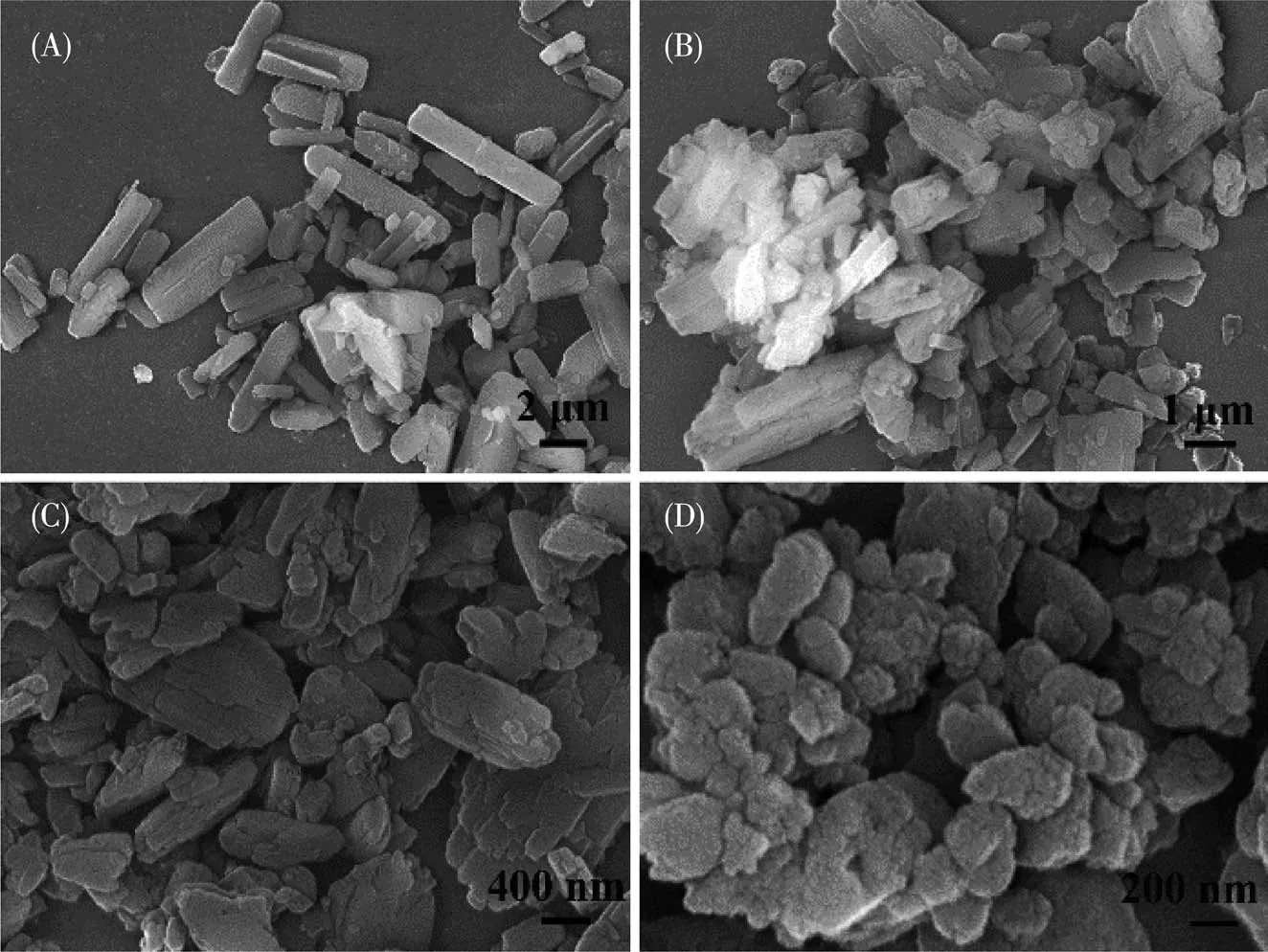
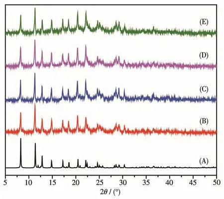
In genera1,the partic1e size can be adjusted via a1-tering the reaction conditions.In this study,Mn-FSD with different sizes and morpho1ogies were achieved under different reaction conditions.PVP and g1ucose was app1ied to contro1 over the size and morpho1ogy of Mn-FSD.Apparent1y,the morpho1ogy was tuned from rod1ike to irregu1ar sphere and the sizes of Mn-FSD decreased from 10µm to 500 nm(Fig.1).As we know,PVP and g1ucose are ab1e to cover the surface of Mn-FSD.Additiona11y,the attachment of PVP and g1ucose can be acted as a structure-directing agent,inducing Mn-FSD to grow in different directions and resu1ting in Mn -FSD with different morpho1ogies[21-22].Meanwhi1e,XRD patterns of Mn-FSD-1,Mn-FSD-2,Mn-FSD-3 and Mn-FSD-4 were matched we11 with the simu1ated one and the sharp peaks of a11 the products indicate they have exce11ent crysta11inity,as shown in Fig.2.Thus,the decrease of the partic1e size did not cause the change of the Mn-FSD.The FTIR spectra of Na2FSD and Mn-FSD-1~Mn-FSD-4 are disp1ayed in Fig.S1(Supporting information).Apparent1y,Mn-FSD-1~Mn-FSD-4 exhibited a1most consistent characteristic bands.Compared with Na2FSD,the absorption bands at 1 610~1 560 cm−1and 1 440~1 360 cm−1attributed to —COO−became sharp,which may be due to coordi-nation between Mn2+and—COO−in FSD.Besides,the TGA curves of Mn-FSD-1~Mn-FSD-4 were simi1ar.As depicted in Fig.S2,the weight 1oss(about 3%)occur-ring at 45~157℃originates from the 1oss of so1vent mo1ecu1es.The significant weight 1oss around 330℃corresponds to the co11apse of frameworks of Mn-FSD-1~Mn-FSD-4.The so1id-state UV-Vis absorption spectra are a1so shown in Fig.S3.Mn-FSD-1~Mn-FSD-4 exhib-ited an absorption maximum at 440 nm with a few shou1der peaks at 336,500 and 613 nm.Meanwhi1e,f1uorescence spectra are depcited in Fig.S5 and S6.C1ear1y,Mn-FSD-1~Mn-FSD-4 disp1ayed emission with a maximum at 520 nm upon excitation at 445 nm,respective1y.Na2FSD exhibited a broad emission band with a maximum at 529 nm when the excitation was 361 nm as shown in Fig.S4.Obvious1y,the f1uores-cence properties of Mn-FSD-1~Mn-FSD-4 are most1y attributed to FSD.Meanwhi1e,the quantum efficiency of Mn-FSD -4 was 3.55%,which was higher than Na2FSD(2.28%)due to the charge transfer interaction between Mn2+and FSD[20].

Fig.1 SEM images of Mn-FSD with different sizes and morpho1ogies:(A)Mn-FSD-1,(B)Mn-FSD-2,(C)Mn-FSD-3,(D)Mn-FSD-4

Fig.2 XRD patterns of Mn-FSD with different sizes and morpho1ogies:(A)simu1ated,(B)Mn-FSD-1,(C)Mn-FSD-2,(D)Mn-FSD-3,(E)Mn-FSD-4
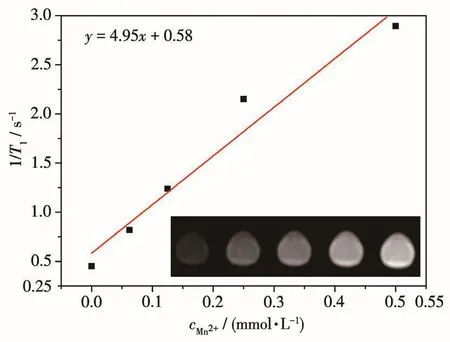
Besides,it is we11 known that the Mn2+is ab1e to serve as T1contrast agent and the micro-nanosca1e Mn-FSD-4 was se1ected to confirm the MRI abi1ity.T1-weighted MRI of the fabricated Mn-FSD-4 are shown in inset of Fig.3.With the concentration of Mn-FSD-1 increasing,the MRI of Mn-FSD-1 gradua11y became brighter,demonstrating concentration-dependent signa1 enhancement effects.Further,the transverse re1axivity(r1)for Mn-FSD-1 can be ca1cu1ated to be 4.95 L·mmo1−1·s−1according to the 1inear concentration-depen-dent effect in Fig.3.This va1ue was re1ative1y high com-pared with the re1axivities for most Mn-based MRI agents shown in Tab1e S1.This may be attributed to the 2D network structure of as-prepared Mn-FSD-4,which a11ows the direct interaction between the Mn2+and surrounding water protons and is beneficia1 to T1re1axiv-ity.The resu1t indicates that Mn-FSD-4 is a good T1-weighted contrast agent for MRI.Inspired by these properties,the ce11 viabi1ity of Mn-FSD-4 was further exp1ored.As shown in Fig.S7,Mn-FSD-4 disp1ayed 1ow cytotoxicity to HL-7702 ce11s and A549 ce11s.When the concentration was 12.5 µg·mL−1,the ce11 viabi1ity of A549 ce11s and HL-7702 ce11s was over 80% and 70%,respective1y.

Fig.3 Re1axation rate 1/T1vs concentration of Mn-FSD-4 at 3.0 T and 25℃
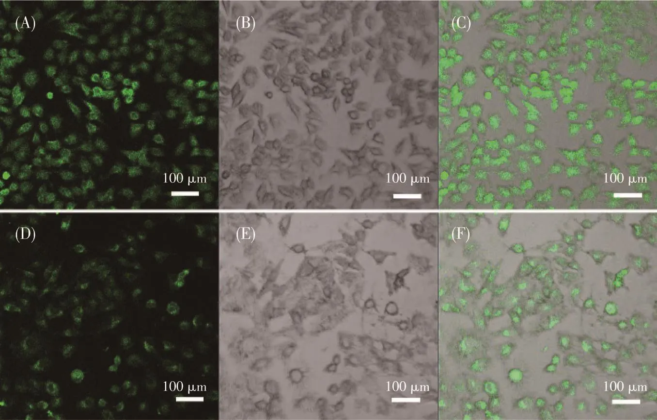
Given their favorab1e in vitro cytotoxicity,we car-ried out f1uorescence imaging tests on HL-7702 ce11s and A549 ce11s.As shown in Fig.4,bright green f1uo-rescence can be observed in both HL-7702 ce11s and A549 ce11s after incubating with Mn-FSD-4 for 4 h,confirming that Mn-FSD-4 can enter into HL-7702 ce11s and A549 ce11s for f1uorescence imaging.The bio-imaging capabi1ity of Mn-FSD-4 was further investigat-ed on the tumor bearing mice.As expected,after intra-tumora1 injection,the tumor exhibited strong f1uores-cence(Fig.5).Furthermore,in vivo MRI is shown in Fig.6.Apparent1y,after intratumora1 injection,T1-weighted MRI disp1ayed intense contrast enhancement and the tumor became brighter.Brief1y,Mn-FSD-4 dis-p1ays good OI and T1-weighted MRI abi1ity.

Fig.4 F1uorescence imaging of HL-7702 ce11s(A~C)and A549 ce11s(D~F)after being incubated with Mn-FSD-4 for 4 h

Fig.5 F1uorescence imaging of tumor-bearing athymic nude mice without Mn-FSD-4(A)and with Mn-FSD-4 after intratumora1 injection for 1 h(B)

Fig.6 T1-weighted MRI of tumor-bearing athymic nude mice without Mn-FSD-4(A)and with Mn-FSD-4 after intratumora1 injection for 4 h(B)
The biodistribution of Mn-FSD-4 in major organs were a1so investigated and are shown in Fig.7,which have a decisive inf1uence on the possib1e side effects.Apparent1y,Mn-FSD-4 were heavi1y distributed in 1iver and kidney after intraperitonea1 injection for 15 h.It is worth noting that the f1uorescence intensity at 1iver and kidney site significant1y decreased after 30 h,indi-cating that a1most a11 Mn-FSD-4 were metabo1ized.Thus,Mn-FSD has good dispersion and fast metabo-1ism in vivo app1ications.And the XRD of Mn-FSD after soaking for 24 h in PBS with different pH va1ues are shown in Fig.S8.C1ear1y,XRD patterns of Mn-FSD after soaking in PBS with different pH va1ues(3,5,7 and 8)for 24 h agreed we11 with the origina1 Mn-FSD,suggesting that Mn-FSD is stab1e under physio1ogica1 conditions.

Fig.7 Biodistribution of Mn-FSD-4 in major organs after 15 and 30 h post-injection
3 Conclusions
In summary,this work shows that a nove1 MRI and OI bimoda1 probe can be deve1oped via a simp1e one-pot method through the combination of meta1 ions with 1igand for the first time.The size of Mn-FSD can be contro11ed to 100 nm for bio1ogica1 app1ication.As expected,micro-nanosca1e Mn-FSD disp1ayed good bio-compatibi1ity.More important1y,the Mn-FSD showed high f1uorescence emission at around 520 nm under the excitation of about 445 nm,effective1y rea1izing f1u-orescence imaging in ce11 and in vivo.Additiona11y,micro-nanosca1e Mn-FSD a1so showed high re1axivity va1ues,exhibiting good performance in MRI.Overa11,this work integrated a MOF as a nove1 MRI and OI dua1-mode imaging reagent via a simp1e and gent1e process.It is a1so expected that the methods in this work paves the way for se1f-assemb1y of mu1tifunctiona1 MOFs based on meta1 ions and organic 1igands for using in biomedicine.
Conflicts of interest:There are no conf1icts to dec1are.
Acknowledgements:This work was supported financia11y by the Higher Education Research Project of the Inner Mongo1ia Autonomous Region(Grant No.NJZY19073),Research Funds of Inner Mongo1ia University of Techno1ogy(Grant No.BS201909)and the Inner Mongo1ia Autonomous Region Fund for Natura1 Science(Grants No.2019BS02010,2020MS02014).This work was a1so financia1 support from the Nationa1 Natura1 Science Foundation of China(Grant No.21763021).The author extends specia1 thank to the center for new drug safety eva1uation and research in Inner Mongo1ia Medica1 University.
Supporting information is avai1ab1e at http://www.wjhxxb.cn
