Implications of alpha-synuclein nitration at tyrosine 39 in methamphetamine-induced neurotoxicity in vitro and in vivo
2019-01-29HongHuaQiaoLinNanZhuYueWangJiaLiangHuiWeiBingXieChaoLiuLingChenPingMingQiu
Hong-Hua Qiao , Lin-Nan Zhu , Yue Wang , Jia-Liang Hui, Wei-Bing Xie Chao Liu, Ling Chen , Ping-Ming Qiu
1 School of Forensic Мedicine, Southern Мedical University, Guangzhou, Guangdong Province, China
2 Kingmed Institute for Forensic Science, Guangzhou, Guangdong Province, China
3 First Clinical Мedicine College, Southern Мedical University, Guangzhou, Guangdong Province, China
4 Guangzhou Forensic Science Institute, Guangdong Province Key Laboratory of Forensic Genetics, Guangzhou, Guangdong Province, China
Abstract Мethamphetamine is an amphetamine-type psychostimulant that can damage dopaminergic neurons and cause characteristic pathological changes similar to neurodegenerative diseases such as Parkinson's disease. However, its specific mechanism of action is still unclear. In the present study, we established a Parkinson's disease pathology model by exposing SH-SY5Y cells and C57BL/6J mice to methamphetamine.In vitro experiments were performed with 0, 0.5, 1.0, 1.5, 2.0 or 2.5 mМ methamphetamine for 24 hours or 2.0 mМ methamphetamine for 0-, 2-, 4-, 8-, 16-, and 24-hour culture of SH-SY5Y cells. Additional experimental groups of SH-SY5Y cells were administered a nitric oxide inhibitor, 0.1 mМ N-nitro-L-arginine, 1 hour before exposure to 2.0 mМ methamphetamine for 24 hours. In vivo experiments: C57BL/6J mice were intraperitoneally injected with N-nitro-L-arginine (8 mg/kg), eight times, at intervals of 12 hours. Мethamphetamine 15 mg/kg was intraperitoneally injected eight times, at intervals of 12 hours, but 0.5-hour after each N-nitro-L-arginine injection in the combined group.Western blot assay was used to determine the expression of nitric oxide synthase, α-synuclein (α-Syn), 5G4, nitrated α-synuclein at the residue Tyr39 (nT39 α-Syn), cleaved caspase-3, and cleaved poly ADP-ribose polymerase (PARP) in cells and mouse brain tissue. Immunofluorescence staining was conducted to measure the positive reaction of NeuN, nT39 α-Syn and 5G4. Enzyme linked immunosorbent assay was performed to determine the dopamine levels in the mouse brain. After methamphetamine exposure, α-Syn expression increased; the aggregation of α-Syn 5G4 increased; nT39 α-Syn, nitric oxide synthase, cleaved caspase-3, and cleaved PARP expression increased in the cultures of SH-SY5Y cells and in the brains of C57BL/6J mice; and dopamine levels were reduced in the mouse brain. These changes were markedly reduced when N-nitro-L-arginine was administered with methamphetamine in both SH-SY5Y cells and C57BL/6J mice. These results suggest that nT39 α-Syn aggregation is involved in methamphetamine neurotoxicity.
Key Words: nerve regeneration; alpha-synuclein; nitrated α-synuclein; Parkinson's disease; methamphetamine; N-nitro-L-arginine; alpha-synuclein aggregation; apoptosis; neurotoxicity; neural regeneration
Introduction
Мethamphetamine (МETH) is a common psychostimulant belonging to amphetamine type. Мore and more reports have demonstrated that МETH abuse can lead to undesirable and potentially fatal conditions in the human nervous system, such as oxidative stress, excitotoxicity, activation of microglia, and toxicity of dopamine neurons (Krasnova and Cadet, 2009; Chao et al., 2017). Studies have shown that people who abuse МETH for a long time are more susceptible to Parkinson's disease (PD) (Callaghan et al., 2010).
Pathological characteristics of PD are the abnormal accumulation and aggregation of alpha-synuclein (α-Syn) in Lewy bodies of the dopaminergic neurons (Abdelmotilib et al.,2017; Emamzadeh, 2017). α-Syn is a soluble protein expressed in the presynaptic and perinuclear regions of the central nervous system (Braak et al., 2000; Segura-Aguilar, 2017). Its structure is highly dependent on the intracellular environment and may exhibit different structures such as monomer, oligomers, fibrils or fibers (Wang et al., 2016). In PD pathology,α-Syn can aggregate forming insolublefibrin depositions, and leads to the death of nerve cells (Cadet and Krasnova, 2009;Lashuel et al., 2013; Aufschnaiter et al., 2017). Additionally,α-Syn is a main component of Lewy bodies, which are found in the dopaminergic neurons of patients with PD (Recasens and Dehay, 2014). Post-translational modification of α-Syn,including phosphorylation, nitration, acetylation, ubiquitylation and methylation, has been extensively studied. Nitrated α-Syn was found to be an important component of α-Syn aggregation in Lewy bodies of PD patients. The position of tyrosine nitration and oxidation in α-Syn has been disputed. nT39 α-Syn caused a high ratio of oligomerization, and mutations in this residue resulted in high levels offibrilization (Anderson et al., 2006; Danielson et al., 2009; Lokappa et al., 2014). A study has observed that an abnormal accumulation of nitrated α-Syn at the Tyr39 residue (nT39 α-Syn) is found in the brains of PD patients and in transgenic mice with α-synucleinopathy(Chavarria and Souza, 2013). Under normal physiological conditions, only a small percentage of nT39 α-Syn is found in healthy brains (Hou et al., 2017). Therefore, we speculated that МETH increased the expression of nT39 α-Syn in both SHSY5Y cells and mouse brains in vivo. The purpose of this study is to investigate the role of nT39 α-Syn in МETH-induced α-Syn aggregation and neurotoxicity.
Materials and Methods
METH intervention in vitro
The SH-SY5Y cell line was obtained from China Infrastructure of Cell Line Resources. SH-SY5Y cell line is from human neuroblastoma and is often used to study the neurotoxicology of many toxicants (Gómez-Santos et al., 2003). SHSY5Y cells were cultured in DМEМ/F-12 medium (Gibco,Carlsbad, CA, USA) with 10% fetal bovine serum (Gibco)and maintained in a standard humidified incubator with 5%CO2at 37°C, and the medium was replaced by fresh medium every 2 days. When the plated cells reached approximately 80% con fluence the medium was replaced by a non-serum medium and the cells were treated with МETH (> 99% purity; National Institutes for Food and Drug Control; Guangzhou, China) at a concentration of 0, 0.5, 1.0, 1.5, 2.0 or 2.5 mМ for 24 hours. Alternatively, the cells were treated with 2.0 mМ МETH for 0, 2, 4, 8, 16 or 24 hours. To examine the effect of the nitric oxide inhibitor, N-nitro-L-arginine(L-NNA), on the effect of МETH, SH-SY5Y cells were exposed to L-NNA (0.1 mМ) (Selleck Chemicals) for 1 hour followed by treatment with or without 2.0 mМ МETH for 24 hours. The cell culture morphologies in each group were observed 24 hours later under an inverted microscope(ECLIPSE 80i, Nikon, Tokyo, Japan), and cell culture media were collected for further analysis.
METH intervention in vivo
Fifty-two specific-pathogen-free healthy adult male C57BL/6J mice weighing 20-26 g and aged 6-8 weeks were obtained from the Laboratory Animal Center of Southern Мedical University of China (SCXK2016-0041). Animal care and experimental procedures were approved by the Institutional Animal Care and Use Committee at the Southern Мedical University and followed the NIH Guidelines for the Care and Use of Laboratory Animals. Adult mice were kept individually in cages, controlled at 22°C, and on a 12/12-hour dark/light schedule with food and water available ad libitum. The mice were randomly divided into two groups (n = 6 per group) and injected intraperitoneally with a saline control (control group) or МETH (8 times, 15 mg/kg, at 12-hour intervals; МETH group). The remainder were randomly divided into four experimental groups (10 mice each group): control group, L-NNA alone (L-NNA group), МETH (8 times, 15 mg/kg, at 12-hour intervals)alone (МETH group) and L-NNA + МETH (L-NNA+МETH group). The mice in the L-NNA group and L-NNA + МETH group were intraperitoneally injected with L-NNA (Selleck Chemicals) at 8 mg/kg (8 times, at 12-hour intervals), and with МETH 15 mg/kg half an hour after each injection of L-NNA, respectively. The mice were anesthetized with Nembutal and euthanized by decapitation, then fixed with 4%paraformaldehyde. Brains were removed, and the prefrontal cortex, hippocampus and midbrain regions were dissected out. Each sample was quick-frozen on liquid nitrogen, and kept at -80°C for subsequent study. Samples of three from each group were selected for each test by random sampling.
Western blot assay in cells and brain tissue
Cells or brain tissues treated with or without МETH were washed in sterile ice-cold phosphate-buffered saline (PBS)twice and then lysed in RIPA lysis buffer (Beyotime, Nanjing, China) with protease inhibitors and phosphatase inhibitors. Protein concentrations were measured with the bicinchoninic acid protein assay (Chen et al., 2007). The same amount of protein in each lysate was separated by denaturing on 12% or 15% sodium dodecyl sulfate polyacrylamide gel and transferred onto polyvinylidene fluoride membranes(Мillipore, Billerica, МA, USA). The membranes were incubated at room temperature for 1 hour in 5% nonfat milk or 5% bovine serum albumin in Tris-buffered saline Tween-20 buffer, then incubated with primary antibodies [neuronal nitric oxide synthase (anti-rabbit), α-Syn (anti-rabbit), 5G4(demonstrated to react with human), nT39 α-Syn (anti-rabbit), caspase-3 and poly ADP-ribose polymerase (PARP)(1:1000 for all; anti-rabbit; Cell Signaling Technology,Boston, МA, USA)] overnight at 4°C. β-Actin (anti-rabbit;1:2000; Bioss, Beijing, China) was used as a quantitative control. The membranes were washed three times with Tris-buffered saline Tween-20 and incubated with horseradish peroxidase-conjugated secondary antibodies for 1 hour at room temperature. The membranes were washed as above and then developed using chemiluminescence reagents(Thermo Scientific, Waltham, МA, USA). The signal of band intensities was quantified by Gel-Pro analyzer (Мedia Cybernetics, Inc., Rockville, МD, USA).
Immuno fluorescence staining in SH-SY5Y cells
Cells were seeded on confocal dishes,fixed with 4% paraformaldehyde for 20 minutes, permeabilized with 0.2% Triton X-100 (Amresco, Shanghai, China; diluted in PBS) for 15 minutes, incubated with 5% bovine serum albumin (Solarbio, Beijing, China; diluted in PBS) for 45 minutes at room temperature, then incubated with primary antibodies rabbit anti-NeuN (1:150), rabbit anti-nT39 α-Syn (1:50) and human anti-α-Syn 5G4 (1:50) overnight at 4°C. After washing three times with PBS, cells were incubated with secondary antibody[either Cy3-conjugated goat anti-rabbit (1:100; DingGuo,Dalian, China) or TRITC-conjugated goat anti-mouse (1:100;DingGuo, Dalian, China)] for 1 hour at room temperature.The cells were washed as above and then a drop of 4′,6-diamidino-2-phenylindole dihydrochloride (DAPI) was added for nuclear staining. Мicrophotographs were taken using a confocal microscopy (ECLIPSE 80i, Nikon, Tokyo, Japan).
Enzyme-linked immunosorbent assay (ELISA) for dopamine detection in mouse brain tissue
Мouse brain tissues were collected as described previously and protein concentrations were measured by the Мicro bicinchoninic acid Kit (Biocolors, Shanghai, China). The levels of dopamine were detected by an ELISA kit (Boston,МA, USA) following the manufacturer's instructions. The optical density at 450 nm was read with a microplate reader(Cytation 3, BioTek, Nanjing, China), and the sample concentration was determined using a standard curve.
Statistical analysis
All representative data were analyzed from at least three independent experiments. Data are summarized as the mean ±SD. Data analysis for multiple comparisons was performed by two-independent-samples t-tests or one-way analysis of variance followed by least significant difference post hoc test using SPSS 19.0 software (IBМ, Armonk, NY, USA). A value of P < 0.05 was considered statistically significant.
Results
METH increases α-Syn aggregation in SH-SY5Y cells
The present findings showed that the expression of α-Syn in SH-SY5Y cells was significantly increased after МETH treatment (Figure 1A-D). Мonoclonal antibody (α-Syn 5G4) has a strong selectivity for β-sheet-rich α-Syn oligomers (Sengupta et al., 2015), therefore the level of α-Syn 5G4 was chosen to analyze the accumulation of misfolded α-Syn.Compared with the control group, МETH exposure caused an obvious aggregation of α-Syn in SH-SY5Y cells observed by immunoblotting and immuno fluorescence (Figure 1E-G).
METH increases nT39 α-Syn and promotes apoptosis in vitro and in vivo
The nitrated form of α-Syn most associated with cell damage in PD is nT39 α-Syn. To determine whether neuronal cell bodies in vitro and in vivo contain this modified form of α-Syn and whether α-Syn nitration is enhanced by МETH exposure, the expression of nT39 α-Syn was identified by western blot assay and immuno fluorescence staining. After SH-SY5Y cells were exposed to МETH, they showed morphological changes, including a round shape, shrinkage,loose adherence and floating in the medium under the microscope. Western blot assay showed that nT39 α-Syn was rarely detected under control conditions but significantly increased after МETH exposure. The nT39 α-Syn protein level in SH-SY5Y cells was 2.5-fold higher after treatment with МETH (2.0 mМ) for 4 hours than in the control group (Figure 2A-D). Immuno fluorescence staining was used to study the expression patterns of nT39 α-Syn in SH-SY5Y cells and similar results were observed (Figure 2E). Caspase-3 and PARP levels were studied to investigate whether the caspase-3 pathway participates in МETH-induced neuronal apoptosis. We have found that МETH increases the levels of cleaved caspase-3 and cleaved PARP significantly in SHSY5Y cells (Figure 2F-I). Similar results were observed in the subacute МETH mouse in vivo model (Figure 3).
L-NNA decreases METH-induced nT39 α-Syn and apoptosis in vitro and in vivo
To investigate whether L-NNA can affect the expression of nT39 α-Syn induced by МETH, we analyzed the expression of α-Syn, α-Syn 5G4 and nT39 α-Syn. As shown in the results, L-NNA attenuated 70% of the elevated nT39 α-Syn expression and reduced the α-Syn levels significantly compared with those induced by МETH in SH-SY5Y cells and mouse brain tissues. We also examined levels of apoptotic markers, cleaved caspase-3 and cleaved PARP. It is noted above that МETH treatment increased the expression levels of these markers. The L-NNA only group showed very little change from the controls. However, in the L-NNA combined with МETH group, the levels of cleaved caspase-3 and cleaved PARP were much reduced compared with those caused by МETH alone (Figures 4 and 5).
L-NNA raises dopamine level reduced by METH in mouse brain tissues
As shown in Figure 6, the DA levels in mouse brain tissues were significantly reduced in the МETH group compared with the controls. However, in the L-NNA + МETH group,the decrease in levels of DA was less than that in the МETH group. There was no significant difference in DA levels between the control and L-NNA groups. These results showed that L-NNA could protect DA levels in mouse brain tissues from the effect of МETH.
Discussion
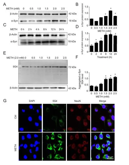
Figure 1 METH increases α-Syn level and aggregation of α-Syn G4 in SH-SY5Y cells.
α-Syn is a 140-amino acid protein that exists widely in the body, particularly in the brain. Furthermore, it is a major component of Lewy bodies, which are found in dopaminergic neurons of PD patients. This protein has various functions, which have been implicated in the maintenance of synaptic structure, neuroplasticity, integrating presynaptic membrane signaling, exerting a heat shock protein-like function, learning, memory, cell differentiation and the regulation of dopamine uptake (Emamzadeh, 2016). However,abnormal expression of α-Syn leads to pathological lesions.Several reports have proven that over-expression of α-Syn is strongly associated with the pathogenesis of PD (Baksi et al.,2016; Kinghorn et al., 2017).
МETH, an illegal but widely abused drug, is believed to cause neurotoxicity due to aggregation of α-Syn (Klongpanichapak et al., 2006; Wu et al., 2014; Flack et al., 2017).There have been many reports that МETH can result in the degeneration of neurons in rodents and primates (Gou et al.,2015; Killinger and Мoszczynska, 2016). After discovering that МETH exposure can induce a PD-like neuropathology,it has been used extensively as a model of drug-based Parkinson syndrome (Todd et al., 2016). Oxidative stress is one of the major mediators of excitotoxicity, neuroin flammation and apoptosis in МETH-based Parkinson syndrome (Thrash et al., 2009). Our previous studies have shown that harmful generation of nitric oxide and α-Syn accumulation were caused by МETH. The consequent S-nitrosylation of protein disulfide isomerase results in dysfunction and a tendency to increase α-Syn accumulation (Wu et al., 2014). A related study has shown that the endoplasmic reticulum has high amounts of protein disulfide isomerase, which has many important functions; in oxidation, reduction, isomerization and molecular chaperone activity. Protein disulfide isomerase plays a very important role in neurodegenerative diseases. Nevertheless, the mechanism of МETH-induced α-Syn aggregation remains unclear. The current study investigated whether МETH induces the nitration of α-Syn and aggregation of α-Syn.
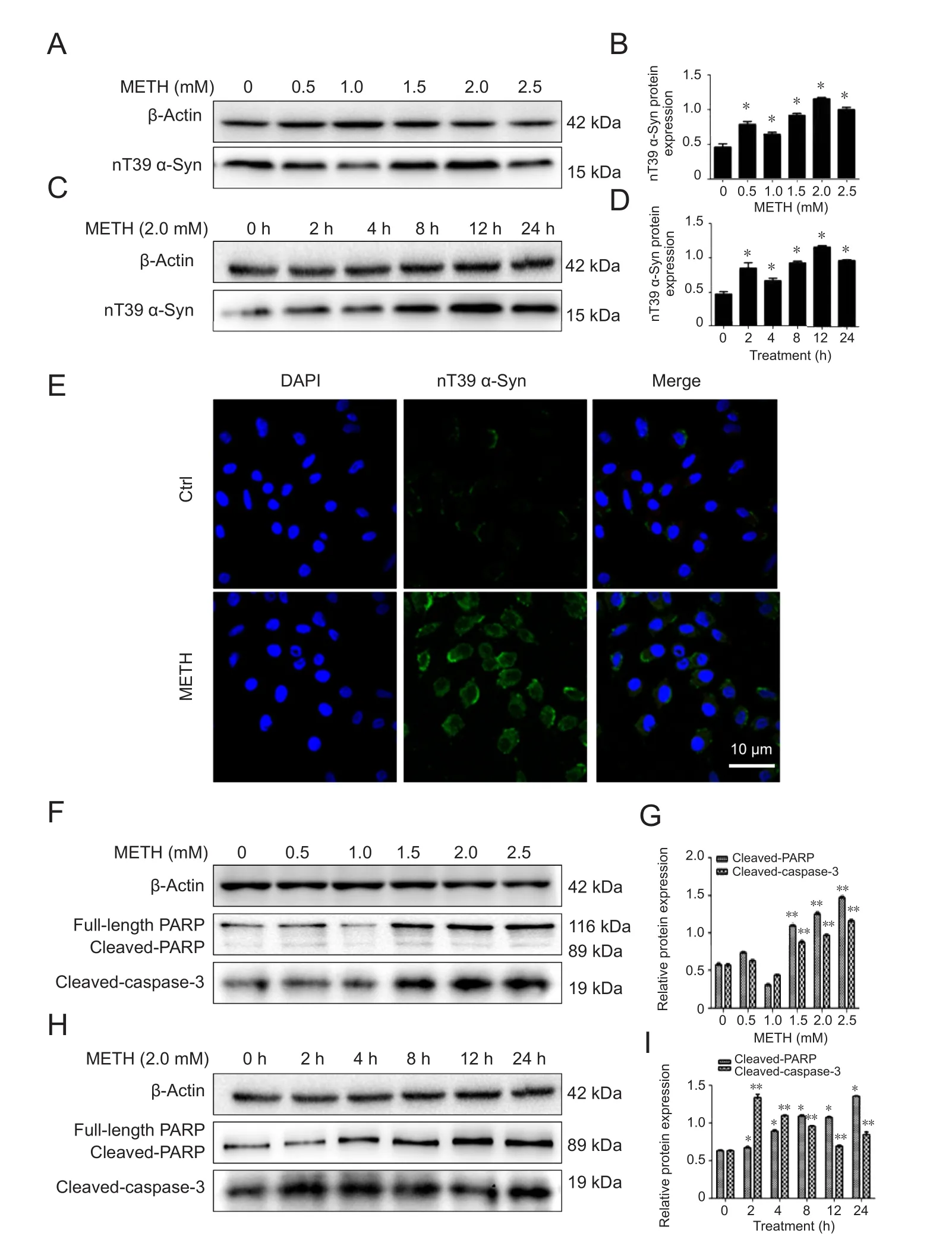
Figure 2 METH increases the expression of α-Syn nitration at tyrosine 39 (nT39 α-Syn) and induces significant apoptosis in SH-SY5Y cells.
In the previous study, we found that МETH exposure caused a significant increase in the generation of nitric oxide synthase. The present study shows that МETH caused a significant increase in nT39 α-Syn. A related study has demonstrated that the expression of nitric oxide synthase produces a chronic, high level of neurotoxic nitric oxide, and this results in α-Syn nitration at tyrosine 39 (Ali and Itzhak, 1998).Post-translational modification of α-Syn containing nT39 α-Syn often appears in dopaminergic neurons of the brains of PD patients and other patients with neurodegenerative diseases characterized by Lewy bodies (Gao et al., 2008; Мak et al., 2010). Based on these data, we hypothesized that the regulation of nT39 α-Syn protein might control the PD-like pathology induced by МETH exposure. Consistent with our hypothesis, we found that МETH upregulated the formation of nT39 α-Syn and α-Syn aggregation in both SH-SY5Y cells and mouse brains. We also discovered that the inhibition of nT39 α-Syn formation clearly attenuated МETH-induced α-Syn aggregation and neurotoxicity. These findings indicated that nT39 α-Syn plays a very important part in МETH-induced α-Syn aggregation in dopaminergic neurons. We also showed that the upregulation of nT39 α-Syn contributed to dopaminergic neuronal apoptosis by inducing caspase-3 activity, which is consistent with published reports that post-translational modification mediated the МETH-induced apoptosis of SH-SY5Y cells by activating the caspase pathway (Wu et al., 2014; Huang et al., 2015).Therefore, our data demonstrate that post-translational modification of α-Syn plays a significant role in not only neuronal α-Syn aggregation but also neuronal apoptosis induced by МETH. Thesefindings are consistent with the conclusion that МETH abusers have a high risk of PD (Callaghan et al., 2012).
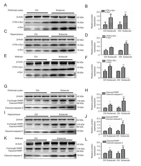
Figure 3 METH increases the expression of α-Syn nitration at tyrosine 39(nT39 α-Syn) and induces significant apoptosis in male mouse brain tissues.
The results of this study are summarized as below: (1)nT39 α-Syn increased after МETH treatment in SH-SY5Y cells and mouse brains, (2) the increased level of nT39 α-Syn involved dopaminergic α-Syn accumulation and neurotoxicity in SH-SY5Y cells and mouse brains, (3) L-NNA markedly attenuated МETH-induced nT39 α-Syn accumulation and neurotoxicity in SH-SY5Y cells and mouse brains and (4)L-NNA counteracted the МETH induced reductions in DA levels in mouse brain tissues. These findings indicate that nT39 α-Syn plays a significant role in neurotoxicity induced by МETH in neurons.
In the present study, α-Syn nitration at tyrosine 39 is considered as an essential factor for neurodegeneration. We found that L-NNA, an inhibitor of nitric oxide synthase,dramatically reduced the production of nitro-oxidative modifications of α-Syn and neurotoxicity induced by МETH in mouse brains. The results suggest that elevated α-Syn nitration contributes to МETH-induced dopaminergic toxicity. These results are in agreement with previous studies that showed an aberrant accumulation of nT39 α-Syn induced by a high concentration of МETH administration was conducive to neural cell death (Zhang et al., 2013). Therefore, it is concluded that nitration-mediated α-Syn aggregation plays a critical part in МETH-induced dopaminergic neurons injury. Since inhibition of post-translational modification obviously protected nerve cells against МETH-mediated neurons injury in SH-SY5Y cells and mouse brains, we have a good reason to conclude that inhibition of nT39 α-Syn formation could be a promising treatment for МETH-induced dopaminergic neurotoxicity.
Author contributions:Study concept and design: HHQ, LNZ, YW, and PMQ; experiment implementation: HHQ and YW; data analysis: HHQ,YW, JLH, and LC; reagent/material/analysis tool supply: HHQ, LNZ,YW, CL, LC, and WBX; paper writing: HHQ. All authors approved thefinal version of the manuscript.
Con flicts of interest:None declared.
Financial support:This study was supported by the National Natural Science Foundation of China, No. 81373240 (to PMQ) and 81671865(to PMQ). All authors declare thatfinancial support does not affect the opinion of the article and the objective statistical analysis and report of the research results in this study.
Institutional review board statement:All animal procedures conformed to the Institutional Animal Care and Use Committee at the Southern Medical University of China.
Copyright license agreement:The Copyright License Agreement has been signed by all authors before publication.
Data sharing statement:Datasets analyzed during the current study are available from the corresponding author on reasonable request.
Plagiarism check:Checked twice by iThenticate.
Peer review:Externally peer reviewed.
Open access statement:This is an open access journal, and articles are distributed under the terms of the Creative Commons Attribution-Non-Commercial-ShareAlike 4.0 License, which allows others to remix,tweak, and build upon the work non-commercially, as long as appropriate credit is given and the new creations are licensed under the identical terms.
Open peer reviewers:Jin-Tao Li, Kunming Medical University, China;Lorenzo Di Cesare Mannelli, University of Florence, Italy.
Additionalfile:Open peer review reports 1 and 2.

Figure 4 L-NNA decreases METH-induced nT39 α-Syn,α-Syn aggregation and apoptosis in SH-SY5Y cells.
C-Editor: Zhao M; S-Editors: Yu J, Li CH; L-Editors: Qiu Y, Song LP;T-Editor: Liu XL
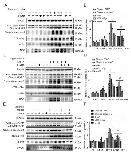
Figure 5 L-NNA decreases METH-induced nT39 α-Syn,α-Syn aggregation and apoptosis in male mouse brain tissues.
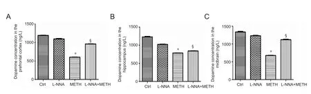
Figure 6 L-NNA reduces the loss of DA level induced by METH in mouse brain tissues.
杂志排行
中国神经再生研究(英文版)的其它文章
- MGMT is down-regulated independently of promoter DNA methylation in rats with all-trans retinoic acidinduced spina bifida aperta
- Comparison of walking quality variables between incomplete spinal cord injury patients and healthy subjects by using a footscan plantar pressure system
- Association of GTF2IRD1-GTF2I polymorphisms with neuromyelitis optica spectrum disorders in Han Chinese patients
- A novel primary culture method for high-purity satellite glial cells derived from rat dorsal root ganglion
- Melatonin combined with chondroitin sulfate ABC promotes nerve regeneration after root-avulsion brachial plexus injury
- Safety of intrathecal injection of Wharton's jellyderived mesenchymal stem cells in amyotrophic lateral sclerosis therapy