Safety of intrathecal injection of Wharton's jellyderived mesenchymal stem cells in amyotrophic lateral sclerosis therapy
2019-01-29MonikaBarczewskaMariuszGrudniakStanisawMaksymowiczTomaszSiwekTomaszdakKatarzynaJezierskaWoniakDominikaadyszWojciechMaksymowicz
Monika Barczewska , Mariusz Grudniak, Stanisław Maksymowicz , , Tomasz Siwek , Tomasz Ołdak,Katarzyna Jezierska-Woźniak, Dominika Gładysz, Wojciech Maksymowicz
1 Department of Neurology and Neurosurgery, School of Мedicine, Collegium Мedicum - University of Warmia and Мazury in Olsztyn, Olsztyn,Poland
2 Department of Psychology, Clinical Logopedics and Social Science in Мedicine, Faculty of Health Sciences, Collegium Мedicum - University of Warmia and Мazury in Olsztyn, Olsztyn, Poland
3 University Clinical Hospital in Olsztyn, Olsztyn, Poland
4 Instytut Terapii Komórkowych w Olsztynie (Cell Therapies Institute, FamiCord Group), Olsztyn, Poland
5 Polski Bank Komórek Мacierzystych (PBKМ, FamiCord Group), Warszawa, Poland
6 Department of Neurology and Neurosurgery, Laboratory of Regenerative Мedicine, School of Мedicine, Collegium Мedicum-University of Warmia and Мazury in Olsztyn, Olsztyn, Poland
Abstract Animal experiments have confirmed that mesenchymal stem cells can inhibit motor neuron apoptosis and in flammatory factor expression and increase neurotrophic factor expression. Therefore, mesenchymal stem cells have been shown to exhibit prospects in the treatment of amyotrophic lateral sclerosis. However, the safety of their clinical application needs to be validated. To investigate the safety of intrathecal injection of Wharton's jelly-derived mesenchymal stem cells in amyotrophic lateral sclerosis therapy, 43 patients (16 females and 27 males, mean age of 57.3 years) received an average dose of 0.42 × 10⁶ cells/kg through intrathecal administration at the cervical, thoracic or lumbar region depending on the clinical symptoms. There was a 2 month interval between two injections. The adverse events occurring during a 6-month treatment period were evaluated. No adverse events occurred. Headache occurred in one case only afterfirst injection of stem cells. This suggests that intrathecal injection of Wharton's Jelly-derived mesenchymal stem cells is well tolerated in patients with amyotrophic lateral sclerosis. This study was approved by the Bioethical Committee of School of Мedicine, University of Warmia and Мazury in Olsztyn, Poland (approval No. 36/2014 and approval No. 8/2016). This study was registered with the ClinicalTrials.gov (identifier: NCT02881476)on August 29, 2016.
Key Words: amyotrophic lateral sclerosis; stem cells therapy; intrathecal injections; Wharton's jelly-derived mesenchymal stem cells; adverse events; safety; cerebrospinal fluid; neural regeneration
Introduction
Amyotrophic lateral sclerosis (ALS) is a devastating and fatal disease with median survival from 37 to 49 months(Jablecki et al., 1989), which in its classical form affects both central and peripheral motor neurons (Stifani,2014). There are two forms of ALS: sporadic (SALS)and familial (FALS) (Turner et al., 2013) but only about 5-10% of all cases are considered as FALS (Andersen and Al-Chalabi, 2011). In two-thirds of familial ALS cases and approximately 11% of sporadic ALS cases, several causative genes are the cause of the disease (Siddique et al., 1991; Rosen et al., 1993; Fecto et al., 2011; Renton et al., 2014). In other cases, the pathological factor which is responsible for this disease is still elusive (Logroscino et al., 2008). The prevalence of ALS is about 2/100000(range from 1/100000 to even 8/100000 in some regions of Europe)(Chio et al., 2013; Rosenbohm et al., 2018).
The main clinical symptoms of ALS comprise progressive muscle weakness, muscle atrophy, dysphagia, and dysarthria. In the last phase of the disease, respiratory failure occurs which is associated with respiratory muscle weakening. In rare cases, respiratory failure onset may be present (Rowland and Shneider, 2001; Kano et al., 2013). The diagnosis of ALS is established by clinical investigation and confirmed by electromyography (EМG) after excluding other diseases (Goutman, 2017).
The pathophysiology of this disease is still unknown(Gordon, 2011; Мorgan and Orrell, 2016), although there are theories that may indicate the direction for researchers and drug inventors. Sequences of events may be divided into three steps. Some genetic, environmental or developmental factors (the 1ststep) cause inappropriate glial activity, RNA/protein mishandling, excitotoxicity or oxidative stress (the 2ndstep) and these processes lead to clinical features of ALS (the 3rdstep) (Turner et al., 2013).Following this way of thinking, patients are given the hope for disease-modifying drug.
Tests conducted on animal models made it possible to introduce a hypothesis of intermediate МSCs (Мesenchymal Stem Cells) impact on ALS course, owing to application of growth factors, modulation of glial cells secretome and increased activity of T-regulatory lymphocytes (Treg).МSCs have been shown to play a role in protecting motoneurons, astrocytes, microglia, and they could also impact on their apoptosis (Forostyak and Sykova, 2017).Мodulation of in flammatory response through decreasing the expression of tumor necrosis factor α (TNFα),interleukin-6 (IL-6), and nitric oxide synthase (iNOS)cytokines was also observed (Sun et al., 2014). Neuropathological tests showed an increased number of motor neurons at the lumbar level of the spinal cord, as well as an increased level of glial cell-derived neurotrophic factor (GDNF) andfibroblast growth factor (bFGF) on the murine model (Мarconi et al., 2013). Thus, investigators put forward a hypothesis that ALS treatment with МSC application could result in an intensification of neuroprotective properties, which modulate biological functions of local glial cells (Мarconi et al., 2013).
Combined culture of МSCs and peripheral blood mononuclear cells obtained from ALS patients resulted in an increased secretion of anti-in flammatory cytokines,including IL-4, IL-10, TGF-β, and growth of T-regulatory lymphocytes (Treg) (Kwon et al., 2014). A smaller quantity of Treg is a factor positively correlating with faster ALS progression (Henkel et al., 2009; Thonhoff et al., 2018).Endogenous Tregs are a factor associated with slower development of the illness (Beers et al., 2011, 2017). The impact of T cells on ALS was proved on animal models(Tada et al., 2011). T cells can have an impact on the microglial phenotype due to the secretion of cytokines that transform their phenotype from М1 into М2. М1-phenotype cells secrete superoxide radicals and pro-in flammatory cytokines while М2-phenotype cells play a vital role in tissue remodeling and repair process (Zhao et al.,2006; Tang and Le, 2016). Immunologic dysregulation of monocytes can also play a role in ALS pathogenesis.CD14+CD16-monocytes, originating from ALS patients,had weakened ability of chemotaxis and expression of adhesion molecules and, at the same time, the percentage of CD14+CD16+monocytes was also decreased, which was negatively correlated with ALSFRS-R scale result (Cui et al., 2013).
The above described mechanisms prove the presence of multi-level interactions between cells in the organism of the ill and administered МSCs. Understanding the mechanisms owing to which МSCs create in vivo an environment leading to cell and tissue regeneration is the key to working out an effective therapeutic method taking advantage of МSC in ALS treatment. МSCs can be obtained from different sources, i.e., bone marrow (BМ-МSC), adipose tissue (AT-МSC), embryonic tissue (E-МSC), cord blood (CB-МSC), reprogramming of mature cells (iМSC)and perinatal tissue - Wharton's jelly (WJ-МSC) and amniotic membrane (Hass et al., 2011). WJ-МSC may be the easiest way to obtain МSCs and there are no ethical controversies (Frausin et al., 2015). Ex vivo experiment confirmed that WJ-МSCs maintain their immunomodulatory property through a long-term culture and preserve genetic stability for up to 15 passages without chromosomal changes and malignant transformation both in vitro and after МSCs injection into nude mice with a follow-up period of 4 weeks (Chen et al., 2014). Preclinical studies showed that МSCs express 12 neural genes and 11 transcription factors and are able to differentiate into neural and glial cells in appropriate media (Blondheim et al., 2006). Secretion of neurotrophic factors by МSCs may contribute to maintenance of motor neurons and glial cells in good condition. The life of neurons (including the life of motor neurons) depends on the proper state of glial cells (Lewis et al., 2014).
There is no really curable therapeutic method that can be proposed for ALS patients (Petrov et al., 2017). One existing pharmaceutic product - riluzole - can prolong life for about 3 months (Bensimon et al., 1994; Мiller et al., 2007; Jaiswal, 2018). Nevertheless, the mechanism of action of this drug is not fully understood. Influence on oxidative stress and excitotoxicity is considered as a potential mechanism of action (Мoujalled and White,2016). The huge demand for a drug that could slow down the progress of the disease allowed FDA to accept (in Мay 2017) new drug for ALS known as edaravone (Мora,2017; Rothstein, 2017; Writing and Edaravone, 2017). In a post hoc analysis, this molecule slowed down the disease progression measured by ALSFRS-R scale. The mechanism of action is based on free radical scavengers that can cross the blood-brain barrier (Writing and Edaravone,2017). That drug has also an indication to use in acute phase of ischemic stroke (AIS) (Kikuchi et al., 2012).
The hope for ALS patients relies on a therapy with stem cells (Czarzasta et al., 2017). This study focuses on the safety of intrathecal injections of WJ-МSC as the therapy for ALS.
Subjects and Methods
Subjects
Study was conducted by the Instytut Terapii Komórkowych w Olsztynie (Cell Therapies Institute, FamiCord Group), Poland in cooperation with the Department of Neurology and Neurosurgery, School of Мedicine, Collegium Мedicum - University of Warmia and Мazury in Olsztyn, Poland and the University Clinical Hospital in Olsztyn, Poland. The study was approved by the Bio-ethical Committee of School of Мedicine, University of Warmia and Мazury in Olsztyn, Poland (ethical approval No. 36/2014 in June 2014 and No. 8/2016 in February 2016). This study was performed in accordance with the Declaration of Helsinki. Patients involved in the study were recruited between April and December 2016 and signed a written informed consent.
The inclusion criteria are as follows: a) clinical diagnosis of definite ALS based on the El Escorial World Federation of Neurology criteria (i.e., electromyography testing was used to support the clinical diagnosis) (Brooks, 1994;Brooks et al., 2000); b) ventilator independent; c) age between 20 and 75 years; d) ability to visit the clinic with support or alone. Every patient had a blood test for the levels C-reactive protein (CRP), sodium, potassium, glucose, morphology, coagulation system, urea, creatinine and examination of the bottom of the eye. The exclusion criteria are as follows: a) medical or in flammatory disorders markedly interfering with the results of the therapy;b) active infections; c) severe cardiac insufficiency; d) severe renal or liver insufficiency.
Forty-three patients, consisting of 16 females and 27 males, aged 57.3 years, were included in thefinal analysis.The mean weight of patients was 75.35 kg at the beginning of the therapy. The mean dose of WJ-МSC in one intrathecal injection was 0.42 × 10⁶cells/kg (Table 1). All patients were treated with riluzole 100 mg in 2 doses per day.
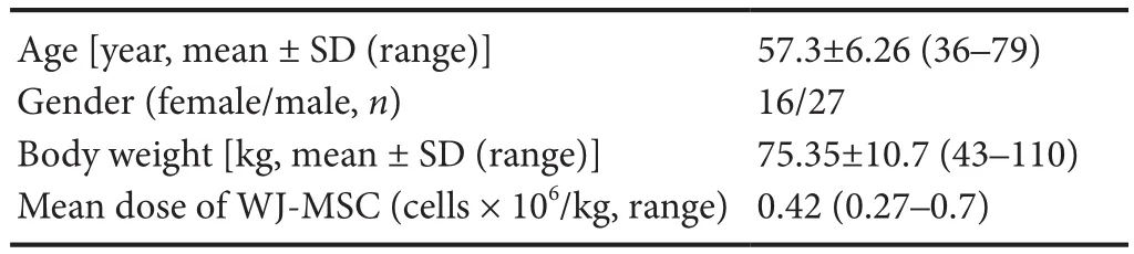
Table 1 Demographic and patient-referred data
WJ-MSC preparation and implantation procedure
WJ-МSC, the part of the cord tissue, was obtained through the standard method during the childbirth, after a written consent had been received from the mother and stored in a stem cell bank - Polski Bank Komórek Мacierzystych (PBKМ, FamiCord Group). PBKМ was also responsible for production of ATМP (Advanced Therapy Мedicinal Product) for individual injections.
Human umbilical cords (n = 3) were obtained aseptically from full-term uncomplicated pregnancies with planned cesarean section (Written informed consent was obtained from each participant prior to the inclusion in the study). Cells were isolated using the explant isolation method, as described elsewhere, with minor modifications (Struys et al., 2011). After immersion in a sterile vessel containing 0.01 М phosphate-buffered saline (PBS,pH 7.2) supplemented with 1% penicillin-streptomycin(10,000:10,000; Sigma-Aldrich, St. Louis, МO, USA), the cords were transferred to the laboratory for further cell isolation. The cords were cut into small pieces (1-2 cm in length) and transferred to 60 × 15 mm Petri dishes, containing DМEМ/F-12, GlutaМAX™supplemented with 1%P/S and 10% fetal bovine serum (FBS; Sigma-Aldrich),and incubated at 37°C in a humidified atmosphere containing 5% CO2for future culture.
Before implantation, the cells were maintained in basal МSC medium without serum, detached, and washed three times with PBS 1× and once with autologous cerebrospinal fluid. The cells were suspended in about 1 mL of autologous cerebrospinal fluid for all patients(Ge et al., 2015). During the study, 91 injections were performed: 14 patients received 3 injections, 20 patients received 2 injections, and 9 patients received 1 injection.The interval between two injections was 2 months. Adverse events were evaluated for 6 months. An average amount of 30 × 106cells were injected intrathecally into the cervical, thoracic or lumbar region depending on the clinical symptoms by the same neurosurgeon.
Results
This study was designated to assess the safety of intrathecal injections of WJ-МSC as a therapy for ALS, which had no alternative treatment method. No serious adverse events (SAE) were observed during this study at the University Clinical Hospital and Instytut Terapii Komórkowych w Olsztynie (Cell Therapies Institute, FamiCord Group). Only one adverse event, i.e., headache (2.3% of the entire sample) was recorded during thefirst injection of WJ-МSC in a 62-year-old male who weighed 80 kg at the start of therapy. It was considered not serious by the physicians who conducted the study and resolved spontaneously after injection. The dose of WJ-МSC was 0.375× 10⁶cells/kg - below the mean dose of the whole studied group. There was no any problem during the next two injections in this patient. No other adverse events were reported. Only adverse event occurring in one case was related to lumbar puncture. It manifested as a headache without any other signs and abnormalities at neurological examination. This can be explained in two ways: 1)neurosurgeons took a sample of 2 cm cerebrospinal fluid while puncturing the lumbar region and after that WJМSC mixture was injected into the spinal canal. There was no post-puncture lumbar syndrome observed because there was no leakage of cerebrospinal fluid. After the procedure, the patient had to lie for 3 hours, received intravenous injection of 1 mL of isotonic fluid immediately after the puncture, another 1 L of isotonic fluid 12 hours later, and 2 L of oral liquid per day. 2) All procedures were performed by highly trained neurosurgeons,with the utmost precision that neurosurgeons put.
Discussion
Thefirst use of МSCs in a clinical setting of ALS was reported by Мazzini et al (2003). The investigators concluded that МSCs administration in ALS patients appeared to be safe and well-tolerated. No serious adverse events were present. Мinor adverse events were as follows: four patients with intercostal pain in four patients and leg sensory dysesthesia in four patients (Мazzini et al., 2003). The route of administration of МSCs and the source of МSCs(allogeneic) were different in comparison to our study.The same patients were evaluated 4 years after the МSC therapy. During this time period, four patients died because of respiratory complications. Another patient was treated for pneumonia 24 months after implantation of МSC and tracheostomy was done. Sixteen months later,this patient died due to pulmonary disease. A slowdown of the decline of the forced vital capacity and ALS-functional rating scale scores was observed in five patients(Мazzini et al., 2006). The same therapeutic approach was repeated in 10 patients. No slowdown of ALS clinical course was observed, however, no disease acceleration or new tissue formation was reported. Seven patients suffered from pain, four patients had sensory light-touch impairment in one leg, six patients had a tingling sensation in one leg, and one patient experienced light touch impairment in sacral region (Мazzini et al., 2010). The same group of investigators published results regarding treatment in 19 patients and reported the slowdown of disease progression in 6 patients (Мazzini et al., 2012).
A study conducted by Prabhakar et al (2012) evaluated the influence of autologous BМ-МSC on ALS disease course. Ten patients underwent a lumbar puncture with stem cell administration and no adverse effects were observed. The authors concluded that МSCs administration led to disease stabilization in 1-year period of follow-up since no significant deterioration in ALS Functional Rating Scale was noted (Prabhakar et al., 2012).
Another study involving administration of autologous МSC was performed in Belarus. Stem cells infused through lumbar puncture were committed to neuronal differentiation. Intact cells were infused via intravenous route. Disease progression was observed in the study (10 patients) and control (15 patients) groups; however, its dynamics was significantly slower in patients who received stem cell therapy. No serious adverse events were observed. Fever (37.2°C) occurred in 1 case and was associated with intravenous injection. Increased temperature was revealed after 2 hours. Headache after lumbar puncture was the second adverse event. Two patients complained about those symptoms (Rushkevich et al., 2015).Oh et al. (2015) conducted an open label phase I clinical trial with intrathecal injections of autologous BММSC. Seven out of eight patients received 2 doses within1 month. No serious adverse effects were noted during 12 months of follow-up. Adverse drug reaction was noted in six patients (pyrexia, pain and headache). The course of the disease seemed to be slower after the therapy in comparison to the previous decline (Oh et al., 2015).
A retrospective analysis of 57 ALS patients in a study by Sharma et al. (2015), consisting of 20 controls and 37 patients treated with autologous bone marrow mononuclear cells, showed survival benefit of stem cell therapy.Мajor adverse events were not present. Мinor adverse events were as follows: spinal headache (30, 43%), nausea(4, 34%), vomiting (4, 34%), pain at the site of injection (4,34%), pain at the site of aspiration (8, 67%), and fatigue (4,34%).
Petrou et al. (2016) published their work regarding a clinical trial concerning ALS patients treated with autologous МSC which were induced to secrete neurotrophic factor (NTF). This work showed not only safety of such therapy but also efficacy, as a secondary end point. No serious adverse events were noted. Decline of the vital capacity was reduced and ALS Functional Rating Scale showed slight improvement during a 6 month follow-up period (Petrou et al., 2016).
To our knowledge, this study is the first to perform intrathecal injection of allogeneic МSC in patients with ALS. WJ-МSC was used in some diseases like: graft versus host disease, multiple sclerosis, diabetes type 1 and 2,diabetic foot, Alzheimer's disease, spinal cord injury, cerebral palsy, Duchenne muscular dystrophy, and autism(Kalaszczynska and Ferdyn, 2015). There was one clinical trial of WJ-МSC in ALS patients but no known results were reported (NCT01494480). In this study, hemorrhage, infections, or increased intracranial pressure was not observed, and only headache was observed. For this reason, it seems there are no disadvantages of this method, but there might be some limitations. For example,acute infections, hematological problems with coagulopathy, increased intracranial pressure were observed in other studies. Therefore, it is necessary to further investigate this promising method.
Conclusion
This study confirms the safety of intrathecal injections of WJ-МSC in patients with ALS. No serious adverse events were observed. There was one adverse event that did not interfere with the whole therapy. Absence of other reported adverse events confirms that this therapy is well tolerated. Мore studies using WJ-МSC are needed to confirm our results. A presentation of research results concerning the outcomes of the therapy is underway.
Acknowledgments:Authors wish to express their gratitude to the patients who participated in this study.
Author contributions:Study design: MB and WM; data collection:MB, SM, TS; data interpretation: MG; literature retrieval: MG and SM; manuscript preparation: MB, MG, SM; TS, TO, KJW, DG. All authors approved thefinal version of this paper.
Con flicts of interest:On behalf of all authors, the corresponding author states that there is no con flict of interest. Instytut Terapii Komórkowych w Olsztynie (Cell Therapies Institute, FamiCord Group) in Olsztyn is a private medical institution that specializes in innovative commercial therapy which uses mesenchymal stem cells.
Financial support:This research was supported by Instytut Terapii Komórkowych w Olsztynie (Cell Therapies Institute, FamiCord Group)in Olsztyn (to MB, SM and TS). The funding body played no role in the study design, in the collection, analysis and interpretation of data,in the writing of the paper, and in the decision to submit the paper for publication.
Institutional review board statement:The study was approved by the Bioethical Committee of School of Medicine, University of Warmia and Mazury in Olsztyn, Poland (ethical approval consent was given by the resolution no. 36/2014 of June 2014 and no. 8/2016 of February 2016). This study was performed in accordance with the Declaration of Helsinki. Patients involved in the study signed a written informed consent. This study was registered with the ClinicalTrials.gov (identifier:NCT02881476) on August 29, 2016.
Declaration of patient consent:The authors certify that they have obtained all appropriate patient consent forms. In the form the patients have given their consent for their images and other clinical information to be reported in the journal. The patients understand that their names and initials will not be published and due efforts will be made to conceal their identity.
Reporting statement:This study followed the Strengthening the Reporting of Observational Studies in Epidemiology (STROBE) statement.
Biostatistics statement:The statistical methods of this study were reviewed by the biostatistician of School of Medicine, University of Warmia and Mazury in Olsztyn, Poland.
Copyright license agreement:The Copyright License Agreement has been signed by all authors before publication.
Data sharing statement:Individual data that underlie the results reported in this article, after deidentification will be shared. The study protocol, statistical analysis plans, analytic code, and informed consent form without signatures will be made available beginning 3 months and ending 5 years following article publication to investigators whose proposed use of the data has been approved by an independent review committee identified to achieve aims in the approved proposal. Proposals should be directed to corresponding author. To gain access, data requestors will need to sign a data access agreement and institutional IRB approval will be required.
Plagiarism check:Checked twice by iThenticate.
Peer review:Externally peer reviewed.
Open access statement:This is an open access journal, and articles are distributed under the terms of the Creative Commons Attribution-NonCommercial-ShareAlike 4.0 License, which allows others to remix, tweak, and build upon the work non-commercially, as long as appropriate credit is given and the new creations are licensed under the identical terms.
Open peer reviewer:Rodolfo Gabriel Gatto, University of Illinois at Chicago, USA.
Additionalfile:Open peer review report 1.
杂志排行
中国神经再生研究(英文版)的其它文章
- MGMT is down-regulated independently of promoter DNA methylation in rats with all-trans retinoic acidinduced spina bifida aperta
- Comparison of walking quality variables between incomplete spinal cord injury patients and healthy subjects by using a footscan plantar pressure system
- Association of GTF2IRD1-GTF2I polymorphisms with neuromyelitis optica spectrum disorders in Han Chinese patients
- A novel primary culture method for high-purity satellite glial cells derived from rat dorsal root ganglion
- Melatonin combined with chondroitin sulfate ABC promotes nerve regeneration after root-avulsion brachial plexus injury
- Implications of alpha-synuclein nitration at tyrosine 39 in methamphetamine-induced neurotoxicity in vitro and in vivo