Anti-schistosomal activities of Echinops kebericho Mesfin root and Hagenia abyssinica (Bruce) J.F Gmel flower part crude extracts in Swiss albino mice
2018-11-14YonasAlemuZelekeMekonnenAhmedZeynudinMotiYohannesAbdissaBiruksewSultanSuleman
Yonas Alemu, Zeleke Mekonnen, Ahmed Zeynudin, Moti Yohannes, Abdissa Biruksew,Sultan Suleman
1School of Medical Laboratory Sciences, Jimma University, P.O. Box: 378, Jimma, Ethiopia
2School of Veterinary Medicine, Jimma University, P.O. Box: 378, Jimma, Ethiopia
3School of Pharmacy, Jimma University, P.O. Box: 378, Jimma, Ethiopia
Keywords:Schistosoma mansoni Echinops kebericho Hagenia abyssinica Crude extract Antischistosomal activity Ethiopia
ABSTRACT Objective: To evaluate the in vivo antischistosomal activities of the crude extracts of Echinops kebericho Mesfin (E. kebericho) root and Hagenia abyssinica (Bruce) J.F. Gmel (H. abyssinica)flower. Methods: Mice were infected with (150 ± 10) Schistosoma mansoni cercariae by paddling technique. Crude extracts were administered orally for five consecutive days at doses of 300, 600 and 1 200 mg/kg/day along with 200 mg/kg/day praziquantel and 3% tween 80 given as a control. Results: E. kebericho root extract showed a statistically significant (P <0.05) reduction in fecal egg count of 64.44%, 42.96% & 26.82% and worm burden of 65.71%,47.86% & 31.43% at treatment doses of 1 200 mg/kg/day, 600 mg/kg/day and 300 mg/kg/day,respectively. Similarly, H. abyssinica flower extracts showed a significant (P < 0.05) reduction in fecal egg count up to 84.57%, 77.06% & 63.89% and worm burden of 91.43%, 81.43%& 70.71% at a respective dose levels. In addition, a significant (P < 0.05) reduction in liver granuloma score was observed in all H. abyssinica administered dose groups and E. kebericho at 1 200 mg/kg/day dose group as compared to infected untreated control group. Conclusions: H.abyssinica and E. kebericho crude extracts show a promising antischistosomal activity.
1. Introduction
Schistosomiasis (bilharziasis) is a chronic and debilitating parasitic disease caused by blood flukes of the genus Schistosoma.Schistosoma mansoni (S. mansoni), Schistosoma haematobium, and Schistosoma japonicum are among the five species of schistosomes that can commonly cause disease in human[1,2]. The disease is endemic in 76 tropical developing countries with 779 million people at risk of infection globally (85% in Africa). It is estimated that annually 207 million people are infected (> 97 % in Africa) and 120 million people suffer from morbidity (20 million being in its severe
form). The annual mortality is 15 000-280 000 and when measured in disability-adjusted life years, the burden of this disease affects approximately 1.7 - 4.5 million people[3-5].
Schistosomiasis prevention mainly depends on community awareness on the modes of transmission, control of snail intermediate host and mass chemotherapy in endemic areas to treat infected individuals for the reduction of egg shedding. Among drugs used for the treatment of schistosomiasis, praziquantel acts against the adult worms of all species of the parasite while the less widely used oxamniquine due to its cost and lower efficacy than praziquantel is effective against only S. mansoni[6,7]. However, the efficacy of praziquantel is becoming less promising than expected which might be due to the reason of tolerance and/or resistance development by the parasites[8] and lack of efficacy against juvenile schistosome worms in vivo and in vitro[9]. Furthermore,praziquantel insusceptibility has been reported in previous studies in Senegal, Egypt and Kenya[10-13]. On the other hand, few efficacy studies carried in Ethiopia showed no evidence of praziquantel insusceptibility[14-17], although not comprehensive.
Nevertheless, schistosomiasis is widespread even in the face of effective but limited anti-schistosomal drug (practically only praziquantel). Thus, it is important to look for alternative way to tackle these diseases and/or to develop new candidate drug for the treatment of schistosomiasis. One such possibility is obtaining new chemotherapeutic agents from indigenous medicinal plants with the evidence of certain medicinal plants showing in vivo antischistosomal activities[18-25]. On the other hand, ethnobotanical surveys revealed the common use of Echinops kebericho Mesfin (E. kebericho) and Hagenia abyssinica (Bruce) J.F. Gmel (H. abyssinica) by traditional healers to treat intestinal helminthiasis especially tape worm in Ethiopia[26,27].
E. kebericho, locally called ‘kebericho’, is an erect massive rootstock bearing perennial herb that grows up to a height of 1.2 m belonging to the genus Echinops[28]. It contains a wide range of chemical constituents comprising alkaloids, polyphenols, saponins, phytosterols,carotenoids, lignans, sesquiterpene alcohols, acetylenic and thiophene compounds and essential oil[29]. Whereas, H. abyssinica named locally Kosso, is a monospecific genus belonging to the family Rosacea. It is a slender tree growing up to 20 m tall, with a short trunk and thick branches of either male or female flowers[30].It is a good source of various phytochemical such as saponins,phlobathanins, flavonoids, anthraquinones, phenols, terpenoids,alkaloids, steroids, glycosides, and tannins[31]. Furthermore, the acute oral administration of H. abyssinica as reported[32] and E.kebericho in the current antischistosomal activity evaluation were found safe up to a dose of 5 000 mg/kg.
The aim of this study was, therefore, to evaluate the in vivo antischistosomal activities of crude hydroalcoholic extracts of E.kebericho root and H. abyssinica flower part in S. mansoni infected Swiss albino mice.
2. Materials and methods
2.1. Plant material and collection
H. abyssinica flower and E. kebericho root parts were collected from Setema and Sigimo woreda (small administrative districts) of Jimma zone (Oromia regional state, Ethiopia) and confirmation was done by a plant taxonomist before storage at Jimma University’s herbarium center under the accession numbers 2349 and 2359, respectively.
2.2. Extraction procedure
The plant materials were air-dried at room temperature with no direct sunlight, reduced into small particles after grinding and powdered using mortar and pistil. Extraction was performed using two techniques namely: 80% methanol maceration and 70% ethanol soxhlet techniques[33]. In 80% methanol maceration technique, H. abyssinica flower powder was soaked in 80% methanol in closed vessel for three days with daily regular shaking and filtered at last. Whereas, 70%ethanol soxhlet extraction technique was performed until the solvent(extracts of E. kebericho root powder in thimble), when heated in the flask, released vapor and condensed, then filled the chamber and droped down the siphon tube, became clear. Finally, extracts were evaporated using Rotavapor, the concentrated layers were further dried in an oven at 40 ℃ and stored at 4 ℃ until use. The extraction percentage yields were calculated 13.3% for H. abyssinica and 14.7% for E. kebericho.
2.3. Experimental animals
Adult Swiss albino mice were procured from a breeding colony in the School of Veterinary Medicine of Jimma University and handled with care in clean cages with water ad libitum and commercial pellet.All applicable institutional guidelines for the care and use of animals were also followed after ethical approval of the study.
2.4. Acute toxicity assessment
Oral acute toxicity assessment of E. kebericho extract was done as per OECD test guideline 425[34]. Twenty-four male mice aged 6-8 weeks weighing 24-38 g were systematically randomized into four treatment groups of 6 mice each. The treatment group was kept in a labeled cage using dose levels and each mouse was marked on the tail for identification. All treatment groups were acclimatized to the laboratory environment for one week prior to conducting the study. Treatment doses of 0.5 mL 3% tween 80 as control, 5 000 mg/kg, 2 500 mg/kg and 1 250 mg/kg of E. kebericho root extract were administered based on body weight after overnight starvation.Individual mice observation was done at least once during the first 30 minutes after dosing and periodically within 6 hours during the first 24 hours (with special attention given during the first 4 hours at 30 minutes interval) and daily thereafter, for a total of 14 days. Parameters for observations were hair erection, walking status, diarrhea, death and body weight measurement (at 1st, 7th, and 14th day). The mice were anesthetized using 60 mg/kg sodium pentobarbital (Nembutal®) via intraperitoneal injection upon termination of the experiment and blood was collected into EDTA test tubes by cardiac puncture technique for the analysis of complete blood count (CBC). Liver and kidney were then collected and preserved in 10% formalin for histopathological examination.
2.5. Antischistosomal activity evaluation
Infected snails were collected from Aniso river of Manna Woreda,Jimma zone in November 2012. The identification of snail species was done based on shell size and morphology[35,36] and shedding of S. mansoni bifurcated tail cercaria was done as previously described[37]. Each mouse was then infected with (150 ± 10) S.mansoni cercariae by the paddling technique[38]. The direct saline wet mount was used for checking egg shedding of mice starting from the fourth week post cercarial exposure. On the 8th-week post infection, five mice per each eight treatment groups were systematically randomized as: E. kebericho and H. abyssinica extract administered groups at dose rates of 1 200 mg/kg/day, 600 mg/kg/day and 300 mg/kg/day, control groups of praziquantel at 200 mg/kg/day (Distocide® EIPICO, Egypt) and 0.2 mL 3% tween 80.Treatment was orally administered daily via oral gavage needle for five consecutive days and S. mansoni ova count was done using Kato-Katz technique[39] before and two weeks post treatment. The percentage of egg reduction rate (ERR) was calculated as:

Two weeks post-treatment, mice were anesthetized using 60 mg/kg sodium pentobarbital (Nembutal®) via intraperitoneal injection for gross pathological observation and worm recovery. Upon mice dissection, changes of liver and spleen were examined for granuloma formation and size enlargement. As a result, examination of liver was given a score of 0: to a liver that had no surface lesion, +1: to a very light mottling lesion on the hepatic surface, +2: to a moderate mottling lesion on the liver and +4: to livers with densely mottled surfaces[40]. Then the recovery of adult worms of S. mansoni from the hepatic portal vein and mesenteric veins were done as per the procedure described[41]. The adult worms were counted based on sex and the percentage of worm recovery (WR) and worm burden reduction (WBR) was calculated as:

2.6. Statistical analysis
Data was analyzed using SPSS version 16.0 and results were expressed as the mean value ± standard deviation. Mann-Whitney and Kruskal Wallis were used for the comparison of measurements across groups whereas, Wilcoxon and Friedman for paired data comparison. Results were considered significant at P< 0.05.
3. Results
3.1. Acute toxicity assessment
The crude extract of E. kebericho in mice was found to be safe up to a dose of 5 000 mg/kg as confirmed by the absence of death (Table 1) indicating LD50to be greater than 5 000 mg/kg. Hair erection and weakness were observed during the first two days in a dose dependent manner which could be due to the extract dose and taste discomfort while no diarrhea was observed across the study groups during the study period. In addition, body weight measurement on day 14 as compared to the first day of E. kebericho root extract oral administration showed a statistically significant increment (P<0.05,Table 1) in the groups administered a dose of 2 500 mg/kg, 1 250 mg/kg and the control groups.
Similarly, a significant difference in the body weight measurement was observed on day 7 as compared to the first day in treatment group of 1 250 mg/kg and control group. However, body weight difference was not significant when comparing the treatment group of 5 000 mg/kg on day 14, day 7 and 2 500 mg/kg on day 7 with the first day which indicate dose dependent effect on the weight of mice.
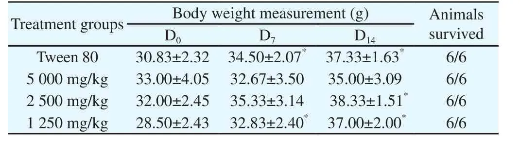
Table 1 Effect of 70% ethanol root extract of E. kebericho Mesfin on body weight measurement and survival of Swiss albino mice.
Whereas, the hematological parameter of CBC measurement showed no statistically significant difference in white blood cell(WBC), red blood cell (RBC), hemoglobin (Hgb), lymphocyte(Lymph), platelet (Plt) and hematocrite (Hct) within the treatment groups (P > 0.05, Table 2).
Also, the histological examinations of the liver of mice treated withdoses of 1 250 mg/kg and 2 500 mg/kg showed normal central vein,and normal surrounding hepatocytes, sinusoid and Kupfer cells. The observed histological appearance of the kidney was also normal and was the same as the control group at these treatment doses. In addition, histological examination of the liver of mice treated with 5 000 mg/kg showed normal cells as the control groups. However,histological examination of the kidney of mice treated with 5 000 mg/kg showed obliterated urinary space different from the control mice.
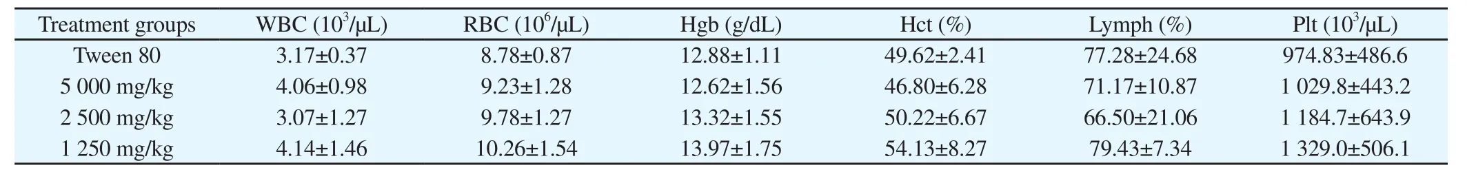
Table 2 Effect of 70% ethanol root extract of E. kebericho on hematological parameters of swiss albino mice.
3.2. Antischistosomal activity evaluation
S. mansoni infection in mice resulted in shedding of eggs in feces starting from the seventh-week post cercarial exposure which indicates the adult worms presence in the portal and mesenteric veins. It is also expected that the rest eggs to be lodged in the tissue of intestine and liver being a reason for granuloma formation.Therefore, liver granuloma was observed in this study and the mean liver granuloma score showed a statistically significant difference(P all < 0.05) for E. kebericho at 1 200 mg/kg/day (2.60 ± 0.55), H.abyssinica at all dose groups (1 200 mg/kg: 1.20 ± 0.45; 600 mg/kg: 1.40 ± 0.55; 300 mg/kg: 1.60 ± 0.55) and praziquantel (1.00 ±0.00) as compared to tween 80 treated control groups (4.60 ± 0.89).Whereas, no statistically significant difference (P > 0.05) of liver granuloma score was observed for E. kebericho administered groups at doses of 600 mg/kg/day (3.00 ± 1.22) and 300 mg/kg/day (3.80 ±1.09) as compared to tween 80 administered control group.
After treatment, the egg count observed both in H. abyssinica and E. kebericho extracts administered mice groups showed a remarkable reduction as compared to the infected but untreated control group(P<0.05, Table 3). Thus, both of the plant extracts reduced the egg burden in a dose-dependent manner even if their potency differs.
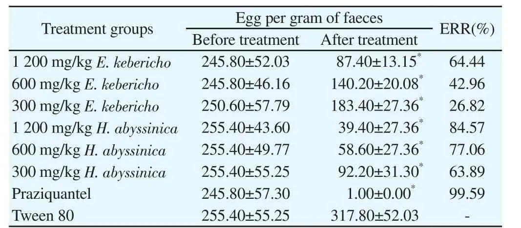
Table 3 Mean S. mansoni egg per gram of feces before and after the treatment of extracts of E. kebericho and H. abyssinica, praziquantel and 3% tween 80.
Furthermore, the worm burden reduction evaluation also showed a statistically significant difference (P all<0.05, Table 4) in male, female and total mean worm count of both plants extract administered dose groups when compared to tween 80 treated control groups. On the contrary, the mean male, female and total worm count of 1 200 mg/kg/day H. abyssinica extract administered dose groups showed no statistically significant difference (P>0.05)as compared to the praziquantel treated control groups indicating its comparable effect on worm count at the higher dose.
Table 4 Mean number of S. mansoni adult worms perfused from mice after the treatment of E. kebericho and H. abyssinica extracts, 3% Tween 80 and Praziquantel.

WBR- worm burden reduction, WR- worm recovery.
4. Discussion
The absence of death up to a dose level of 5 000 mg/kg in the acute oral toxicity assessment of ethanolic E. kebericho root extract indicates the plant is safe. This is due to the fact that substances with LD50higher than 5 000 mg/kg by oral route are regarded as safe or practically nontoxic[42]. Similarly, haematological indices in animals are also important to determine the toxicity risk since the changes in the blood system have a higher predictive value for human toxicity[43]. Therefore, haematological parameters studied revealed that the extract administration couldn’t bring any alteration on CBC measurements.
Moreover, as liver is known to be a key organ in the metabolism and detoxification of xenobiotics and kidney, also due to their high rate of perfusion and ability to concentrate a range of substances in the tubular lumen, is vulnerable to damage induced by a huge variety of chemicals[44,45]. But, the histological examination of liver and kidney showed that the plant extracts had no effect on the organs of the animals at 2 500 mg/kg and 1 250 mg/kg dose level although obliterated urinary space of kidney seen at dose level of 5 000 mg/kg might be due to the toxic effects of the plant extract.
Whereas, the major pathology of schistosomiasis is a granulomatous inflammation which is observed due to a cellular immune response to antigens secreted by those schistosomes’ ova trapped in organs as lung, liver, and spleen[46]. Therefore, the reduction of hepatomegaly,splenomegaly and granuloma after treatment might suggest a possible anti-inflammatory role of our study plant extracts.
Nevertheless, the reduction in the egg density and worm burden in treated mice was considered by several authors as a strong evidence of the efficiency of anti-schistosomal drugs[47,48]. Therefore, the egg reduction rate observed in mice treated with praziquantel was the highest indicating the potency of the drug. Additionally, both of our study plant extracts showed a significant reduction in egg count after treatment even if their potency differs. The fact that the egg load reduction observed in the feces of treated mice may be attributed to either the reduction in the worm burden as a result of treatment,the low productivity of the female already present and/or the active destruction of the few eggs produced by the host’s tissue reaction[48].
On the other hand, the total worm burden recovered from the untreated control group gave an idea of the rate of experimental infection. Thus, the mean total of 28.00 worms recovered in our study represents an infection rate of 18.67%. This study also showed a higher proportion of adult male worms from the total adult worms across the treatment groups indicating the proportions of adult female worms recovered were lower in number. This could be due to the fact that male-female interactions could take an important place in the establishment of higher male proportion due to the development of a lot of genotypes of the parasites, inducing a strong competition between females and thus an increase in the male proportion[49,50].
Consequently, the high egg reduction observed in this study could emanate either from the lower worm recovery due to death of the adult worms or reduced fecundity of female worms already present.As a result, the lowest worm recovery and highest worm burden reduction in praziquantel treated group was expected and can be attributed to the fact that praziquantel has good efficacy against the adult S. mansoni worm. Additionally, our finding also indicates the extract of H. abyssinica possesses better potency in reducing the total adult worms including female worms which could contribute for reduced fecundity.
Generally, it has been shown that anti-microbial and antiparasitic properties of plant extracts are assigned to some chemical compounds as tannins, terpenes, flavonoids, phenols, and alkaloids present in plants extract[51-53]. Activity against Plasmodium falciparum and gram-positive bacteria of indoloquinoline alkaloids extracted from Sida acuta[54, 55], analgesic and anti-inflammatory activities of alkaloids, flavones[56] and activity against Schistosoma japonicum of sesquiterpene lactones isolated from Vernonia amygdalina[57] were also reported as responsible chemicals.Therefore, the polar constituents present in these plant extracts could be responsible for their bioactivity as shown by the reduction in the egg count, worm burden and liver granuloma. Therefore, hydroalcoholic crude extracts of both H. abyssinica flower and E. kebericho root parts possessed a promising antischistosomal activity as shown with a significant reduction in egg production, liver granuloma and worm burden in all treatment groups.
Conflict of interest statement
The authors declare that they have no conflict of interest.
杂志排行

Asian Pacific Journal of Tropical Medicine的其它文章
- Acute kidney injury in leptospirosis: Overview and perspectives
- In vitro antiproliferative and apoptosis inducing effect of a methanolic extract of Azadirachta indica oil on selected cancerous and noncancerous cell lines
- Efficacy of voriconazole on leishmaniasis by Leishmania major: An in vitro and in vivo study
- Calcium carbonate supplementation causes memory impairment in mice
- In vitro anticancer activity of polysaccharide extracted from red alga Jania rubens against breast and colon cancer cell lines
- Investigation of cryptic diversity and occurrence of echinostome metacercariae infection in Anentome helena (von dem Busch, 1847)