山楂酸对肺癌95D细胞增殖、凋亡的影响
2017-07-31白雪,张月,李慧,何平
白 雪,张 月,李 慧,何 平
山楂酸对肺癌95D细胞增殖、凋亡的影响
白 雪,张 月,李 慧,何 平*
目的 探讨山楂酸对肺癌细胞95D增殖及凋亡产生的影响。方法 将山楂酸作用于人的肺癌细胞系95D,通过四甲基偶氮唑蓝(MTT)法检测山楂酸对细胞增殖的影响;通过流式细胞仪Annexin V/PI双染法检测细胞凋亡;通过荧光显微镜检测细胞核的形态变化;采用Western blot方法检测凋亡相关蛋白的影响。结果 随着山楂酸浓度增大,95D细胞的增殖率明显降低,早期及晚期凋亡率逐渐增加,与对照组比较差异有统计学意义;荧光电子显微镜观察山楂酸作用后的细胞,可见染色体边集、核碎裂等典型的凋亡改变,而对照组没有相应的变化;Western blot显示,随着山楂酸浓度的增加,Bcl-2蛋白表达逐渐降低,Bax蛋白表达逐渐升高。结论 山楂酸可以抑制肺癌95D细胞增殖,且具有浓度依赖性,山楂酸抑制肺癌95D细胞增殖是通过诱导细胞凋亡实现的。
山楂酸;肺癌;增殖;凋亡
0 引言
肺癌是目前世界上发病率和死亡率最高的恶性肿瘤之一,尽管近年来肺癌的诊疗技术有了一定程度的提高,肺癌的5年生存率仍较低[1-2]。近年来,中药抗肿瘤作用及机制已成为国内外研究的热点[3-4]。相关研究表明,中药抗肿瘤的作用机制包括:诱导肿瘤细胞凋亡、抑制肿瘤血管生成、调节细胞信号传导、诱导肿瘤细胞分化、细胞毒作用、调节机体免疫功能、逆转肿瘤细胞多药耐药作用等[5-7]。
本研究选取的药物—山楂酸(Maslinic acid),是一种三萜类天然有机化合物,存在于多种天然植物,特别是山楂、红枣、枇杷叶和油橄榄中,对人体的安全性较高,具有抗癌、抗氧化、抗艾滋病、抗菌、抗糖尿病等多种生物活性[8-10]。近年研究发现,山楂酸对人结肠癌HT29细胞[11]、人前列腺癌 DU145细胞[12]、小鼠黑色素瘤细胞[13]等有较强的抗肿瘤活性,其中部分作用机制与诱导肿瘤细胞凋亡相关。本实验主要研究山楂酸对非小细胞肺癌95D细胞的增殖及凋亡的影响。
1 材料与方法
1.1 材料、试剂与仪器 人肺癌细胞系95D购于美国典型培养物保藏中心(American Type Culture Collection,ATCC)。山楂酸购于上海纯优生物科技有限公司,RPMI-1640培养基购于Gibco公司,胎牛血清购于Hyclone公司,胰蛋白酶购于以色列Biological Industries公司,四甲基偶氮唑蓝(MTT)、Hoechst 33342购于美国Sigma公司,AnnexinV/PI双染试剂盒购于BD公司,超净工作台(SW-CJ-1FD)购于蚌埠净化设备厂,酶标仪(BIOTEK-ELX808)购于美国赛默飞世尔公司,流式细胞仪(FACSCalibur)购于美国BD公司,倒置显微镜(Olympus CKX31)购于日本奥林巴斯公司,荧光电子显微镜(Nikon C1)购于日本尼康公司,离心机购于德国Eppendorf公司。
1.2 方法
1.2.1 细胞培养 人肺癌95D细胞培养于含10%胎牛血清的RPMI-1640培养基中,置于37 ℃、饱和湿度、5%二氧化碳的培养箱中培养。细胞呈单层贴壁生长,接种细胞过夜进入对数生长期后,将细胞浓度调整为5×10⁵/mL,开始进行药物干预。
1.2.2 MTT实验 山楂酸对95D细胞增殖的抑制作用:消化收集对数增殖期细胞,将细胞悬液浓度调整为1×10⁵/mL后接种于96孔板,每孔100 μL,每个浓度设3个复孔,同时设立含RPMI-1640全培养基的空白对照和未予药物作用的阴性对照。实验组加入终浓度依次为0、2、4、6、8、10 μg/mL的山楂酸溶液,将96孔板置于37 ℃、饱和湿度、5%二氧化碳的培养箱中培养24 h,每孔加入MTT 20 μL,继续培养4 h,培养结束后吸去上清,每孔加入DMSO 150 μL,振荡混匀10 min。酶标仪检测波长570 nm的每孔光密度值(OD值)。增殖抑制率按如下公式计算:抑制率=(1-加药组OD值/对照组OD值)×100%。实验重复3次。
1.2.3 流式细胞仪(FCM)AnnexinV/PI双染法检测山楂酸诱导95D细胞凋亡 95D细胞以5×10⁵/mL接种于6孔板中,分别加入终浓度为0、2、4、6、8、10 μg/mL的山楂酸溶液,并设空白对照组(加等量RPMI-1640培养基),于37 ℃、饱和湿度、5%二氧化碳的培养箱中培养24 h后胰酶消化收获细胞。4 ℃、1 500 r/min离心5 min后收集细胞,弃去培养基,冷PBS洗涤、离心细胞2次。用500 μL Binding Buffer悬浮细胞,浓度约为1×10⁶/mL,依次加入5 μL AnnexinV-FITC、5 μL PI混匀,2~8 ℃避光条件下孵育15 min,流式细胞仪检测,分析凋亡的百分比。每组实验重复3次。
1.2.4 荧光显微镜观察凋亡 将预处理过的盖玻片置于六孔板内,95D细胞以5×10⁵/mL接种于盖玻片中培养过夜,加入终浓度为8 μg/mL的山楂酸溶液,设空白对照组(加等量RPMI-1640培养基),于37 ℃、饱和湿度、5%二氧化碳的培养箱中培养24 h。PBS洗2次,吹干,固定15 min,加入Hoechst 33342荧光染料(5 mg/L) 1 mL,37℃避光孵育15 min,含10%甘油的PBS封片,PBS洗2次,于荧光显微镜下观察、照相。每组实验重复3次。
1.2.5 Western blot检测Bcl-2、Bax的表达 95D细胞培养过夜,加入终浓度为4、6、8 μg/mL的山楂酸溶液,并设空白对照组(加等量RPMI-1640培养基),于37 ℃、饱和湿度、5%二氧化碳的培养箱中培养24 h后胰酶消化,收获细胞。裂解,低温离心10 min (12 000 r/min,4 ℃)取上清液,加入上样缓冲液,煮沸5 min,冷却。加样,Marker,行SDS-PAGE电泳,浓缩胶80 V电泳约30 min,分离胶100 V、90 min电泳至底部。转移、封闭后,加入一抗,本实验所用一抗为Bcl-2、Bax、GAPDH (1∶200;Santa Cruz;USA),4 ℃过夜孵育。应用羊抗鼠IgG-HRP(1∶3 000)标记二抗,孵育2 h。PVDF膜上均匀滴加适量发光液,行ECL底物发光。

2 结果
2.1 山楂酸对95D细胞生长抑制作用 MTT法检测细胞增殖表明,随着山楂酸作用浓度的增加,对95D细胞的增殖有明显的抑制作用,呈现剂量效应关系,与对照组比较差异有统计学意义(P<0.05)。见图1。
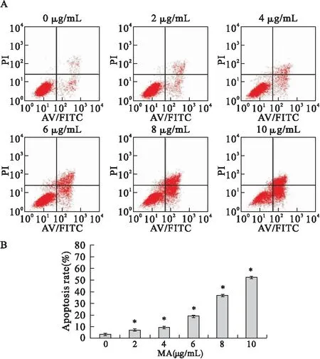
2.2 山楂酸对95D细胞凋亡的诱导作用 流式细胞仪AnnexinV/PI双染法检测表明,山楂酸作用24 h,随山楂酸浓度的增高,早期凋亡和晚期凋亡细胞的比例均逐渐增加,与对照组比较差异有统计学意义(P<0.05)。见图2。

图1 山楂酸(MA)对肺癌95D细胞增殖的抑制作用注:*与MA 0 μg/mL比较,P<0.05

图2 流式细胞仪AnnexinV/PI双染法检测MA作用于肺癌 95D细胞的凋亡率注:*与MA 0 μg/mL比较,P<0.05
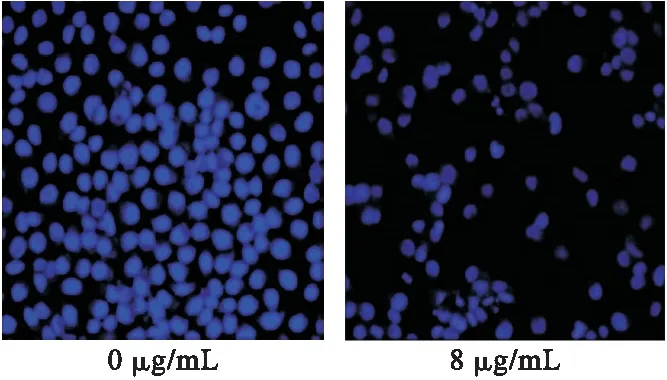
2.3 Hoechst33342染色荧光显微镜观察细胞核形态 8 μg/mL山楂酸作用于95D细胞24 h后,细胞形态出现显著变化,呈现染色质边集、核碎裂等改变。随着药物浓度增加,细胞数量明显减少,凋亡的细胞明显增加,凋亡改变明显。见图3。

图3 山楂酸作用于95D细胞后引起细胞核形态改变(400×)
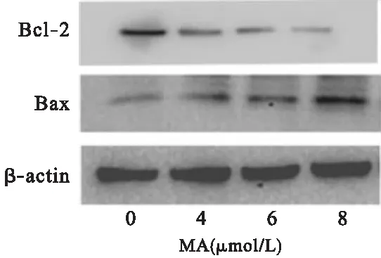
2.4 山楂酸对凋亡相关蛋白Bcl-2、Bax表达的影响 Western blot方法检测结果显示,随着山楂酸作用浓度的增加,Bcl-2蛋白表达逐渐降低,Bax蛋白表达逐渐增高。见图4。

图4 MA作用于肺癌95D细胞后对Bcl-2、Bax表达的影响
3 讨论
目前,许多抗肿瘤药物通过促进肿瘤细胞发生凋亡来实现抗肿瘤作用[14-17]。Bcl-2家族是细胞凋亡的关键调节因子,在线粒体凋亡途径的调控中起重要作用,其中,Bcl-2、Bax在目前研究最为广泛。当细胞接受死亡信号刺激,Bax被激活,从细胞浆转移到线粒体膜上,进而形成同源二聚体,促进细胞凋亡,而Bcl-2则在Bax诱发凋亡过程中对其功能进行限制。当细胞内Bcl-2表达增多时,细胞凋亡受到抑制,Bax表达增多时,细胞凋亡受到促进,Bcl-2/Bax比例对细胞凋亡的发生意义重大[18]。
近年来,中药及其有效成分因其抗肿瘤作用确切,在肿瘤治疗中的地位日趋受到重视。研究发现,从中药中提取的一些萜类化合物,尤其是三萜类化合物,具有潜在的抗肿瘤作用,其抗肿瘤机制与促进肿瘤细胞凋亡有关[19-24]。
本研究表明,山楂酸具有较强的抗氧化、抗炎、抗病毒、抗肿瘤等多种活性作用,研究显示,山楂酸对肝癌、乳腺癌、胰腺癌、结肠癌细胞有很强的抑制作用[25-29]。Fernando等[30]通过山楂酸对人结肠癌癌细胞HT29的实验证明,山楂酸通过直接激活线粒体凋亡通路导致人结肠癌癌HT29细胞死亡。
本实验MTT结果显示,山楂酸对95D细胞具有明显的增殖抑制作用,并且存在剂量依赖关系。AnnexinV/PI双染法结果显示,山楂酸对95D细胞的增殖抑制作用是由诱导细胞凋亡来实现的,且凋亡率随着药物浓度的增加而升高,呈剂量依赖性。通过荧光显微镜,从形态学上证实,山楂酸可以诱导95D细胞凋亡。Western blot检测结果显示,随着山楂酸作用浓度的增加,Bcl-2蛋白表达逐渐降低,Bax蛋白表达逐渐增高。
综上所述,山楂酸可以有效地抑制人肺癌95D细胞的增殖,且呈浓度依赖性,山楂酸抑制肺癌95D细胞的增殖是通过诱导其凋亡来实现的,凋亡通路之一可能是线粒体凋亡通路。
[1] Blumenthal GM,Karuri SW,Zhang H,et al.Overall response rate,progression-free survival,and overall survival with targeted and standard therapies in advanced non-small-cell lung cancer:US Food and Drug Administration trial-level and patient-level analyses[J].J Clin Oncol,2015,33(9):1008-1014.
[2] Li Y,Jiang L,Liu XG,et al.Efficacy and survival rate analysis of lung cancer with spinal metastases[J].Beijing Da Xue Xue Bao,2014,46(1):138-143.
[3] Xia L,Plachynta M,Liu T,et al.Pro-inflammatory effect of a traditional Chinese medicine formula with potent anti-cancer activity in vitro impedes tumor inhibitory potential in vivo[J].Mol Clin Oncol,2016,5(6):717-723.
[4] Xu J,Song Z,Guo Q,et al.Synergistic effect and molecular mechanisms of traditional chinese medicine on regulating tumor microenvironment and cancer cells[J].Biomed Res Int,2016,2016:1490738.
[5] Guo Q,Li J,Lin H.Effect and molecular mechanisms of traditional chinese medicine on regulating tumor immunosuppressive microenvironment[J].Biomed Res Int,2015,2015:261620.
[6] Liu M,Jia LQ.Preliminary clinical experience of Prof.Li Pei-wen in treating complications of tumor by external treatment of traditional Chinese medicine[J].Zhongguo Zhong Xi Yi Jie He Za Zhi,2014,34(11):1390-1391.
[7] Zhang YH,Wang Y,Yusufali AH,et al.Cytotoxic genes from traditional Chinese medicine inhibit tumor growth both in vitro and in vivo[J].J Integr Med,2014,12(6):483-494.
[8] Sommerwerk S,Heller L,Kuhfs J,et al.Urea derivates of ursolic,oleanolic and maslinic acid induce apoptosis and are selective cytotoxic for several human tumor cell lines[J].Eur J Med Chem,2016,119:1-16.
[9] Siewert B,Pianowski E,Csuk R.Esters and amides of maslinic acid trigger apoptosis in human tumor cells and alter their mode of action with respect to the substitution pattern at C-28[J].Eur J Med Chem,2013,70:259-272.
[10]Li C,Yang Z,Zhai C,et al.Maslinic acid potentiates the anti-tumor activity of tumor necrosis factor alpha by inhibiting NF-kappaB signaling pathway[J].Mol Cancer,2010,9:73.
[11]Peragón J,Rufino-Palomares EE,Muoz-Espada I,et al.A New HPLC-MS Method for Measuring Maslinic Acid and Oleanolic Acid in HT29 and HepG2 Human Cancer Cells[J].Int J Mol Sci,2015,16(9):21681-21694.
[12]Park SY,Nho CW,Kwon DY,et al.Maslinic acid inhibits the metastatic capacity of DU145 human prostate cancer cells:possible mediation via hypoxia-inducible factor-1alpha signalling[J].Br J Nutr,2013,109(2):210-222.
[13]Mokhtari K,Rufino-Palomares EE,Pérez-Jiménez A,et al.Maslinic acid,a triterpene from olive,affects the antioxidant and mitochondrial status of B16F10 melanoma cells grown under stressful conditions[J].Evid Based Complement Alternat Med,2015,2015:272457.
[14]Zheng X,Jin X,Li F,et al.Inhibiting autophagy with chloroquine enhances the anti-tumor effect of high-LET carbon ions via ER stress-related apoptosis[J].Med Oncol,2017,34(2):25.
[15]Sun D,Mou Z,Li N,et al.Anti-tumor activity and mechanism of apoptosis of A549 induced by ruthenium complex[J].J Biol Inorg Chem,2016,21(8):945-956.
[16]Li W,Tian YH,Liu Y,et al.Platycodin D exerts anti-tumor efficacy in H22 tumor-bearing mice via improving immune function and inducing apoptosis[J].J Toxicol Sci,2016,41(3):417-428.
[17]Wang L,Zhang J,Yuan Q,et al.Separation and purification of an anti-tumor peptide from rapeseed (Brassica campestris L.) and the effect on cell apoptosis[J].Food Funct,2016,7(5):2239-2248.
[18]Brenner C,Lemoine A.Mitochondrial proteins (e.g.,VDAC,Bcl-2,HK,ANT) as major control points in oncology[J].Front Oncol,2014,15(4):365-366.
[19]Bai ST,Zhu GL,Peng XR,et al.Cytotoxicity of Triterpenoid Alkaloids from Buxus microphylla against Human Tumor Cell Lines[J].Molecules,2016,21(9).E1125.
[20]Zong J,Wang R,Bao G,et al.Novel triterpenoid saponins from residual seed cake of Camellia oleifera Abel.show anti-proliferative activity against tumor cells[J].Fitoterapia,2015,104:7-13.
[21]Dasgupta A,Sawant MA,Lavhale MS,et al.AECHL-1,a novel triterpenoid,targets tumor neo-vasculature and impairs the endothelial cell cytoskeleton[J].Angiogenesis,2015,18(3):283-299.
[22]Xia Q,Mao W.Anti-tumor effects of traditional Chinese medicine give a promising perspective[J].J Cancer Res Ther,2014,10(Suppl 1):1-2.
[23]Zhou J,Zhou T,Jiang M,et al.Research progress on synergistic anti-tumor mechanisms of compounds in traditional Chinese medicine[J].J Tradit Chin Med,2014,34(1):100-105.
[24]Ling CQ,Yue XQ,Ling C.Three advantages of using traditional Chinese medicine to prevent and treat tumor[J].J Integr Med,2014,12(4):331-335.
[25]Fukumitsu S,Villareal MO,Fujitsuka T,et al.Anti-inflammatory and anti-arthritic effects of pentacyclic triterpenoids maslinic acid through NF-kappaB inactivation[J].Mol Nutr Food Res,2016,60(2):399-409.
[26]Hung YC,Yang HT,Yin MC.Asiatic acid and maslinic acid protected heart via anti-glycative and anti-coagulatory activities in diabetic mice[J].Food Funct,2015,6(9):2967-2974.
[27]Kapoor S.Maslinic acid and its in vitro anti-neoplastic effects[J].Nat Prod Res,2013,27(23):2210-2211.
[28]Rufino-Palomares EE,Reyes-Zurita FJ,García-Salguero L,et al.Maslinic acid,a triterpenic anti-tumoural agent,interferes with cytoskeleton protein expression in HT29 human colon-cancer cells[J].J Proteomics,2013,83:15-25.
[29]Huang L,Guan T,Qian Y,et al.Anti-inflammatory effects of maslinic acid,a natural triterpene,in cultured cortical astrocytes via suppression of nuclear factor-kappa B[J].Eur J Pharmacol,2011,672(1-3):169-174.
[30]Reyes-Zurita FJ,Pachón-Pea G,Lizárraga D,et al.The natural triterpene maslinic acid induces apoptosis in HT29 colon cancer cells by a JNK-p53-dependent mechanism[J].BMC Cancer,2011,11:154.
Effect of maslinic acid on the proliferation and apoptosis of lung cancer 95D cells
BAI Xue,ZHANG Yue,LI Hui,HE Ping*
(Shengjing Hospital of China Medical University,Shenyang 110004,China)
Objective To research the effect of maslinic acid on the proliferation and apoptosis of lung cancer 95D cells.Methods The effects of maslinic acid on proliferation of 95D cells was determined by MTT assay,and the apoptosis was analyzed by Annexin V/PI flow cytometric analysis.The morphology of apoptotic cell was detected by fluorescent microscopy.The effect of apoptosis-related proteins was detected by Western blot.Results Maslinic acid inhibited the proliferation of 95D cells in a dose-dependent manner significantly,and the proportion of Annexin V positive cells in drug-treated cells were increased in a dose-dependent manner,there were significant differences compared with control group (P<0.05).The observation of fluorescent microscopy showed that treatment with maslinic acid for 24 h could significantly induce the apoptotic cell death with chromatin condensation and nuclear fragmentation,while no corresponding change was found in control group.Western blot showed that the expression of Bcl-2 protein decreased gradually and the expression of Bax protein increased gradually with the increase of maslinic acid′s concentration.Conclusion Maslinic acid could inhibit the proliferation of 95D cell lines in a dose-dependent manner,which is achieved by induction of apoptosis.
Maslinic acid;Lung cancer;Proliferation;Apoptosis
2017-02-11
中国医科大学附属盛京医院,沈阳 110004
*通信作者
10.14053/j.cnki.ppcr.201707002
