受体相互作用蛋白3对神经胶质瘤U251细胞增殖的影响
2017-06-01于泽奇武慧丽涂悦衣泰龙杨小飒张
于泽奇+武慧丽+涂悦+衣泰龙+杨小飒+张赛+程世翔


[摘要] 目的 探讨受体相互作用蛋白3(RIP3)在神经胶质瘤细胞中的表达及其对U251细胞增殖的影响。 方法 采用实时定量PCR(real-time RT-PCR)法分别检测神经胶质瘤A172、U251、U373和U87细胞中RIP3的表达水平,随后应用U251稳定过表达RIP3(U251-RIP3)细胞模型,分别采用MTT法和克隆形成实验检测细胞增殖能力,流式细胞仪评价细胞周期变化。结果 RIP3 mRNA在4株胶质瘤细胞(A172、U251、U373、U87)中均有表达,并在U251细胞中表达最低。real-time RT-PCR结果表明U251-RIP3细胞中稳定过表达RIP3,与阴性对照(空载)(NC)相比,RIP3过表达能够抑制U251细胞增殖能力;能够降低U251细胞克隆形成能力,培养11 d后细胞集落数目(67±2)較NC(75±3)减少(P < 0.05);能够延缓细胞周期进程,U251-RIP3细胞的S期比例较NC组(16.0±0.4)%升至(18.2±1.0)%(P < 0.05),同时G0/G1期比例较NC组(55.7±0.5)%降至(57.4±0.4)%(P < 0.01)。 结论 RIP3过表达后可抑制神经胶质瘤U251细胞增殖,有望成为靶向控制胶质瘤细胞增殖的潜在治疗靶点。
[关键词] 受体相互作用蛋白3;胶质瘤;细胞增殖;可控性坏死
[中图分类号] R739.41 [文献标识码] A [文章编号] 1673-7210(2017)04(a)-0004-04
[Abstract] Objective To investigate the expressive changes of RIP3 in all sorts of glioma cell lines and the effects on the cell proliferation in the U251 cells. Methods The expression levels of RIP3 mRNA in the four glioma cell lines (A172, U251, U373 and U87) were firstly selected by real-time RT-PCR. Then, U251 cells were engineered to overexpress RIP3 for further study. Lastly, the MTT, colony formation, and flow cytometry assays were used to detect the effects of RIP3 on the cell proliferation, clone formation, and cell cycle of U251 cells, respectively. Results The expression levels of RIP3 mRNA was detected in all four glioma cell lines, and the lowest level of it was discovered in U251 cell line. Then, real-time RT-PCR assay showed that RIP3 was successfully overexpressed in U251-RIP3 cells compared with negative control (NC) cells with empty vector. The overexpressed RIP3 altered the cell viability in U251 cells, Additionally, the overexpressed RIP3 also reduced the colony formation of U251 cells: after 11 d of cell culture, the colony number of U251-RIP3 cells (67±2) was significantly decreased compared with that of NC cells (75±3) (P < 0.05). Correspondingly, the proportion of U251-RIP3 cells in S phase arrest was higher than that in NC cells [from (16.0±0.4)% to (18.2±1.0)% ] (P < 0.05). Meanwhile, that in G0/G1 phase was reduced compared with that in NC cells [from (57.4±0.4)% to (55.7±0.5)%] (P < 0.01). Conclusion Studies suggested that the overexpression of RIP3 could confer inhibition to the cell proliferation in glioma U251 cells; and it indicated that RIP3 may be as a potential target for glioma chemotherapy.
[Key words] Receptor-interacting protein 3; Glioma; cell proliferation; Regulated necrosis
脑肿瘤占中枢神经系统肿瘤的90%以上[1],而神经胶质瘤(gliomas)是最常见的脑肿瘤。近20年来,胶质瘤患者中位生存期无明显改善,其高复发率成为治疗的最大难题[2]。随着医学的飞速发展,使我们对肿瘤的发生机制有了更深入的了解,同时也提供了肿瘤治疗的新策略和新手段,其中基因治疗被认为是前景广阔的一种治疗方式,已成为神经科学中最富挑战而又亟待解决的课题。
受体相互作用蛋白3(receptor-interacting protein 3,RIP3)是受体相互作用蛋白家族(RIPs)的重要成员。激活状态下的RIP3 能够调控细胞可控性坏死以及炎症的发生[3]。研究表明,RIP3在丘脑、黑质、胼胝体、壳核、延髓和脊髓中呈现高表达水平,提示RIP3可能在神经系统发育和疾病进程中扮演着重要角色[4]。近年来,RIP3在多种肿瘤(例如肝癌、肺癌、胶质瘤等)中发挥的重要作用受到广泛关注[5-7],然而,RIP3对肿瘤细胞的功能影响尚存在争议。本研究旨在探讨RIP3在各种神经胶质瘤细胞中的表达情况及其对胶质瘤细胞生物学功能的影响,为其在胶质瘤治疗中的应用提供实验依据。
1 材料与方法
1.1 细胞株
胶质瘤细胞株A172、U251、U373、U87购自美国ATCC公司,常规用含10%胎牛血清的DMEM培养基,于37℃、5% CO2培养箱中常规培养。
1.2 仪器与试剂
CO2恒温三气培养箱(150i,美国Thermo),倒置显微镜(TS100-LED MV,日本Nikon),Real time PCR 仪器(LightCycler 480 II,德国Roche),酶标仪(1510,美国Thermo),流式细胞仪(FACSCallibur,美国BD);Trizol试剂(美国Invitrogen),M-MLV、dNTPs(美国Promega),oligo dT(上海生工),RNAase Inhibitor(美国Promega),Primer(上海生工),SYBR?誖 Premix Ex Taq(大连宝生物工程有限公司),MTT、DMSO、Giemsa染色液、PI(美国Sigma),RNase A(康为世纪公司),凋亡试剂盒(美国eBioscience)。
1.3 引物设计和RIP3重组载体构建
RIP3基因上游引物:5′-TGG CGG TCA AGA TCG TAA ACT C-3′,下游引物5′-TTC TGG TCG TGC AGG TAA AAC A-3′,长度265 bp;GAPDH基因上游引物:5′-TGA CTT CAA CAG CGA CAC CCA-3′,下游引物5′-CAC CCT GTT GCT GTA GCC AAA-3′,长度121 bp。采用Age I和EcoR I酶切目的片段和慢病毒载体(GeneChem),通过T4 DNA连接酶将其在NE Buffer中16℃连接过夜,转化DH5α感受态并涂板。PCR鉴定阳性重组子,DNA测序验证后,转染293T细胞48 h。收集上清液,经浓缩过滤后获得浓缩病毒液,分装,-80℃保存。
1.4 real-time RT-PCR法检测RIP3 mRNA表达水平
用Trizol试剂盒提取四株胶质瘤细胞总RNA,使用M-MLV试剂盒将其反转录成cDNA,反应条件:42℃水浴反应1 h,70℃水浴10 min使逆转录酶失活,将得到的cDNA置于-20℃保存备用。再以此cDNA为模板进行real-time RT-PCR,扩增条件:95℃预变性5 min,95℃ 45 s,55℃ 45 s,72℃ 1 min,40次循环,72℃ 10 min。RIP3基因相对表达量用2-ΔΔCt 方法计算。
1.5 U251细胞稳定转染
取对数生长期的U251细胞,按(3~5)×104个/mL接种于细胞培养瓶中,待融合度约为40%时更换含有RIP3慢病毒载体的维持液,维持液中感染复数(MOI)为10、16 h后将各孔中细胞收集到干净的1.5 mL EP管中,2000 r/min离心2 min,去掉上清液,更换为完全培养基,轻轻混匀后继续培养。感染后72 h更换含浓度为2 μg/mL的嘌呤霉素培养基筛选细胞,细胞存活即为阳性感染。实验分设U251-RIP3和阴性对照(空载)(NC)。
1.6 MTT法检测细胞增殖
取对数生长期的U251细胞,按每孔2000个接种于96 孔板中,置于细胞培养箱中持续培养1、2、3、4、5 d。检测时间点终止前4 h加入20 μL 5 mg/mL的MTT于孔中,无需换液。4 h后完全吸去培养液,加100 μL DMSO溶解甲瓒颗粒。振荡器振荡2~5 min,酶标仪490 nm检测OD值。
1.7 细胞克隆形成实验
取对数生长期的U251细胞,按每孔细胞1000个接种于6孔板中,待细胞贴壁后2 h,在孔内分别加入等体积的DMEM培养基,于培养箱内孵育11 d后,用PBS洗3次,加入4%多聚甲醛固定细胞60 min,洗涤后每孔加入Giemsa染液500 μL,染色30 min,洗涤晾干后拍照计数。
1.8 细胞周期分析
收集各组U251细胞,1000 r/min离心5 min后,棄上清,加入预冷PBS液4 mL,反复吹打,1000 r/min再离心5 min。反复冲洗3次后,用75%冰乙醇固定细胞,-20℃过夜。将固定好的细胞用PBS洗涤离心后,再用500 μL PBS将细胞重悬,分别加入50 μg/mL碘化丙啶(PI)和100 μg/mL RNase A。37℃避光孵育30 min后,用流式细胞仪进行检测,每组实验重复3次。
1.9 统计学方法
采用Graphpad Prism 6.0软件进行数据分析,正态分布的计量资料以均数±标准差(x±s)表示,组间数据的比较采用非配对样本t检验,以P < 0.05为差异有统计学意义。
2 结果
2.1 U251-RIP3稳定转染细胞模型的建立
采用real-time RT-PCR法检测了RIP3 mRNA在4株人脑胶质瘤细胞(A172、U251、U373和U87)中的表达水平,结果显示(图1A),U373细胞中RIP3 mRNA表达水平最高,而U251细胞表达最低。为此,本实验选择U251细胞为模型,通过构建的RIP3过表达重组载体稳定转染U251细胞,结果表明(图1B),与NC比较,U251-RIP3组中RIP3表达显著升高(P < 0.01),提示U251-RIP3稳定转染细胞系构建成功。
2.2 过表达RIP3降低U251细胞增殖能力
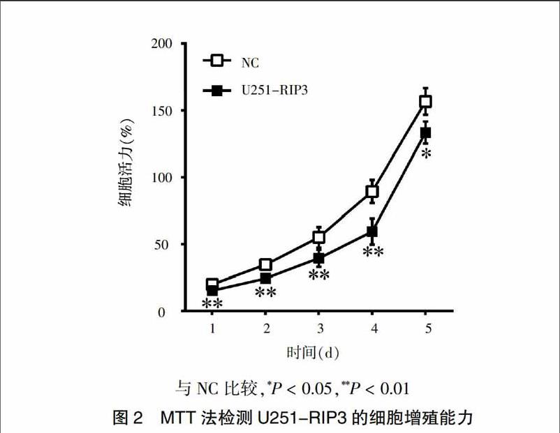
采用MTT法检测RIP3对U251细胞增殖能力的影响,结果显示:在检测的各个时间点,U251-RIP3 细胞生长速度明显低于NC[1 d:(0.15±0.00)比(0.20±0.00),2 d:(0.24±0.01)比(0.35±0.01,3 d:(0.40±0.05)比(0.565±0.06),4 d:(0.59±0.08)比(0.89±0.07),5 d:(1.33±0.07)比(1.57±0.08); P < 0.01或P < 0.05。见图2],提示RIP3能够抑制U251细胞增殖。
2.3 过表达RIP3降低U251细胞克隆形成能力
采用细胞克隆形成实验检测RIP3对U251细胞克隆形成能力的影响。结果表明,培养11 d后,U251-RIP3 细胞集落数目(67±2)较NC(75±3)减少(P < 0.05,图3A),而且形成的集落中细胞数目减少(图3B),提示RIP3能够降低U251细胞的克隆形成能力。
2.4 过表达RIP3延缓U251细胞周期进程
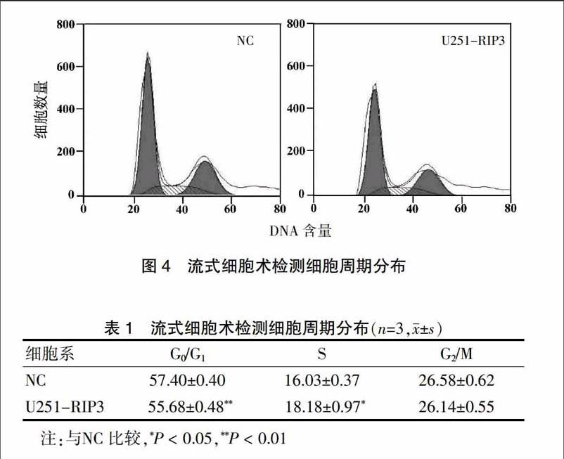
采用流式细胞术评价RIP3对U251细胞周期的影响(图4)。与NC比较(表1),U251-RIP3组细胞可降低G0/G1期细胞比例,由(57.4±0.4)%降至(55.7±0.5)%(P < 0.01),同时升高S期细胞比例,由(16.0±0.4)%升至(18.2±1.0)%(P < 0.05),而G2/M期变化无差异,提示RIP3使U251细胞阻滞于S期,延缓细胞周期进程,抑制U251细胞增殖。
3 讨论
近年来研究研究表明,肿瘤的发生、进展和耐药与肿瘤细胞的可控性坏死密切相关[3]。传统观点认为,坏死是在强烈的外界刺激下发生的被动病理过程。后续研究证实,细胞坏死可受死亡信号通路的调控,2005年Degterev等[8]首次将这种可受调控的细胞坏死称为坏死性凋亡(necroptosis),其中RIP3是坏死性凋亡通路中的核心分子。
RIP3是受体相互作用蛋白家族的成员之一,具有特异的丝氨酸/苏氨酸激酶活性,其独特的C端能够感受细胞内环境变化,精细调控细胞死亡和存活[9]。另有文献报道,RIP3在脑、心、肝、肾等正常组织中均有广泛表达[10],而在肝癌、肺癌等内都是低表达状态[11-12],而且RIP3多定位于肿瘤相关的染色体14q11.2区[13],这提示RIP3可能与肿瘤细胞的生物学功能联系密切。为此,本实验首先利用慢病毒载体构建RIP3稳定过表达U251细胞株。该方法具有较高的转导效率,能感染分裂期或非分裂期细胞,可将外源RIP3基因整合入U251细胞中得到稳定表达,是基因治疗中强有力的工具。
目前研究认为,神经胶质瘤的发生发展与细胞增殖与死亡平衡功能失调有关,研究发现RIP1-RIP3复合体能够调控放疗诱导的胶质瘤细胞可控性坏死的发生[14]。然而,另有报道显示,RIP1、RIP3却在乳腺癌细胞增殖中扮演了促进角色[15],这提示RIP3介导肿瘤细胞坏死性凋亡可能在不同类型肿瘤中发生“双刃剑”作用。为此,本实验旨在探讨RIP3在神经胶质瘤细胞增殖中所发挥的作用。结果表明,过表达RIP3能够降低U251细胞增殖能力、降低细胞克隆形成、延缓细胞周期进程。
综上所述,本实验首次证实RIP3在不同胶质瘤细胞中的表达水平,并深入探討了过表达RIP3在U251细胞中的生物学功能,这为临床靶向治疗胶质瘤提供了新的理论和实验依据。
[参考文献]
[1] McNeill KA. Epidemiology of brain tumors [J]. Neurol Clin. 2016; 34(4):981-998. DOI:10.1016/j.ncl.2016.06.014.
[2] Mohammadzadeh A,Mohammadzadeh V,Kooraki S,et al. Pretreatment evaluation of glioma [J]. Neuroimaging Clin N Am. 2016,26(4):567-580. DOI:10.1016/j.nic.2016.06.006.
[3] Moriwaki K,Balaji S,Chan FK. Border security:the role of RIPK3 in epithelium homeostasis [J]. Front Cell Dev Biol,2016,4:70. DOI:10.3389/fcell.2016.00070.
[4] H?ckendorf U,Yabal M,Herold T,et al. RIPK3 restricts myeloid leukemogenesis by promoting cell death and differentiation of leukemia initiating cells [J]. Cancer Cell,2016, 30(1):75-91. DOI:10.1016/j.ccell.2016.06.002.
[5] Suda J,Dara L,Yang L,et al. Knockdown of RIPK1 markedly exacerbates murine immune-mediated liver injury through massive apoptosis of hepatocytes,independent of necroptosis and inhibition of NF-κB [J]. J Immunol,2016,197(8):3210-3129. DOI:10.4049/jimmunol.1600690.
[6] Dillon CP,Green DR. Molecular cell biology of apoptosis and necroptosis in cancer [J]. Adv Exp Med Biol,2016, 930:1-23. DOI:10.1007/978-3-319-39406-0_1.
[7] Fukasawa M,Kimura M,Morita S,et al. Microarray analysis of promoter methylation in lung cancers [J]. J Hum Genet,2006,51(4):368-374. DOI:10.1007/s10038-005-0355-4.
[8] Degterev A,Huang Z,Boyce M,et al. Chemical inhibitor of nonapoptotic cell death with therapeutic potential for ischemic brain injury [J]. Nat Chem Biol,2005,1(2):112-119. DOI:10.1038/nchembio711.
[9] Meylan E,Tschopp J. The RIP kinases:crucial integrators of cellular stress [J]. Trends Biochem Sci,2005,30(3):151-159. DOI:10.1016/j.tibs.2005.01.003.
[10] Newton K,Sun X,Dixit VM. Kinase RIP3 is dispensable for normal NF-kappa Bs,signaling by the B-cell and T-cell receptors,tumor necrosis factor receptor 1,and Toll-like receptors 2 and 4 [J]. Mol Cell Biol,2004,24(4):1464-1469.
[11] Koppe C,Verheugd P,Gautheron J,et al. IκB kinaseα/β control biliary homeostasis and hepatocarcinogenesis in mice by phosphorylating the cell-death mediator receptor-interacting protein kinase 1 [J]. Hepatology,2016,64(4):1217-1231. DOI:10.1002/hep.28723.
[12] Diao Y,Ma X,Min W,et al. Dasatinib promotes paclitaxel-induced necroptosis in lung adenocarcinoma with phosphorylated caspase-8 by c-Src [J]. Cancer Lett.,2016, 379(1):12-23. DOI:10.1016/j.canlet.2016.05.003.
[13] Gary M. Kasof,Judith C,et al. The RIP-like kinase,RIP3,induces apoptosis and NF-kappaB nuclear translocation and localizes to mitochondria [J]. FEBS Lett,2000,473(3):285-291.
[14] Das A,McDonald DG,Dixon-Mah YN,et al. RIP1 and RIP3 complex regulates radiation-induced programmed necrosis in glioblastoma [J]. Tumour Biol,2016,37(6):7525-7534. DOI:10.1007/s13277-015-4621-6.
[15] Liu X,Zhou M,Mei L,et al. Key roles of necroptotic factors in promoting tumor growth [J]. Oncotarget,2016,7(16):22219-22233. DOI:10.18632/oncotarget.7924.
(收稿日期:2017-01-15 本文編辑:苏 畅)
