The relationship between color vision discrimination ability and depth perception among university students
2016-08-08NimetUnayGundoganBelkisSalmanKotekinAyseGulKoakAltinta
Nimet Unay Gundogan, Belkis Salman Koçtekin, AyseGul Koçak Altinta
The relationship between color vision discrimination ability and depth perception among university students
Nimet Unay Gundogan1, Belkis Salman Koçtekin2, AyseGul Koçak Altinta3
1Department of Physiology, Baskent University Faculty of Medicine, Ankara 06815, Turkey
2Department of Physiology, Antalya Education and Research Hospital, Health Sciences University, Antalya 07100, Turkey
3Department of Ophthalmology, Ulucanlar Education and Research Hospital, Health Sciences University, Ankara 06240, Turkey
Correspondence to:Nimet Unay Gundogan. Department of Physiology, Baskent University Faculty of Medicine, Ankara 06815, Turkey. nimetg@yahoo.com
Received: 2016-03-08Accepted: 2016-05-13
目的:评价色觉正常大学生的色觉辨别能力与深度知觉的关系。
方法:选取巴什肯特大学医学专业学生52例,其中男性33例(63.46%),女性19例(36.54%),平均年龄21.18±2.52岁。参与学生视力正常(20/20),且经Ishihara假同色测试法显示无先天性色觉缺陷。运用Gundogan方法确定主视眼(DE)。通过法-孟二氏100色度试验(FM100HT)检验色觉辨别能力,包括左右眼及双眼(TE)的总误差分(TES)、部分误差分(LES)。误差分分为三组:双眼、DE及非主视眼(NDE)。应用TNO检查双眼视觉与立体感,并根据480-15 arc/s范围的立体感水平分为四组。
结果:FM100HT的误差分显示无性别差异的TES,蓝黄LES和红绿LES分别为61.22±30.32(58.50)[mean±SD],35.80±19.32(36.50)和25.42±14.65(24.00)。男性受试者(n=31)分别为67.45±29.95(61.00),40.25±18.83(39.00)和27.19±14.30(24.00)。女性受试者(n=19)分别为51.05±28.84(47.00),25.52±18.32(28.00)和22.52±31.13(23.00)。根据FM100HT的误差分,得出女性颜色视觉辨别能力高于男性。通过FM100HT将色觉辨别能力分为较高(6%,TES=0~20),中等(86%,TES=20~100)和较低(8%,TES>100),中等水平最为常见(P<0.05)。DE和NDE的红绿LES分别为24.12±14.70和32.20±14.21,DE和NDE的蓝黄LES分别为34.68±18.95和36.24±17.56。女性(n=19)DE和NDE的红绿色LES分别为21.89±15.06和31.00±22.42;男性(n=31)则为25.48±14.55和32.93±17.31。女性(n=19)DE和NDE蓝黄色LES分别为29.63±18.62和33.42±17.38,男性(n=31)则为37.77±18.78和37.96±17.73。所有学生的TE,DE及NDE的TES和立体视觉水平均进行比较,差异均无统计学意义(P=1)。研究表明色觉辨别能力和双眼深度知觉无关。
结论:FM100HT的TES显示:正常人不同个体立体视觉水平无差异,基于色觉分离的TNO检测的深度直觉与色觉辨别能力不相关。在之前的研究中,DE色觉辨别能力优于NDE。但目前研究表明在深度知觉方面DE并非优于NDE。
引用:Gundogan NU, Koçtekin BS, AltintaAGK.大学生色觉辨别能力与深度知觉的相关性分析.国际眼科杂志2016;16(8):1412-1418
Abstract
•AIM:To evaluate the relationship between color discrimination ability (CDA) and depth perception among university students with normal color vision.
•METHODS:A total of 52 students, 33 males (63.46%) and 19 females (36.54%) from Baskent University Faculty of Medicine, aged 21.18±2.52y included in this study. Subject has normal visual acuity (20/20) and without congenital color vision deficiency (CCVD). They were evaluated by Ishihara Pseudoisochromatic Plate Test (IPPT). Dominant eye (DE) was determined using the Gundogan Method. The CDA was examined by Farnsworth-Munsell 100 Hue Test (FM100HT) test for detecting total error score (TES) and local error score (LES) for two eyes (TE) open, the right eye (RE) open and the left eye (LE) open. The error scores were divided into the three groups: for the TE, for the dominant eye (DE) and for the non-dominant eye (NDE). The presence of the binocularity and stereoscopic sensitivity (SS) were investigated by TNO test which were also divided into four groups according to the level of SS within a range of 480-15 arc/s.
•RESULTS:The error scores of FM100HT without gender difference for TES, blue/yellow(b/y) LES and red/green(r/g) LES were found 61.22±30.32(58.50), 35.80±19.32 (36.50) and 25.42±14.65 (24.00) respectively. In male subjects (n=31) were found 67.45±29.95 (61.00), 40.25±18.83 (39.00) and 27.19±14.30 (24.00) respectively. In female subjects (n=19) were found 51.05±28.84 (47.00), 25.52±18.32 (28.00) and 22.2±31.13 (23.00) respectively. Females had higher CDA than males according FM100HT error scores. CDA classification according to FM100HT were found to be higher (6%, TES=0-20), medium (86%, TES=20-100), lower (8%, TES>100), the medium level was statistically more frequently observed (P<0.05). The r/g color LES for DE and NDE were 24.12±14.70 and 32.20±14.21, b/y color LES for DE and NDE were 34.68±1.95 and 36.24±17.56 respectively. In female (n=19) r/g color LES for DE and NDE were 21.89±15.06 and 31.00±22.42; in male (n=31) 25.48±14.55 and 32.93±17.31. In female (n=19) b/y color LES for DE and NDE were 29.63±18.62 and 33.42±17.38; in male (n=31) 37.77±18.78 and 37.96±17.73 respectively. All students’ TES of TE, DE and NDE subgroups and stereopsis level of were compared, the differences were not statistically significant (P=1). According to our research CDA and binocular depth perception does not effect on each other.
•CONCLUSION: In normal subjects TES of FM100HT showed that no difference in the subject with different stereopsis level and the depth perception evaluated by TNO test which is based on color dissociation and the ability to color discrimination was not correlated. Our previous study DE’s CDA was found superior to the NDE’s. But in the presenting study showed that DE was not superior to NDE in terms of depth perception.
KEYWORDS:•color vision; color discrimination; depth perception; Farnsworth Munsell 100 Hue Test; dominant eye Gundogan Method; TNO test
Citation:Gundogan NU, Koçtekin BS, AltintaAGK. The relationship between color vision discrimination ability and depth perception among university students.GuojiYankeZazhi(IntEyeSci) 2016;16(8):1412-1418
INTRODUCTION
Normal color vision has been explained with the trichromatic theories as Young-Helmholtz-Maxwell Theory and Herring’s Opponent Color Theory[1-4]. According to these theories cone photoreceptors absorb electromagnetic energy and convert into neural signals which are essential for normal color vision. At the end of serial neuronal process color information reaches to the area of color vision in the primary visual cortex[2,5-10].
The ability to discriminate color vision can be defective due to either congenital or acquired cases. It can also has physiological differences in healthy individuals[11-13]. Even Ishihara Pseudoisochromatic Plate Test (IPPT), has been widely used for color vision, it is effective only for detecting for red/green(r/g) congenital color vision deficiency (CCVD)[14-17]. The Farnsworth-Munsell 100 Hue Test (FM100HT) is one of the preferred methods in which total and partial error scores were calculated not only red green but also blue yellow color spectrum according to the resulting rankings. In healthy subjects, according to the total error score (TES), the color vision ability can be graded as; upper (<20), medium (20-100) and lower (>100). In subjects who have high levels of TES (>100); mixed r/g or blue/yellow (b/y) color zones could be evaluated for detailed examination in order to fine local error scores (LES)[13,18-23].
Recent studies have reported that in binocular vision condition, eyes have unequal functional roles. The dominant eye (DE) was found to determine the perception and superior than the non-dominant eye (NDE) in visual functions like visual acuity and contrast sensitivity[24-26]. According to monocular stimulated imaging studies, while only the DE was stimulated, the bilateral activation area of the brain was found to be greater than when only one NDE was stimulated. In healthy subjects, brain combines the separate images from each eye and converts them into one image[24-27].
Binocular vision occurs only when both eyes see the same image. There are three stages of binocular vision; simultaneous perception, fusion and depth perception that are also called stereopsis. Stereopsis is the highest stage of binocular vision which occurs due to subtle differences between the right and left eyes observation. The presence of binocularity and the degree of stereopsis can be determined by tests which have been developed according to different principles[28-29]. TNO is random dot stereoscopic vision test based on color dissociation which consist of colored tables grades the stereopsis into six levels between 480-15 arc/s.
Color vision ability and its effect on depth perception is a controversial issue, as far as we know their relationship has not fully been explained yet. In order to clarify this question, we examined the relationship between color vision ability and depth perception in healthy subjects by determining the DE, using the TNO and FM100HT.
SUBJECTS AND METHODS
SubjectsThis study was carried out in Baskent University Faculty of Medicine Department of Physiology. This study was approved by Baskent University Institutional Review Board and Ethics Committee (KA09/14). All procedures were performed in accordance with the ethical standards with the principles outlined in the Declaration of Helsinki (2008) for human subjects.
RecruitmentAll voluntary students were informed in writing and verbally about the aim and method of the study and informed consent was obtained. Subjects with a history of ocular surgery, ocular disease such as strabismus, nystagmus, retinal pathology which could affect visual acuity or which could affect vision, a systemic disease such as diabetes mellitus, hypertension also excluded from the study[30-34].
MethodsEach subject underwent a complete ophthalmological examination included visual acuity evaluation, examination ocular motility[15,17]. IPPT was applied to the volunteers for detecting CCVD[14-17]. Healthy individuals without having CCVD were re-examined by the FM100HT for determination of color vision discrimination performance[13,18-23]. The presence of binocularity and level of stereoscopic sensitivity (SS) were evaluated by TNO test which has color based ocular dissociation[28-29].
To investigate the relationship between level of stereo-acuity and eye dominance color perception degree, each subjects’ dominant eyes were evaluated by Gundogan Method[28-29].
Color Vision Discrimination with FM100HTTo standardize the test conditions the FM100HT apparatus was located on a black floor in suitable position which received daylight from the north; the test was not applied in cloudy weather. Each test performed within the same time interval (between 01:00-02:00 pm) with the reading distance of 50 cm[20].
To eliminate the color artifact of eye glasses, subjects were allowed to wear only colorless ones during the test. The participant was asked to place blocks in order according to color tones in a horizontal line. The test was administered for three times; with TE, with only RE and with only LE. Each test was done separately at two daily intervals so artifacts came out of trying was role out. The TES and LES were calculated in Microsoft Excel (Microsoft Inc., USA).
According to TES, the color discrimination ability of individuals was classified as high (TES<20), medium (TES between 20-100), and low (TES>60)[18].
Dominant Eye DetectionDominant eyes of all subjects were determined by Gundogan Method which is based on the near-far alignment test[35-36]. The subjects have to align two reference points in the horizontal eye-level plane. The line running through two targets intersects the intraocular axis at midpoint between the eyes. The intersection can be imagined as fictive vantage point from which the two targets appear in the same direction. The two equal size black round shaped reference points were used as it was reported by Gundogan previously[35]. After the DE and NDE detections FM100HT error scores were reevaluate according to eye dominance.
Depth Perception Detection with TNO TestTNO test was performed under the same test conditions to investigate presence of binocularity and depth perception[37-38]. Special test eye glasses of TNO with red and green non-permeable for specific wavelengths filters were used. Filters of test eye glasses were analyzed of FM100HT by Hacettepe University Faculty of Engineering, Department of Physics. A sensitivity peak wavelength of green lens was 526 nm and the red one was 668 nm.
The test plates were made up 7 colored tables. Individuals who saw in three-dimensional vision in the first 3 tables were determined as having binocularity.
Test Table 4 was then used to determine the presence or absence of suppressed eye. Subjects who had suppression were then eliminated from further investigation. Each colored tables from 5 to 7 had two depth perception levels which were used with the remaining participants to determine level of SS in between 480-15 arc/s. To eliminate observational error, the TNO tests were then carried out with colorless glasses similar to FM100HT performance.
Statistical AnalysisThe compliance of continuous variables with normal distribution was checked using the Shapiro-Wilk test. The homogeneity of variances was analyzed with the Levene’s test. Because the assumptions of parametric tests were not fulfilled, the Mann-Whitney test was used to compare two independent groups. Dependent two-group comparisons were performed with the Wilcoxon test. In order to compare three or more dependent groups, the Friedman test and then the Bonferroni-Dunn test, which is one of the multiple comparison methods, were used. The results of the tests were expressed as the number of observation (n), mean±standard deviation and median. Categorical variables were statistically evaluated by Fisher’s exact test. The results of the tests were expressed as the number of observations (n) and percentages (%). Data sets were evaluated using SPSS software (SPSS version 17.0; SPSS Inc., Chicago IL, USA).Pvalue less than 0.05 were considered to be statistically significant.
RESULTS
Local Region and Total Error Scores of FM100HTA total of 50 healthy subjects who have binocular single vision and normal color vision: 31 males (62%, 21.03±2.12y) and 19 females (38%, 21.42±3.11y), aged 21.18±2.52y were included in this study.
For all subjects without gender difference TES average was 61.22±30.32 (58.50), b/y LES average 35.80±19.32 (36.50), r/g LES average was 25. 42±14.65 (24.00) respectively.
In male subjects TES average 67.45±29.95 (61.00), b/y LES average 40.25±18.83 (39.00)z, r/g LES average 27.19±14.30 (24.00), respectively was observed.
In female subjects TES average 51.05±28.84 (47.00), b/y LES average 25.52±18.32 (28.00), r/g LES average 22.52±31. 13 (23.00) was observed respectively (Table 1). The color vision perception in female subjects in both b/y and r/g spectrum were higher than that of male subjects (P> 0.05).
Classification of the individual’s CDA according to TES results in FM100HT: three students (6%) had high, 43 students (86%) had moderate and 4 students (8%) had low CDA was found (Table 2). Females color sensitivity level according numeric values of error scores were showed that females color vision sensitivity level superior than male healthy subjects.
To investigate the relationship between color vision perception and eye dominance the total and both r/g-b/y color spectral regions error scores were compared by evaluating the difference of CDA between DE and NDE. In male subjects there was a statistically significant difference in r/g LES between the mean group values of DE (25.48±14.55) and NDE (32.93±17.31) (P=0.014). But there was no statistically significant difference in b/y LES and TES between the DE and NDE values (P=0.943,P=0.153 b/y and total error scores respectively). In female subjects there was also statistically significant difference in r/g LES between the mean group values of DE 21.89±15.06 and NDE 31.00±22.42 (P=0.043). On the other hand, it was not found any statistically significant difference in both b/y LES and TES between the mean group values of DE and NDE (P=0.533,P=0.074 b/y LES and TES respectively).
For all subjects (n=50) evaluation, the TES of DE was (58.80±29.92) significantly lower than NDEs’ TES (P=0.002). According to subgroup analyses r/g LES of DE and NDE were 24.12±14.70 and 32.20±19.21 respectively and DEs were statistically significantly superior than NDE in terms of r/g perception. (P=0.025).
Table 1TES and LES of FM100HT in healthy subjects

±s, median values)
TES: Total error scores; LES: Local error scores; FM100HT: Farnsworth-Munsell 100 Hue test.
Table 2Evaluation of the CDA in normal subjects
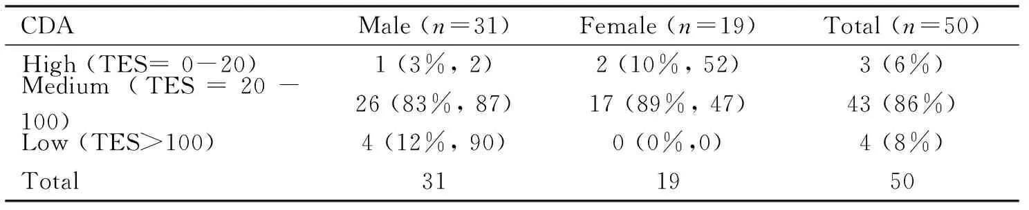
CDAMale(n=31)Female(n=19)Total(n=50)High(TES=0-20)1(3%,2)2(10%,52)3(6%)Medium(TES=20-100)26(83%,87)17(89%,47)43(86%)Low(TES>100)4(12%,90)0(0%,0)4(8%)Total311950
TES: Total error scores, CDA: Color discrimination ability.
Table 3Results according TES of FM100HT for comparing CDA evaluating the difference between DE and NDE

±s, median values)
TES: Total error scores; LES: Lokal error scores; FM100HT: Farnsworth-Munsell 100 Hue Test; CDA: Color discrimination ability; DE: Dominant eye; NDE: Non dominant eye;aP<0.05vsr/g local color spectral region error scores between DE and NDE in male and female;bP<0.01vstotal color spectral region error scores between DE and NDE in all subjects.
Table 4TNO hidden images seen in relation to the proportion of students were tested are shown

ParametersSSM(n=33)F(n=19)Total(n=52)ColoredTable1-431(93%)19(100%)50(96.15%)ColoredTable5SS1+(480arc/s)30(90%)19(100%)49(94.23%)SS2+(240arc/s)29(87%)19(100%)48(92.30%)ColoredTable6SS3+(120arc/s)29(87%)17(89.47%)46(88.46%)SS4+(60arc/s)28(84%)16(84.21%)44(84.61%)ColoredTable7SS5+(30arc/s)1(3%)01(1.9%)SS6+(15arc/s)000
SS: Stereoscopic sensitivity.
But it was not found statistically significant differences in b/y color spectral region for LES between the mean group values of DE and NDE (P=0.739) (Table 3).
Examining The Relationship Between Depth Perceptionand Color VisionAccording to TNO test results, it was determined that all female subjects had binocular vision. According to the investigation, 2 of the 19 females had SS of 240 arc/s while one subject had 120 arc/s and rest of 16 had 60 arc/s.
However, none of the female students had 30 arc/s or better binocular vision. Two of the male students did not see any of the tests that evaluated visual depth perception, and thus were determined not to have depth perception. Another male student had 480 arc/s while two students had 120 arc/s of stereopsis. Only one male subject had 60 arc/s and another one had 30 arc/s and these two participants were determined to have a very high level of stereopsis (Table 4).
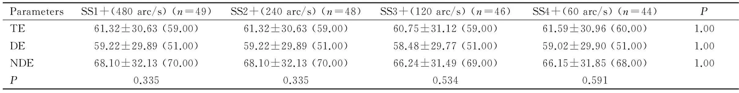
The TES of the students who participated in the TNO test and could see each colored table; two eyes, were compared with the DE and NDE’s FM100HT (Table 5).
The students who had SS1+, SS2+, SS3+, SS4+ were not determined to show a significant difference with regard to TES in the TE, DE and in the NDE groups (P=0.335,P=0.335,P=0.534,P=0.591). Therefore, the comparison of depth perception, were made by comparing the FM100HT error scores between SS1+ (480 arc/s) and SS4+ m (60 arc/s) sensitivities. When we compared the SS1+ and SS4+ sensitivity using TES for all students in the TE, DE and NDE groups, we didn’t find a statistically significant difference (P=1.000).
DISCUSSION
In presenting study, after we found r/g color perception difference in DE and NDE we investigate the relation of color vision level and stereopsis degree, measured by TNO test which is widely used in clinical practice that functions based on color discrimination. To obtain numeric score of CDA, FM100HT were preferred but its error scores are highly related with race, age, and even education[20,34,39]. Macular pigments such as lutein and zeaxanthin are playing an important role for CDA. The CDA was found superior in the Caucasians due to higher concentration of macular lutein and zeaxanthin pigmentation in Caucasians’ macula comparing to other races[18,20,34,36-40]. On the other hand, age is another important factor for CDA which is also related to lutein and zeaxanthin concentrations, both of them decrease progressively by aging[40-42]. In presenting observational study to prevent demographic side effects, young Turkish Caucasians with the mean age of 21.18±2.52y whom were high educated in the Baskent University Faculty of Medicine were included.
Table 5TES of FM100HT for comparing of all students who participated in the TNO test
ParametersSS1+(480arc/s)(n=49)SS2+(240arc/s)(n=48)SS3+(120arc/s)(n=46)SS4+(60arc/s)(n=44)PTE61.32±30.63(59.00)61.32±30.63(59.00)60.75±31.12(59.00)61.59±30.96(60.00)1.00DE59.22±29.89(51.00)59.22±29.89(51.00)58.48±29.77(51.00)59.02±29.90(51.00)1.00NDE68.10±32.13(70.00)68.10±32.13(70.00)66.24±31.49(69.00)66.15±31.85(68.00)1.00P0.3350.3350.5340.591
TES: Total error scores; TE: Two eyes; DE: Dominant eye; NDE: Non-dominant eye; FM100HT: Farnsworth-Munsell 100 Hue Test; SS: Stereoscopic sensitivity.
In literature there are some different opinions about relationship between gender and CDA. Even it has been reported no gender difference in terms of CDA, recently little but significant difference were presented by Abramovetal[43]. They found that female subjects have superior CDA than males but they could not clarify the mechanism. Even no evidence based explanation was presented, according to different studies, effects of hormonal variations on CDA was reported. Giuffreetal[44]were found higher CDA during the ovulation comparing with beginning and later stage of menstrual cycle. In our study both genders were included and female subject were evaluated in between 5-20thdays of menstrual cycle. In research conducted by Orbanetal[45], it was observed that error scores were significantly higher during pregnancy comparing to non-pregnant women.
According to similar studies about CDA in a wide spectrum of the enlightenment which were in between 200-1000 lux were used. As far as we know even the tests were performed under the same illumination in the same studies any standard illumination level was not given in any reports[18,20,44]. One of the study which conducted under the artificial illumination Karacaetal[41], were used two fluorescein lamb with the power of 1300 lux to obtain reference total error score of FM100HT.
In presented study before the evaluation of depth perception, FM100HT were applied initially observing with RE following the LE. The mean TES of RE was higher than that of LE’s CDA and LE was seems to be significantly superior to that of the RE. But these differences may have explained by an indirect evidence of learning and adaptation effect obtained by the REs, which were tested before the following eye. The eye difference were not observed in Mantjarv’s[20]study, performed under the artificial illumination in which Macbeth Easel lamb with 1000 lux. Similarly, Giuffreetal[44]had not observed any differences between the TES of the RE and the LE in FM100HT under the artificial illumination stereoscopic sensitivity of 250 lux. In presented study, each test was performed under the natural light source which recommended in the literature as an optimal test condition[18,46]. In addition to illumination difference between our research and the other studies, learning effect may cause the difference between RE and the LE’s results comparing to other studies. Because the only homogenous highly educated subjects were included in our research therefore learning and adaptation effect may have more prominent comparing to other studies.
Even it has been reported that the contrast sensitivity has effect on visual perception in binocular vision, but the effect of CDA on in binocular vision is still not well understood and in subject of controversy. Even preciously widely accepted idea is colors are not effect on binocular vision, recent studies of last decade found different results. Due to Roe and Ts’o[47], Landsman and Ts’o[48]studies in Makah monkey they postulate that there are special neurons in V2 area for adjusting both binocular vision and color difference. Oudenetal’s[49]reported that colors are used to binocular corresponding on visual system and determine the amount of depth perception. It postulated that multiplex color and disparity cells, located in the V2 area effect on binocular corresponding. While the numbers of the studies are increasing that support theory of the correlation between color perception and the depth perception, still this probable correlation level could not well explain. Therefore, aim of the presented study is evaluate the correlation between these two factors using with FM100HT and TNO test. The comparison of TES in subjects with different level of binocular vision, FM100HT was performed in different situations such as vision either with TE, DE and NDE with the method previously reported[49-51].
In the presented study the TNO test preferred to other binocular single vision tests such as Titmus or Lang tests both are black/white and function with the polarization principle. In addition other test such as Titmus or Lang are contour tests that both which could be seen even in monocular condition. But TNO test is a random dot test which is the only one and based on color discrimination compering of TNO and Titmus test result in subject with color deficiency[37]. In TNO’s test booklet half of the image is made of green while the rest part is red. Binocular single vision could only be observed by a special goggle with red and green filter in each side[38,52].
In each TE, DE and NDE test condition groups, there was no statistically significant difference found between the mean TES in each subgroup of depth perception level of SS1+ (480 arc/s), SS2+ (240 arc/s), SS3+ (120 arc/s) and SS4+ (60 arc/s) (P=1.000). In addition, according to comparison of TES in each subgroup with the same depth perception while testing with TE, DE and NDE the difference was not significant either (P=0.335,P=0.335,P=0.534,P=0.591 respectively). According to these results, our research showed that CDA and binocular depth perception does not effect on each other.
For evaluating the effect on eye dominance on CDA by comparison with DE’s and NDE’s TES, it was not found any differences. In our previous study about the correlation between depth perception and eye dominance it was found that depth perception was not affected by the TE, DE and NDE either[36]. Aslankurtetal[51]compared the DE and NDE effect’s on depth perception by TNO test and Titmus tests and they did not find any difference between DE and NDE. Based on this finding they reported that eye dominance does not effect on depth perception but inter-pupillary difference was effect on it.
In the presented study physiologic factors which could be effect on color vision were excluded as much as possible. Therefore, Turkish Caucasians with the similar age and similar high education level were evaluated. As a result of present study in normal subjects TES of FM100HT showed no difference in different stereopsis level and the depth perception evaluated by TNO test which is based on color dissociation was not correlated. But in further evaluation when subjects were divided into 3 subgroups such as high, medium and low CDA according to the result of TES, presence of lower number of subject in each high and low CDA is one of the restricted parts of this study. On the other hand, we hadn’t change to work with Nagel’s anemoscopes which is considered the gold standard for color vision testing in clinical research; however, it is an expensive instrument requiring an experience examiner’s skills[52]. We hope that if the researcher could able to work with more sensitive instruments for analyzing relationship between CDA and depth perception with the high number of subjects in further studies will enlighten in this research field, the subject. We would also like to note that comparison of stereopsis tested by Titmus or other methods in the subjects with and without color vision deficiency may give more explanations than today.
REFERENCES
1 Pridmore RW. Cone photoreseptör sensitivities and unique hue chromatic res-ponses: correlation and causation imply the physiological basis of unique hues.PloSOne2013;8(10),e77134
2 Conway BR, Chatterjee S, Field GD, Hortwitz GD, Johnson EN, Koida K, Mancuso K. Advances in color science: from retina to behavior.JNeurosci2010;30(45):14955-14963
3 Valberg A. Unique hues: an old problem for a new generation.VisionRes2001;41(21):2811
4 Horiguchi H, Winawer J, Dougherty RF, Wandell BA. Human trichromacy revisited.ProcNatlAcadSciUSA2012,110(3):E260-269
5 Sung C, Chuang J. The cell biology of vision. J.CellBiol2010;190(6):953-963
6 Mustafi D, Engel AH, Palczewski K. Structure of cone photoreceptors.ProgRetinEyeRes2009;28(4):289-302
7 Livitz G, Yazdanbakhsh A, Eskew RT Jr, Mingolla E. Perceiving opponent hues in color induction displays.SeeingPerceiving2011;24(1):1-17
8 Shapley R, Hawken MJ. Color in the cortex: single- and double-opponent cells.VisionRes2011;51(7):701-717
9 Conway BR. Color vision, cones, and color-coding in the cortex.Neuroscientist2009;15(3):274-290
10 Costa TL, Nagy BV, Barboni MT, Boggio PS, Ventura DF. Transcranial direct current stimulation modulates human color discrimination in a pathway-specific manner.FrontPsychiatry2012;3:78
11 Nathans J, Piantanida TP, Eddy RL, Shows TB, Hogness DS. Molecular genetics of inherited variation in human color vision.Science1986;232(4747):203-210
12 Neitz J, Neitz M. The genetics of normal and defective color vision.VisionRes2011;51(7):633-651
13 Gupta A, Laxmi G, Nittala MG, Raman R. Structural and functional correlates in color vision deficiency.Eye(Lond) 2011;25(7):909-917
14 Spalding JA. Colour vision deficiency in the medical profession.BrJGenPract1999;49(443):469-475
15 Birch J. Identification of red-green colour deficiency: sensitivity of the Ishihara and American Optical Company (Hard, Rand and Rittler) pseudo-isochromatic plates to identify slight anomalous trichromatism.OphthalmicPhysiolOpt2010;30(5):667-671
16 Thiadens AA, Hoyng CB, Polling JR, Bernaerts-Biskop R, van den Born LI, Klaver CC. Accuracy of Four Commonly Used Color Vision Tests in the Identification of Cone Disorders.OphthalmicEpidemiol2013;20(2):114-122
17 Gundogan NÜ, Durmazlar N, Gümüt K, Ozdemir PG, Altintao AG, Durur I, Acaroglu G. Projected color slides as a method for mass screening test for color vision deficiency (a preliminary study).IntJNeurosci2005;115(8):1105-1117
18.Kowser Z, Shaj B, Ramya D, Vaitheeswaran K, Deepa V, rajiv R, Tarun S. Effect of Illumination on Colour Vision Testing with Farnsworth-Munsell 100 Hue Test: Customized ColourVision Booth versus Room Illumination.KoreanJOphthalmol2010;24(3):159-162
19 Smith VC, Pokorny J, Pass AS. Color-axis determination on the Farnsworth-Munsell 100- Hue test.AmJOphthalmol1985;100(1):176-182
20 Mantyjarvi M. Normal test scores in the Farnsworth-Munsell 100 hue test.DocOphthalmol2001;102(1):73-80
21 Gundogan FC, Tas A, Altun S, Oz O, Erdem U, Sobaci G. Color vision versus pattern visual evoked potentials in the assessment of subclinical op-tic pathway involvement in multiple sclerosis.IndianJOphthalmol2013;61(3):100-103
22 Salamone G, Lorenzo C, Mosti S, Lupo F, Cravello L, Palmer K, Musicco M, Caltagirone C, Color Discrimination Performance in Patients with Alzhei-mer’s Disease.DementGeriatrCognDisord2009; 27:501-507
23 Koçak-Altintas AG, Satana B, Koçak I, Duman S. Visual acuity and color vision deficiency in amblyopia.EurJOphthalmol2000;10(1):77-81
24 Shneor E, Hochstein S. Eye dominance effects in future search.VisionRes2006;46(25):4258-4269
25 Erdoǒan AR, Ozdikici M, Aydyn MD, Aktat O, Dane S. Right and left visüel cortex areas in healty subjects with right and left-eye dominance.IntJNeurosci2002;112(5):517-523
26 Oishi A, Tobimatsu S, Arakawa K, Taniwaki T, Kira J. Ocular dominancy in conjugate eye movements at reading distance.NeurosciRes2005;52(3):263-268
27 Rombouts SA, Barkhof F, Sprenger M, Valk J, Scheltens P. The functional ba-sis of ocular dominance: functional MRI (fMRI) findings.NeurosciLeft1996;221(1):1-4
28 Nongpiur ME, Sharma P. Horizontal lang two-pencil test as a screening test for stereopsis and binocularity.IndianJophthalmol2010;58(4):287-290
29 Ferrer-Blasco T, Madrid-Costa D, Garcia-Lazaro S, Cervino A and Montes-Mico R. Stereopsis in bilaterally multifocal pseudophakic patients.GraefesArchClinExpOphthalmol2011;249(2):245-251
30 Shaygannejad V, Golabchi K, Dehghani A, Asthtari F, Haghighi S, Mirzendeh-del M, Ghasemi M. Color blindness among multible sclerosis patients in Isfa-han.JResMedSci2012;17(3):254-257
31 Shah A, Hussain R, Fareed M, Afzal M. Prevalence of red-green color vision defects among muslim males and females of Manipur, India.IranJPublicHealth2013;42(1):16-24
32 Barboni MTS, Nagy BV, Araujo Moura AL, Damico FM, Costa MF, Kremers J. ON and OFF electroretinography and Contrast Sensitivity in DuchenneMuscu-lar Dystrophy.InvestOphthalmolVisSci2013;54:3195-3204
33 Qian YS, Chu RY, He JC, Sun XH, Zhou XT, Zhao NQ, Hu DN, Hoffman MR, Dai JH, Qu XM, Pao KE. Incidence of myopia in high school students with and without red-green color vision deficiency.InvestOphthalmolVisSci2009;50(4):1598-1605
34 Siaudvytyte L. Mitkute D. Balciuniene J. Quality of life in patients with age-related macular degeneration.Medicina(Kaunas) 2012;48(2):109-111
35 Gundogan NU. Yazycy AC, Simsek A. Study on dominant eye measurement.IntJOphthalmol2009;2(3):271-277
36 Koctekin B, Gundogan NU, AltyntatAK, Yazycy AC.Relationof eye dominancy with color vision discrimination performance ability in normal subjects.IntJOphthalmol2013;6(5):733-738
37 Momeni-Moghadam H, Kundart J, Ehsani M, Ghlami K.The comparison of stereopsis with TNO and Titmus test result in symptomatic and asymptomatic university students.JournalofBehavioralOptometry2011;23(2),35-39
38 TNO test for stereoscopic vision, Lameris Instrumentenb.v., biltstraat 449, 3572 aw Utrecht Schairer-Ophthal-Technic, Alexander str.55-70182 Eighth edition Stuttgart, 1972
39 Kinnear PR, Sahraie A. New Farnsworth-Munsell 100 hue test norms of normal observers for each year of age 5-22 and forage decades 30-70.BrJOphthalmol2002;86(12):1408-1411
40 Woo GC, Lee MH. Are ethnic differences in the F-M 100 scores related to ma-cular pigmentation?ClinExpOptom2002;85(6):372-377
41 Karaca A,Saatçi AO, Kaynak C. The result of Farnsworth-Munsell 100 Hue test in Turkish population.Ret-Vit2005;13(2):119-123
42 Loskutova E, Nolan J, Howard A, Beatty S. Macular Pigment and Its Contri-bution to Vision.Nutrients2013;5(6):1962-1969
43 Abramov I, Gordon J, Feldman O, Chavarga A. Sex and vision II: color appearance of monochromatic lights.BiolSexDiffer2012;3(1):21
44 Giuffre G, Di Rosa L, Fiorino F. Changes in colour discrimination during the menstrual cycle.Ophthalmologica2007;221(1):47-50
45 Orbán LL, Dastur FN. Shifts in color discrimination during early pregnancy.EvolPsychol2012;10(2):238-252
46 Melamud A, Hagstrom S, Traboulsi E. Color vision testing.OphthalmicGenet2004;25(3):159-187
47 Roe AW, Ts’o DY. Specificity of color connectivity between primate V1 and V2.JNeurophysiol1999;82(5):2719-2730
48 Landisman CE, Ts’o DY. Color processing in macaque striate cortex: electrophysiological properties.JNeurophysiol2002;87(6):3138-3151
49 denOuden HE, van Ee R, de Haan EH. Colour helps to solve the binocular matching problem.JPhysiol2005;567(Pt 2):665-671
50 Johansson J, Pansell T, Ygge J, Seimyr GÖ. The effect of contrast on monocular ver-sus binocular reading performance.JournalofVision2014;14(5):8
51 Aslankurt M, Aslan L, Aksoy A, Özdemir M, Dane a, Lateralty does not affect the depht perception but interpupillary distance.JOphthalmol2013; 2013:485059
52 Pandey N, Chandrakar AK, Garg ML. Tests for color vision deficiency: It time to revize the standard?IndianJOphthalmol2015;63(9):753-753
(作者单位:106815土耳其安卡拉巴什肯特大学医学专业生理学系;207100 土耳其安塔利亚健康科学大学教育研究院生理学系;3 06240 土耳其安卡拉健康科学大学教育研究院眼科)
通讯作者:Nimet Unay Gundogan.nimetg@yahoo.com
DOI:10.3980/j.issn.1672-5123.2016.8.04
关键词:色觉;色觉辨色能力;深度知觉;法-孟二氏100色度试验;主视眼Gundogan方法;TNO测试
大学生色觉辨别能力与深度知觉的相关性分析
Nimet Unay Gundogan1, Belkis Salman Koçtekin2, AyseGul Koçak Altinta3
摘要