心肺运动试验评价食管癌患者化疗后整体功能变化的临床研究
2016-07-20孙兴国CurtisHightowerWilliamStringer葛万刚
孙兴国,Curtis Hightower,刘 方,William W.Stringer,葛万刚
·论著·
心肺运动试验评价食管癌患者化疗后整体功能变化的临床研究
孙兴国,Curtis Hightower,刘 方,William W.Stringer,葛万刚
100037北京市,国家心血管病中心 中国医学科学院阜外医院 心血管疾病国家重点实验室 国家心血管疾病临床医学研究中心,北京协和医学院(孙兴国,刘方,葛万刚);德州大学安德森癌症中心麻醉科(Curtis Hightower);美国加州大学洛杉矶分校医学中心,洛杉矶生物医学研究院,圣约翰心血管研究中心(William W.Stringer)
【摘要】背景化疗对人体整体功能具有显著损害,但目前尚缺乏对化疗患者整体功能变化进行客观定量评估的可靠方法。目的探讨心肺运动试验(CPET)对食管癌患者化疗前后整体功能变化的评估作用。方法选取2006—2007年度在德州大学安德森癌症中心进行术前化疗的白种人男性食管癌患者6例,分别在化疗前后进行CPET检查,按照美国加州大学洛杉矶分校医学中心标准连续递增功率方案完成症状限制性极限运动,通过对数据标准化分析计算其核心指标,从系统软件导出静息状态、热身状态、无氧阈状态、极限状态时的循环指标及呼吸指标。结果6例食管癌患者化疗后,除1例患者无氧阈略有升高外,其余患者的峰值摄氧量、无氧阈、峰值氧脉搏、摄氧效率平台、峰值负荷功率和递增功率运动时间均降低;化疗后,除1例患者的二氧化碳通气当量斜率略有降外,其余所有患者的二氧化碳通气当量斜率和二氧化碳通气当量最低值均升高。食管癌患者化疗前后无氧阈(L/min、ml·min-1·kg-1、%pred)、二氧化碳通气当量斜率(slope、%pred)、递增功率运动时间(min)比较,差异均无统计学意义(P>0.05);化疗后峰值摄氧量(L/min、ml·min-1·kg-1、%pred)、峰值氧脉搏(ml/次、%pred)、摄氧效率平台(比值、%pred)、峰值负荷功率(W)较化疗前降低,二氧化碳通气当量最低值(比值、%pred)较化疗前升高(P<0.05)。食管癌患者化疗后极限状态时摄氧量较化疗前降低(P<0.05);静息状态时心率较化疗前升高,收缩压较化疗前降低(P<0.05);热身状态时舒张压、平均动脉压较化疗前降低(P<0.05);4个状态时氧脉搏较化疗前降低(P<0.05);静息状态、热身状态、极限状态时摄氧通气效率较化疗前降低(P<0.05);极限状态时二氧化碳通气当量较化疗前升高(P<0.05)。食管癌患者化疗后热身状态时潮气末二氧化碳分压较化疗前降低(P<0.05);静息状态、热身状态、极限状态时潮气末氧分压较化疗前升高(P<0.05);极限状态时每呼吸摄氧量较化疗前降低(P<0.05);无氧阈状态、极限状态时每呼吸二氧化碳排出量较化疗前降低(P<0.05)。结论化疗使得食管癌患者循环和细胞代谢功能显著降低,而呼吸受限相对较轻,且处于相对代偿反应。CPET检查可以对食管癌患者化疗前的整体功能进行客观定量评估,同时也可以对化疗所造成的整体功能改变进行评估。
【关键词】食管肿瘤;抗肿瘤联合化疗方案;心肺运动试验
孙兴国,Hightower C,刘方,等.心肺运动试验评价食管癌患者化疗后整体功能变化的临床研究[J].中国全科医学,2016,19(17):2046-2052.[www.chinagp.net]
Sun XG,Hightower C,Liu F,et al.Clinical study of cardiopulmonary exercise testing in the evaluation of the changes of holistic function of patients with esophagus cancer after chemotherapy[J].Chinese General Practice,2016,19(17):2046-2052.
食管癌是常见的人类消化道恶性肿瘤之一,其亚型食管腺癌是近年来发病率增长最快的恶性肿瘤[1],由于该疾病一般发现晚[2-3],缺乏有效治疗措施,故预后极差,病死率较高,为了提高手术切除率及减少术后复发率,术前化疗是食管癌较好的选择[4]。术前化疗即新辅助化疗(neoadjuvant chemotherapy),首先是由Frei[5]作为肿瘤综合治疗的一部分而提出。术前化疗可以降低部分肿瘤患者的临床分期,进而提高手术切除率[6-9],但由于化疗药物的毒副作用,如骨髓抑制、消化道症状以及肝损害等[10],使部分肿瘤患者不能耐受,从而导致术前化疗在临床的开展受到了一定的限制。化疗在肿瘤的综合治疗中占有重要地位,但常规化疗药物均为细胞毒性药物,不仅能促进肿瘤细胞坏死和诱导肿瘤细胞凋亡,还可杀伤机体正常增殖和分化的细胞,从而抑制患者的免疫功能,削弱机体免疫细胞抗肿瘤的能力[11]。所以术前化疗必定会对人体造成一定的损害,从微观角度来看是细胞功能下降,从宏观角度看则是人体整体功能下降,但是目前尚没有特定的方法对化疗患者整体功能的下降进行评估。
心肺运动试验(cardiopulmonary exercise testing,CPET)作为一种无创伤、客观、定量、连续、可重复的临床检测方法,是目前唯一的人体整体功能学检测方法,通过运用整体整合生理学医学新理论体系对心肺运动期间呼吸、循环、血液、代谢等多系统功能的连续动态变化进行整合分析[12-14],在临床上广泛应用于心血管、呼吸、代谢系统等疾病的诊断、功能及疾病严重程度评估、治疗效果评价、预后预测及指导康复治疗等[15-16]。从整体整合角度分析肿瘤患者化疗前后CPET检查指标动态变化,也必然可以为化疗影响人体整体功能找到一种客观定量检测方法。本研究通过分析食管癌患者化疗前后CPET检查指标,探讨整体功能的客观定量和稳定可靠的评估方法。
1对象与方法
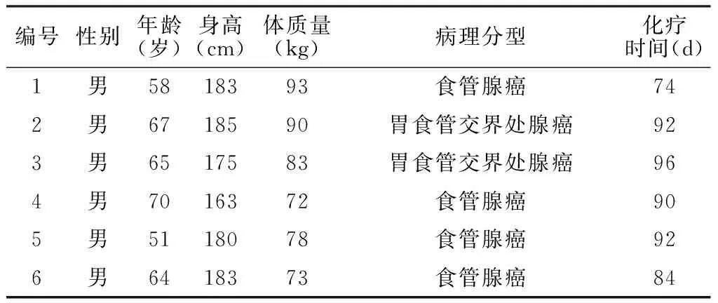
1.1研究对象选取2006—2007年度在德州大学安德森癌症中心进行术前化疗的6例食管癌美国白种人患者为研究对象,均为男性;年龄51~70岁,平均年龄(62.5±6.3)岁;身高163~185 cm,平均身高(178±8)cm;体质量72~93 kg,平均体质量(82±8)kg;病理分型为:食管腺癌4例,胃食管交界处腺癌2例(见表1)。

表1 6例食管癌患者一般资料
1.2CPET检查患者分别于化疗前及化疗后手术之前在德州大学安德森癌症中心签署知情同意书后完成CPET检查[17],按照美国加州大学洛杉矶分校医学中心标准方法[17-22]对两次CPET检查数据进行分析判读,计算出各生理学指标。在美国本试验数据的回顾性再分析立项获得国家心血管病中心阜外医院立项委员会的批准同意(2013-ZX29)。采用MedGraphics Ultima系统Breeze5.2软件记录12导联心电图、无创血压、氧饱和度、各项肺通气和气体交换等指标[17]。按照美国加州大学洛杉矶分校医学中心标准连续递增功率方案完成症状限制性极限运动[15,18-20]:先记录静息3 min;然后,以60 r/min 蹬车速率无负荷热身3 min;根据患者年龄、性别和估计的功能状态预设功率自行车功率递增速率为10~30 W/min,使患者在6~10 min达到症状限制性最大极限运动,获得最大运动功率,继续记录恢复期5~10 min。
1.3CPET检查数据标准化分析从Breeze 5.2软件系统导出所有测定指标,如峰值摄氧量(L/min、ml·min-1·kg-1、%pred)、峰值氧脉搏(ml/次、%pred)、摄氧效率平台(比值、%pred)、二氧化碳通气当量斜率(slope、%pred)、二氧化碳通气当量最低值(比值、%pred)、峰值负荷功率(W)、递增功率运动时间(min)的每次呼吸数据,经每秒分切之后,再用10 s平均数据来制图和进行结果分析[15,18-22]。将10 s的二氧化碳排出量与摄氧量数据做图用V-slope法测得无氧阈(L/min、ml·min-1·kg-1、%pred)[22-23]。记录静息状态、热身状态、无氧阈状态、极限状态时CPET检查中循环指标(摄氧量、心率、收缩压、舒张压、平均动脉压、氧脉搏、摄氧通气效率、二氧化碳通气当量)和呼吸指标(分钟通气量、潮气量、呼吸频率、呼吸交换比值、潮气末二氧化碳分压、潮气末氧分压、每呼吸摄氧量、每呼吸二氧化碳排出量),所有静息状态时的指标以120 s数据计算,热身状态和极限状态时的指标以30 s数据计算,无氧阈状态的指标以无氧阈之前(氧气)和无氧阈之后(二氧化碳)60 s数据计算[24-27]。
1.4食管癌患者术前化疗方案6例食管癌患者给予平均(88.0±7.9)d的术前化疗。化疗方案为:顺铂(DDP,20 mg/m2,1~5 d)+5-氟尿嘧啶(5-FU,20 mg/m2,1~5 d),或紫杉醇(PTX,175 mg/m2,1 d)+ DDP(80 mg/m2,1 d)+5-FU(750 mg/m2,1~5 d)。

2结果
2.16例食管癌患者化疗前后体质量比较化疗前食管癌患者体质量为(81.6±8.2)kg,化疗后为(77.7±8.0)kg,差异有统计学意义(t=4.420,P=0.007)。
2.26例食管癌患者化疗前后CPET检查核心功能指标比较6例食管癌患者化疗后,除1例患者无氧阈略有升高外,其余患者的峰值摄氧量、无氧阈、峰值氧脉搏、摄氧效率平台、峰值负荷功率和递增功率运动时间均降低;化疗后,除1例患者的二氧化碳通气当量斜率略有降低外,其余所有患者的二氧化碳通气当量斜率和二氧化碳通气当量最低值均升高(见图1)。
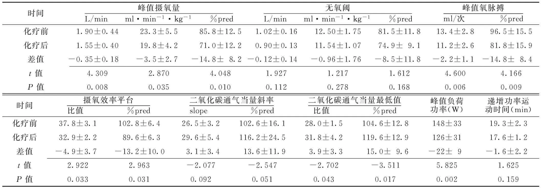
食管癌患者化疗前后无氧阈(L/min、ml·min-1·kg-1、%pred)、二氧化碳通气当量斜率(slope、%pred)、递增功率运动时间(min)比较,差异均无统计学意义(P>0.05);化疗后峰值摄氧量(L/min、ml·min-1·kg-1、%pred)、峰值氧脉搏(ml/次、%pred)、摄氧效率平台(比值、%pred)、峰值负荷功率(W)较化疗前降低,二氧化碳通气当量最低值(比值、%pred)较化疗前升高,差异有统计学意义(P<0.05,见表2)。

图1 6例食管癌患者化疗前后CPET检查核心指标的个体化变化情况

时间峰值摄氧量L/min ml·min-1·kg-1 %pred无氧阈L/min ml·min-1·kg-1 %pred峰值氧脉搏ml/次 %pred化疗前1.90±0.4423.3±5.585.8±12.51.02±0.1612.50±1.7581.5±11.813.4±2.896.5±15.5化疗后1.55±0.4019.8±4.271.0±12.20.90±0.1311.54±1.0774.9±9.111.2±2.681.8±15.9差值-0.35±0.18-3.5±2.7-14.8±8.2-0.12±0.14-0.96±1.76-8.5±11.8-2.2±1.1-14.8±8.4t值4.3092.8704.0481.9271.2171.6124.6004.166P值0.0080.0350.0100.1120.2780.1680.0060.009时间摄氧效率平台比值 %pred二氧化碳通气当量斜率slope %pred二氧化碳通气当量最低值比值 %pred峰值负荷功率(W)递增功率运动时间(min)化疗前37.8±3.1102.8±6.426.5±3.2102.6±16.128.0±1.5104.6±12.8148±3319.3±2.3化疗后32.9±2.289.6±6.329.6±5.4116.2±24.531.8±4.2119.6±12.9126±3117.6±1.2差值-4.9±3.7-13.2±10.03.1±3.413.6±11.93.9±3.315.0±9.6-22±9-1.6±2.2t值2.9222.963-2.077-2.547-2.702-3.5115.8251.625P值0.0330.0310.0920.0510.0430.0170.0020.159
注:%pred=实测值/预计值×100%
2.3食管癌患者化疗前后不同状态时CPET检查中循环指标比较食管癌患者化疗后极限状态时摄氧量较化疗前降低,差异有统计学意义(P<0.05);静息状态、热身状态、无氧阈状态时摄氧量与化疗前比较,差异无统计学意义(P>0.05)。食管癌患者化疗后静息状态时心率较化疗前升高,收缩压较化疗前降低,差异有统计学意义(P<0.05);热身状态、无氧阈状态、极限状态时心率、收缩压与化疗前比较,差异无统计学意义(P>0.05)。食管癌患者化疗后热身状态时舒张压、平均动脉压较化疗前降低,差异有统计学意义(P<0.05);静息状态、无氧阈状态、极限状态时舒张压、平均动脉压与化疗前比较,差异无统计学意义(P>0.05)。食管癌患者化疗后4个状态时氧脉搏较化疗前降低,差异有统计学意义(P<0.05)。食管癌患者化疗后静息状态、热身状态、极限状态时摄氧通气效率较化疗前降低,差异有统计学意义(P<0.05);无氧阈状态时摄氧通气效率与化疗前比较,差异无统计学意义(P>0.05)。食管癌患者化疗后极限状态时二氧化碳通气当量较化疗前升高,差异有统计学意义(P<0.05);静息状态、热身状态、无氧阈状态时二氧化碳通气当量与化疗前比较,差异无统计学意义(P>0.05,见表3)。
2.4食管癌患者化疗前后不同状态时CPET检查中呼吸指标比较食管癌患者化疗前后4个状态时分钟通气量、潮气量、呼吸频率、呼吸交换比值比较,差异无统计学意义(P>0.05)。食管癌患者化疗后热身状态时潮气末二氧化碳分压较化疗前降低,差异有统计学意义(P<0.05);静息状态、无氧阈状态、极限状态时潮气末二氧化碳分压与化疗前比较,差异无统计学意义(P>0.05)。食管癌患者化疗后静息状态、热身状态、极限状态时潮气末氧分压较化疗前升高,差异有统计学意义(P<0.05);无氧阈状态时潮气末氧分压与化疗前比较,差异无统计学意义(P>0.05)。食管癌患者化疗后极限状态时每呼吸摄氧量较化疗前降低,差异有统计学意义(P<0.05);静息状态、热身状态、无氧阈状态时每呼吸摄氧量与化疗前比较,差异无统计学意义(P>0.05)。食管癌患者化疗后无氧阈状态、极限状态时每呼吸二氧化碳排出量较化疗前降低,差异有统计学意义(P<0.05);静息状态、热身状态时每呼吸二氧化碳排出量与化疗前比较,差异无统计学意义(P>0.05,见表4)。
3讨论
3.1食管癌患者化疗后功能损害与功能评估方法食管癌患者特别是肿瘤过大难以手术者,化疗是常规的辅助治疗方法,虽然化疗降低机体功能的毒副作用确实存在,但客观定量功能评估的临床研究报道较少。近几年Gutschow等[28]通过功能量表和症状得分等方法对化疗食管癌患者的健康状况进行评估,结果提示化疗患者的功能状态较健康人明显降低。早在20世纪末Dimeo等[29]开始对化疗肿瘤患者进行康复运动,可降低患者化疗毒副作用。然而该研究的方法是主观评估,可信度较低。2014年Ruiz-Casado等[30]应用身体锻炼的强度及时间对食管癌患者化疗后的功能状态进行评估,但也缺乏客观定量评估方法。
3.2CPET客观定量评估整体功能CPET作为目前唯一人体整体功能学检测方法,其利用检测外呼吸测定气体交换将外呼吸与细胞呼吸联系起来量化人体整体细胞呼吸的状态和时间经过,反映细胞呼吸的功能变化,反映人体最大有氧代谢能力和心肺储备能力,能够比较安全、客观地从呼吸、循环、细胞代谢等方面评估食管癌患者化疗前后整体功能的改变。本研究根据CPET检查的峰值摄氧量、无氧阈、摄氧效率平台、心率、血压等循环指标及呼吸指标的变化,从患者化疗前后心功能、循环运氧能力、肺换气功能以及细胞代谢方面,反映化疗后患者整体功能的变化。

表3 食管癌患者化疗前后不同状态时CPET检查中循环指标比较±s,n=6)

表4 食管癌患者化疗前后不同状态时CPET检查中呼吸指标比较±s,n=6)
3.3食管癌患者化疗后整体功能变化
3.3.1食管癌患者化疗后整体功能变化——循环系统本研究结果显示,食管癌患者化疗后静息状态时心率明显升高,同时患者静息状态时摄氧量几乎不变,即患者静息状态时的基础代谢率不变,提示化疗后患者每搏量降低,每搏输氧能力下降,心率则代偿性升高;静息状态时舒张压变化无差异,即患者心脏的前后负荷均无明显增加,提示患者化疗后心脏的收缩功能降低。除了极限状态外,其他3个状态时摄氧量均有下降趋势,但是无统计学差异,可能与本研究样本量较小有关,本课题组将进一步增加样本量,继续观察。食管癌患者化疗后4个状态时氧脉搏较化疗前降低,静息状态、热身状态、极限状态时摄氧通气效率较化疗前降低,极限状态时二氧化碳通气当量较化疗前升高,提示患者心脏射血能力、循环运氧能力及整体细胞代谢率较化疗前降低。
3.3.2食管癌患者化疗后整体功能变化——呼吸系统本研究结果显示,食管癌患者化疗前后分钟通气量、呼吸频率、呼吸交换比值均无变化,化疗后仅热身状态时潮气末二氧化碳分压较化疗前降低;而潮气末氧分压,除无氧阈状态时,其他3个状态时均明显升高,提示患者出现过度通气状态,同时可以得出,短期化疗对患者肺功能的损伤相对较心功能损伤低,心脏受损后肺功能有所代偿。静息状态、热身状态、无氧阈状态时每呼吸摄氧量与化疗前无差异,静息状态、热身状态时每呼吸二氧化碳排出量与化疗前无差异,亦提示循环和代谢的抑制显著高于呼吸(潮气量)的抑制。
3.3.3食管癌患者化疗后整体功能变化——循环系统和呼吸系统匹配本研究结果显示,食管癌患者化疗后极限状态时二氧化碳通气当量较化疗前升高,其他3个状态时虽无统计学差异但也有所上升;化疗后静息状态、热身状态、极限状态时摄氧通气效率较化疗前降低,提示患者每次呼吸运氧能力明显下降,反映化疗后患者的循环运氧和细胞代谢能力明显下降,而呼吸处于相对代偿性过度通气状态。
3.4本研究的不足本研究的不足之处在于样本量小,可能存在统计偏倚,许多升高或降低的趋势无统计学差异;选取的病例为美国白种人男性患者,在我国患者适用情况如何?本课题组将继续进行该方面的研究,加大样本量。
综上所述,CPET检查可以从呼吸、循环、代谢等方面指标的变化客观、定量地评估食管癌患者化疗前后整体功能的变化。食管癌患者化疗后循环运氧能力和细胞代谢功能呈现显著抑制,而相对而言,呼吸抑制较轻,且呈现明显地代偿性反应。CPET检查评估及其指导下个体化高强度精准运动康复可以用于肿瘤患者化疗后功能评估和康复手段,值得深入研究。
作者贡献:孙兴国、Curtis Hightower、刘方、William W.Stringer、葛万刚进行课题设计与实施、资料收集整理、撰写论文、成文并对文章负责;孙兴国、刘方、葛万刚进行课题实施、评估、资料收集;孙兴国进行质量控制及审校。
本文无利益冲突。
参考文献
[1]李婧文,房殿春.食管腺癌相关危险因素的研究进展[J].现代消化及介入诊疗,2014(2):94-96.
[2]Brenner B,Ilson DH,Minsky BD.Treatment of localized esophageal cancer[J].Semin Oncol,2004,31(4):554-565.
[3]Zhuang JL,Ming DS,Xu RY,et al.Study on differentially expressed genes in esophageal carcer by cDNA microarray[J].Chinese Journal of Experimental Surgery,2002,19(6):527-528.(in Chinese)
庄建良,明德松,许荣誉,等.应用微矩阵表达谱基因芯片检测食管癌的基因改变[J].中华实验外科杂志,2002,19(6):527-528.
[4]Shi Y,Qin R,Wang ZK,et al.Nanoparticle albumin-bound paclitaxel combined with cisplatin as the first-line treatment for metastatic esophageal squamous cell carcinoma[J].Onco Targets Ther,2013,6:585-591.
[5]Frei E 3rd.Clinical cancer research:an embattled species[J].Cancer,1982,50(15):1979-1992.
[6]Hermann RM,Horstmann O,Haller F,et al.Histomorphological tumor regression grading of esophageal carcinoma after neoadjuvant radiochemotherapy:which score to use?[J].Dis Esophagus,2006,19(5):329-334.
[7]Triboulet JP,Mariette C.Esophageal squamous cell carcinoma stage Ⅲ:state of surgery after radicchemotherapy[J].Cancer Radiother,2006,10:456-461.
[8]Sujendran V,Wheeler J,Baron R,et al.Effect of neoadjuvant chemotherapy on circumferential margin positivity and its impact on prognosis in patients with resectable esophageal cancer[J].Br J Surg,2008,95(2):191-194.
[9]Matsuyama J,Doki Y,Yasuda T,et al.The effect of neoadjuvant chemotherapy on lymph node micrometastases in squamous cell carcinomas of the thoracic esophagus[J].Surgery,2007,141(5):570-580.
[10]Andc N,Iizuka T,Idc H,et al.Surgery plus chemotherapy compared with surgery alone for localized squamous cell carcinoma of the thoracic esophagus:a Japan Clinical Oncology Group Study——JCOG9204[J].J Clin Oncol,2003,21(24):4592-4596.
[11]张传芳,杨伟青,徐明跑,等.替加氟栓术前化疗对结肠癌病人免疫功能的影响[J].浙江临床医学,2008,10(5):629.
[12]孙兴国.整体整合生理学医学新理论体系:人体功能一体化自主调控[J].中国循环杂志,2013,28(2):88-92.
[13]Sun XG.New theoretical system of holistic control and regulation for life and cardiopulmonary exercise testing[J].Medicine & Philosophy,2013,34(5):22-27.(in Chinese)
孙兴国.生命整体调控新理论体系与心肺运动试验[J].医学与哲学,2013,34(5):22-27.
[14]谭晓越,孙兴国.从心肺运动的应用价值看医学整体整合的需求[J].医学与哲学,2013,34(5):28-31.
[15]Wasserman K,Hansen J,Sue D,et al.Principles of exercise testing and interpretation [M].5th ed.Philadelphia:Lippincott Williams & Wilkins,2011.
[16]孙兴国.心肺运动试验在临床心血管病学中的应用价值和前景[J].中华心血管病杂志,2014,42(4):347-351.
[17]Hightower CE,Riedel BJ,Feig BW,et al.A pilot study evaluating predictors of postoperative outcomes after major abdominal surgery:physiological capacity compared with the ASA physical status classification system[J].Br J Anaesth,2010,104(4):465-471.
[18]Sun XG,Hansen JE,Oudiz RJ,et al.Exercise pathophysiology in patients with primary pulmonary hypertension[J].Circulation,2001,104(4):429-435.
[19]Sun XG,Hansen JE,Oudiz RJ,et al.Gas exchange detection of exercise-induced right-to-left shunt in patients with primary pulmonary hypertension[J].Circulation,2002,105(1):54-60.
[20]Wasserman K,Sun XG,Hansen JE.Effect of biventricular pacing on the exercise pathophysiology of heart failure[J].Chest,2007,132(1):250-261.
[21]孙兴国.更为强化心肺代谢整体功能的心肺运动试验新9图图解[J].中国应用生理学杂志,2015,31(4):369-373.
[22]孙兴国.心肺运动试验的规范化操作要求和难点——数据分析图示与判读原则[J].中国应用生理学杂志,2015,31(4):361-365.
[23]Beaver WL,Wasserman K,Whipp BJ.A new method for detecting the anaerobic threshold by gas exchange[J].J Appl Physiol(1985),1986,60(6):2020-2027.
[24]Sun XG,Hansen JE,Beshai JF,et al.Oscillatory breathing and exercise gas exchange abnormalities prognosticate early mortality and morbidity in heart failure[J].J Am Coll Cardiol,2010,55(17):1814-1823.
[25]Sun XG,Hansen JE,Stringer WW.Oxygen uptake efficiency plateau:physiology and reference values[J].Eur J Appl Physiol,2012,112(3):919-928.
[26]Sun XG,Hansen JE,Garatachea N,et al.Ventilatory efficiency during exercise in healthy subjects[J].Am J Respir Crit Care Med,2002,166(1):1443-1448.
[27]Sun XG,Hansen JE,Stringer WW.Oxygen uptake efficiency plateau best predicts early death in heart failure[J].Chest,2012,141(5):1284-1294.
[28]Gutschow CA,Hölscher AH,Leera J,et al.Health-related quality of life after Ivor Lewis esophagectomy[J].Langenbecks Arch Surg,2013,398(2):231-237.
[29]Dimeo F,Fetscher S,Lange W,et al.Effects of aerobic exercise on the physical performance and incidence of treatment-related complications after high-dose chemotherapy[J].Blood,1997,90(9):3390-3394.
[30]Ruiz-Casado A,Verdugo AS,Solano MJ,et al.Objectively assessed physical activity levels in Spanish cancer survivors[J].Oncol Nurs Forum,2014,41(1):E12-20.
(本文编辑:陈素芳)
Clinical Study of Cardiopulmonary Exercise Testing in the Evaluation of the Changes of Holistic Function of Patients With Esophagus Cancer After Chemotherapy
SUNXing-guo,CurtisHightower,LIUFang,etal.
NationalCenterforCardiovascularDisease,FuwaiHospital,ChineseAcademyofMedicalSciences,StateKeyLaboratoryofCardiovascularDisease,NationalCenterforCardiovascularDiseaseClinicalMedicineResearch,PekingUnionMedicalCollege,Beijing100037,China
【Abstract】BackgroundChemotherapy has a serious damage on the holistic function of human body,while there is a lack of reliable methods for the objective and quantitative assessment of the changes of holistic function of patients after chemotherapy.ObjectiveTo investigate the role of cardiopulmonary exercise testing (CPET) in the evaluation of the changes of holistic function of patients with esophagus cancer after chemotherapy.MethodsFrom 2006 to 2007,we enrolled 6 Caucasian male patients with esophagus cancer who underwent preoperative chemotherapy in MD Anderson Cancer Center of University of Texas and conducted CPET examinations on them before and after chemotherapy.According to standard continuously increasing power scheme of UCLA Medical Center,we completed symptom-limited extreme sports and calculated the core indexes by standardized data analysis.The circulation indexes and respiration indexes in a quiescent state,a warm-up state,an anaerobic threshold(AT) state and an extreme state were derived by system software.ResultsOf the 6 esophagus cancer patients,only one patient had increase in AT and the rest patients all saw decrease in peak VO2,AT,peak oxygen pulse,oxygen uptake efficiency plateau,peak load power and increasing power movement time after chemotherapy;only one patient had decrease in the slope of ventilatory equivalent for carbon dioxide and the rest patients all had increase in the slope of ventilatory equivalent for carbon dioxide and the minimum ventilatory equivalent for carbon dioxide.The AT(L/min,ml·min-1·kg-1,%pred),slope of ventilatory equivalent for carbon dioxide(slope,%pred),and increasing power movement time (min) after chemotherapy of the patients were not significantly different from those before chemotherapy (P>0.05);the peak VO2(L/min,ml·min-1·kg-1,%pred),peak oxygen pulse (ml/time,%pred),oxygen uptake efficiency plateau (ratio,%pred),and peak load power (W) were lower than those before chemotherapy,while the minimum ventilatory equivalent for carbon dioxide (ratio,%pred) were higher than those before chemotherapy (P<0.05).After chemotherapy,the patients had lower oxygen uptake in an extreme state than that before chemotherapy(P<0.05);the heart rate in a quiescent state was higher than that before chemotherapy,and the systolic pressure in a quiescent state was lower than that before chemotherapy (P<0.05);the diastolic pressure and mean arterial pressure in a warm-up state were lower than those before chemotherapy (P<0.05);the oxygen pulse in the four states were lower than those before chemotherapy (P<0.05);the oxygen uptake efficiency in a quiescent state,a warm-up state,and an AT state after chemotherapy were lower than those before chemotherapy (P<0.05);the ventilatory equivalent for carbon dioxide after chemotherapy was higher than that before chemotherapy(P<0.05).After chemotherapy,the ETCO2 in a warm-up state after chemotherapy was lower than that before chemotherapy (P<0.05);the PETO2 in a quiescent state,a warm-up state,and an extreme state after chemotherapy were higher than those before chemotherapy (P<0.05);the oxygen intake of every breath in an extreme state was lower than that before chemotherapy (P<0.05);the carbon dioxide output of every breath in an AT state and extreme state after chemotherapy were lower than those before chemotherapy (P<0.05).ConclusionChemotherapy makes circulation and the metabolic function of cells significantly deteriorate,but causes only slight limit on respiration which is relative compensatory response.CPET can be an objective and quantitative assessment not only for holistic functional status of cancer patients before chemotherapy,but also for physiological changes in holistic function caused by chemotherapy.
【Key words】Esophageal neoplasm;Antineoplastic combined chemotherapy protocols;Cardiopulmonary exercise testing
基金项目:国家自然科学基金医学科学部面上项目(81470204);中国医学科学院国家心血管病中心科研开发启动基金(2012-YJR02)
通信作者:孙兴国,100037北京市,国家心血管病中心 中国医学科学院阜外医院 心血管疾病国家重点实验室 国家心血管疾病临床医学研究中心,北京协和医学院;E-mail:xgsun@labiomed.org
【中图分类号】R 735.1
【文献标识码】A
doi:10.3969/j.issn.1007-9572.2016.17.013
(收稿日期:2016-03-21;修回日期:2016-05-22)
·专题研究·
