反义miR-30d对乳腺癌细胞侵袭迁移能力的影响
2014-08-08赵瑞君谢春伟
陈 戈 骆 萍 龚 宇 周 瑶 赵瑞君 谢春伟
反义miR-30d对乳腺癌细胞侵袭迁移能力的影响
陈 戈 骆 萍 龚 宇 周 瑶 赵瑞君 谢春伟△
目的分析miR-30d在乳腺癌组织中的表达情况,体外研究反义miR-30d寡核苷酸(miR-30d ASO)对乳腺癌细胞侵袭和迁移的影响。方法运用荧光定量PCR检测108例乳腺癌组织及对应正常癌旁组织中miR-30d的表达;通过miR-30d ASO降低乳腺癌细胞中miR-30d的表达,采用Transwell实验和划痕实验观察miR-30d ASO对乳腺癌细胞侵袭和迁移能力的影响,运用Western blot方法检测侵袭相关蛋白基质金属蛋白酶(MMP)-2和MMP-9蛋白的表达变化。结果乳腺癌组织中miR-30d基因表达水平明显高于对应癌旁组织(P<0.05);与空白对照组和无义干扰组相比,miR-30d ASO可以显著降低乳腺癌细胞miR-30d的表达(P<0.05);Transwell实验和划痕实验结果显示转染miR-30d ASO后,乳腺癌细胞的侵袭及迁移能力明显下降,同时MMP-2和MMP-9表达下降(P<0.05)。结论miR-30d在乳腺癌组织中表达上调,降低miR-30d的表达可有效抑制乳腺癌细胞的侵袭和迁移。miR-30d有可能成为乳腺癌侵袭转移调控的新靶点。
乳腺肿瘤;癌;肿瘤侵润;细胞运动;miR-30d;反义寡核苷酸
MicroRNAs(miRNAs)是一种内源性的非编码小分子RNA,其可以在转录后水平调节蛋白质的合成,广泛分布于生物细胞体内。大量研究已经证实miRNAs在恶性肿瘤细胞的发生、发展中发挥重要作用,其中包括调控细胞的增殖、分化、凋亡、侵袭和转移[1-2]。近年miR-30d在恶性肿瘤发生发展中的作用已受到广泛关注。研究报道miR-30d在前列腺癌、成神经管细胞瘤以及肝癌等恶性肿瘤中表达上调[3-5]。过表达的miR-30d可以促进肝癌细胞的侵袭和转移[5]。然而miR-30d在乳腺癌中的表达及其对乳腺癌细胞侵袭迁移能力的影响尚少见报道。本研究采用荧光定量PCR检测miR-30d在乳腺癌组织中的表达;利用反义寡核酸技术(antisense oligonucleotide,ASO)抑制乳腺癌细胞中miR-30d的表达,分析miR-30d对乳腺癌细胞侵袭迁移能力的影响。同时检查侵袭相关蛋白基质金属蛋白酶(MMP)-2和MMP-9蛋白的表达变化。
1 材料与方法
1.1 材料
1.1.1 患者资料 收集2008年9月—2012年5月我院乳腺外科108例乳腺癌患者的乳腺癌组织及其正常癌旁组织手术标本,所有标本均经病理学检查确诊。患者均为女性,年龄16~68岁,术前均未接受放化疗。
1.1.2 主要试剂和仪器 乳腺癌MCF-7和MDA-MB-231细胞由本实验室保存;DMEM高糖培养基(美国Gibco公司)、胎牛血清(美国Hyclone公司)、脂质体LipfectamineTM2000(美国Invitrogen公司);反义miR-30d寡核苷酸(miR-30d ASO)购自大连宝生物公司;总蛋白提取试剂盒(北京普利莱)、兔抗人MMP-2和MMP-9单克隆抗体(美国santa cruz公司)、βactin二抗(北京中杉金桥公司);Transwell Chamber(美国Chemicon公司),Matrigel(美国BD公司);TaqMan miRNA分析试剂盒、荧光定量PCR分析仪7500(美国ABI公司)。
1.2 方法
1.2.1 实时荧光定量PCR检测miR-30d的表达 采用RNA提取试剂盒提取癌组织及对应正常组织的总RNA,测定浓度后-80℃保存。运用miRNA分析试剂盒检测miR-30d的表达。首先取2 μg总RNA为反应模板与3 μL逆转录酶混合,反应体系为20 μL,反应条件为:16 ℃ 30 min,45 ℃ 30 min,85℃5 min。反应结束后,收集cDNA。采用SYBR Green法定量检测,将质粒稀释品梯度稀释为1×10⁻¹、1×10⁻²、1×10⁻³、1×10⁻⁴、1×10⁻⁵、1×10⁻⁶、1×10⁻⁷和1×10⁻⁸,用于建立标准曲线。反应条件:95℃ 15 min;95℃ 30 s,60℃ 30 s,72℃ 30 s,共45个循环;最后72℃延伸7 min。在每个循环的最后增加溶解曲线。阈值定义为基础荧光信号的10个标准差,即时循环数为Ct,依据标准曲线计算目的基因的mRNA量。实验重复3次。
1.2.2 miR-30d ASO序列设计 获取人miR-30d的序列(http://www.sanger.ac.uk/software/Rfam/mirna),设计其反义寡核苷酸序列,miR-30d:正义 5'-AGUCACUAGUGGUUCCGUUUA-3',反义5'-TAAACGGAACCACTAGTGACT-3',运用NCBI BLAST检索程序以排除其他的同源序列。同时设计1条无义干扰阴性对照序列:正义5'-UCUUCCGAACGUGUCACGUTT-3',反义:5'-AAGUGACACGUUCGGAGAATT-3',送上海英骏生物技术公司合成。
1.2.3 细胞培养和反义寡核苷酸转染 将MCF-7和MDAMB-231乳腺癌细胞接种于含10%胎牛血清的DMEM培养基,37℃、5%CO2条件下培养。运用LipfectamineTM2000转染试剂盒进行转染,miR-30d ASO终浓度分别为:50、100、150、200和250 nmol/L。本实验组筛选出最佳终干扰浓度为150 nmol/L。转染后培养时间分别为24、48、72 h,初步筛出最佳作用时间为48 h。上述操作重复3次。
1.2.4 观察miR-30d ASO对miR-30d表达的影响 转染终浓度为150 nmol/L的miR-30d ASO 48 h后,提取总RNA,测定浓度,逆转录为cDNA(反应条件同1.2.1),测定cDNA浓度。运用microRNA检测试剂盒检测miR-30d的表达(具体条件同1.2.1)。上述操作重复3次。
1.2.5 Transwell侵袭实验 实验分为空白对照(MOCK)组、无义干扰(NC)组和干扰miR-30d(miR-30d ASO)组,下面的分组也是如此。Transwell小室的上下室之间以孔径为8 μm的聚碳酸酯微膜孔分隔开,滤膜上层铺盖Matrigel。实验开始前将Transwell小室复温至37℃,在内室中加入300 μL的DMEM培养液,置于培养箱中孵育2 h,使Matrigel水化。用胰酶消化细胞成单细胞悬液,800 r/min离心5 min,收集细胞,用PBS洗涤细胞2次,最后用无血清的DMEM培养基重悬细胞,调整细胞数为1×10⁶个/mL。吸出内室中培养液,吸取500 μL含10%血清的DMEM于外室中作为趋化因子,加300 μL细胞悬液于内室中,37℃、5%CO2培养箱中培养24 h。取出Transwell小室吸除培养基,利用棉签擦净Transwell小室滤膜上层的Matrigel及未穿过滤膜的细胞。将小室浸入细胞染色液中染色20 min,采用蒸馏水冲洗,风干。实验中每组每次同时做3个重复小室,显微镜观察计数,取平均值。
1.2.6 Transwell迁移实验 具体过程与Transwell侵袭实验基本一致,不同的是不需要铺Matrigel。
1.2.7 划痕实验 采用Marker笔在6孔板背面均匀划横线,约0.5~1 cm宽,横穿过孔,每孔至少穿过5条线。在孔中加入约5×10⁵个细胞,掌握为过夜细胞能铺满整孔。第2天用10 μL枪头依横线划痕,注意枪头垂直。PBS洗细胞3次,去除划下的细胞,加入培养基。置于37℃、5%CO2培养箱培养。0 h、48 h取样,用显微镜拍照测量各组划痕愈合率情况。每个样本至少重复3次。
1.2.8 Western blot检测各组细胞中MMP-2和MMP-9蛋白的表达 运用总蛋白提取试剂盒提取MCF-7和MDA-MB-231细胞总蛋白,经10%SDS-PAGE电泳后转膜,将膜放在含5%脱脂奶粉的TBST缓冲液中37℃封闭2 h,加一抗稀释液1∶400稀释兔抗人MMP-2和MMP-9单克隆抗体在4℃孵育过夜,1×TBST缓冲液3次(每次10 min),加辣根过氧化物酶标记的山羊抗兔IgG(1∶5 000稀释),置于37℃孵育2 h,1×TBST缓冲液3次(每次10 min),运用ECL化学发光法检测目的蛋白条带。以β-actin作为内参。
1.3 统计学方法 采用SPSS 17.0统计软件进行分析。计量资料以均数±标准差(±s)表示,癌及癌旁组织比较采用配对样本t检验,多组均数之间比较采用单因素方差分析,两两比较采用LSD-t检验,P<0.05为差异有统计学意义。
2 结果
2.1 乳腺癌及癌旁组织miR-30d的表达情况 乳腺癌组织中miR-30d的表达明显高于相应的正常癌旁组织(t=31.32,P < 0.05),见图1。

Figure 1 Comparison of the expression level of miR-30d between breast cancer and adjacent normal tissues图1 乳腺癌及对应癌旁组织的miR-30d表达水平比较
2.2 抑制miR-30d对miR-30d表达的影响 MCF-7细胞中,miR-30d ASO组的miR-30d相对表达量较MOCK组和NC组明显降低(F=391.66,P <0.05);MDA-MB-231细胞实验结果与MCF-7细胞结果一致(F=488.42,P < 0.05),见图2。

Figure 2 Comparison of the expression level of miR-30d between three groups(*compared with the miR-30d ASO group,P < 0.05)图2 3组细胞中miR-30d的表达水平比较(*与miR-30d ASO组比较,P <0.05)
2.3 Transwell侵袭实验检测抑制miR-30d的表达对乳腺癌细胞侵袭能力的影响 miR-30d ASO组的MCF-7细胞侵袭能力低于MOCK组及NC组(F=671.28,P<0.05),而MOCK组与NC组之间无明显差别(P > 0.05),见图3、4。MDA-MB-231细胞实验结果与MCF-7细胞一致(F=741.18,P < 0.05),见图5、6。
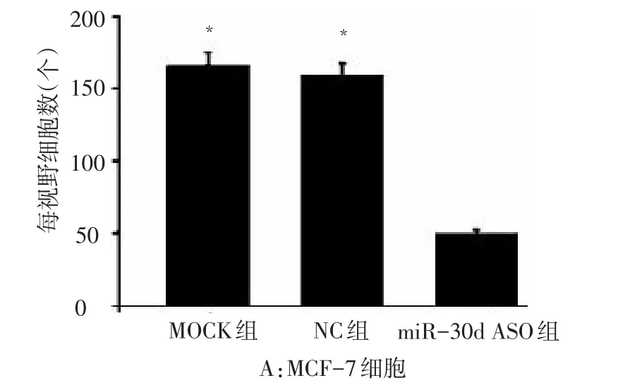
Figure 3 Comparison of the invasion ability of MCF-7 cells between three groups(*compared with the miR-30d ASO group,P < 0.05)图3 3组MCF-7细胞的侵袭能力比较(*与miR-30d ASO组比较,P <0.05)
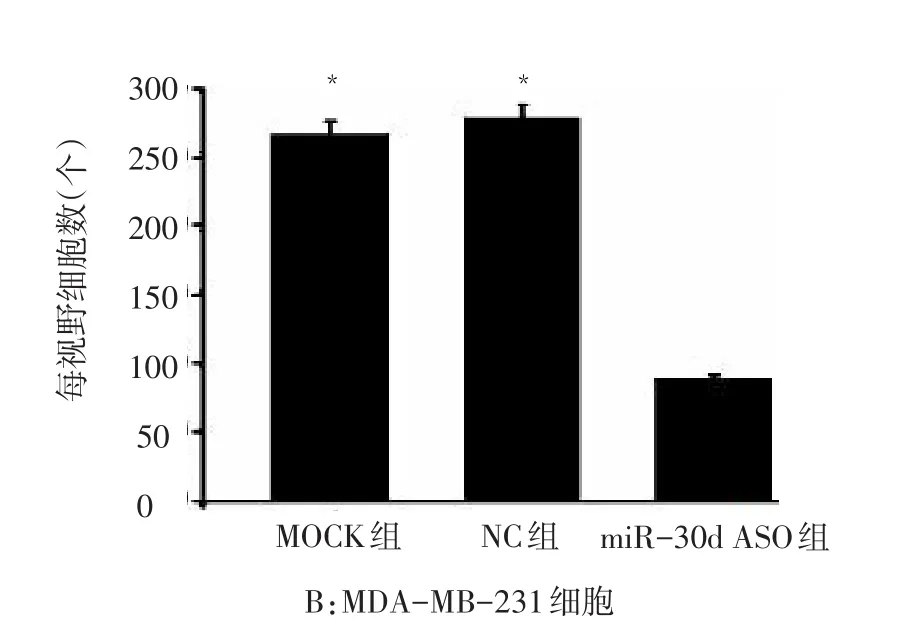
Figure 5 Comparison of the invasion ability of MDA-MB-231 cells between three groups(*compared with the miR-30d ASO group,P<0.05)图5 3组MDA-MB-231细胞的侵袭能力比较(*与miR-30d ASO组比较,P<0.05)
2.4 Transwell迁移实验检测抑制miR-30d的表达对乳腺癌细胞迁移能力的影响 转染miR-30d ASO组的MCF-7细胞迁移能力低于MOCK组及NC组(F=472.57,P < 0.05),而MOCK组与NC组无明显差别(P > 0.05),见图7、8。MDA-MB-231细胞实验结果与MCF-7细胞一致(F=289.34,P < 0.05),见图9、10。
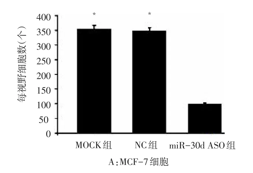
Figure 7 Comparison of the migration ability of MCF-7 cells between three groups(*compared with the miR-30d ASO group,P < 0.05)图7 3组MCF-7细胞的迁移能力比较(*与miR-30d ASO组比较,P<0.05)
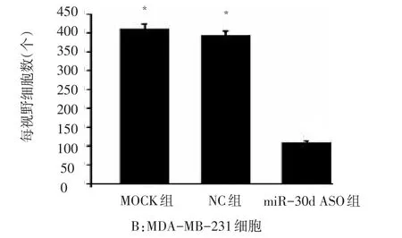
Figure 9 Comparison of the migration ability of MDA-MB-231 cells between three groups(*compared with the miR-30d ASO group,P < 0.05)图9 3组MDA-MB-231细胞的迁移能力比较(*与miR-30d ASO组比较,P<0.05)
2.5 划痕实验检测降低miR-30d基因表达对乳腺癌细胞迁移能力的影响 转染48 h后,对于MCF-7细胞,miR-30d ASO组划痕的愈合率明显低于MOCK组和NC组(F=743.24,P <0.05);MDA-MB-231细胞与MCF-7结果一致(F=564.18,P<0.05),见表1、图11。
Table 1 Comparison of the wound healing ability of breast cancer cells between three groups表1 3组乳腺癌细胞划痕的愈合率比较 (±s)

Table 1 Comparison of the wound healing ability of breast cancer cells between three groups表1 3组乳腺癌细胞划痕的愈合率比较 (±s)
**P<0.01;a与(3)组比较,P < 0.05
组别MOCK组(1)NC组(2)miR-30d ASO组(3)F n333 MCF-7细胞0.55±0.09a0.59±0.08a0.23±0.03 743.24**MDA-MB-231细胞0.61±0.11a0.67±0.09a0.15±0.02 564.18**
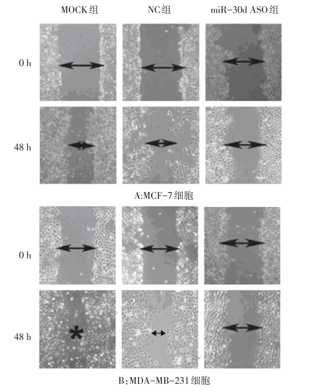
Figure 11 Comparison of the wound healing ability between three groups图11 3组细胞的划痕愈合能力比较
2.6 抑制miR-30d的表达对MMP-2和MMP-9蛋白表达的影响 对于MCF-7细胞和MDA-MB-231细胞,miR-30d ASO组的MMP-2和MMP-9相对表达量明显低于MOCK组和NC组(P <0.05),见表2、图12。
Table 2 Comparison of the expression levels of MMP-2 and MMP-9 between three groups表2 各组细胞MMP-2和MMP-9蛋白表达水平比较(n=3±s)
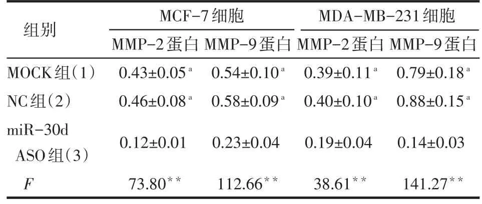
Table 2 Comparison of the expression levels of MMP-2 and MMP-9 between three groups表2 各组细胞MMP-2和MMP-9蛋白表达水平比较(n=3±s)
**P < 0.01;a与(3)组比较,P < 0.05
组别MOCK组(1)NC组(2)miR-30d ASO组(3)F MCF-7细胞MMP-2蛋白0.43±0.05a0.46±0.08a0.12±0.01 73.80**MMP-9蛋白0.54±0.10a0.58±0.09a0.23±0.04 112.66**MDA-MB-231细胞MMP-2蛋白0.39±0.11a0.40±0.10a0.19±0.04 38.61**MMP-9蛋白0.79±0.18a0.88±0.15a0.14±0.03 141.27**
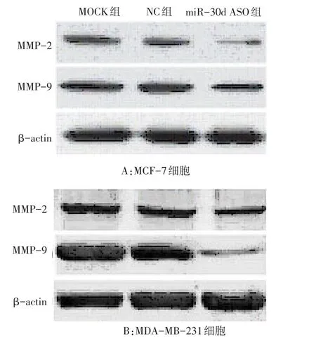
Figure 12 The expression levels of MMP-2 and MMP-9 in three groups图12 MMP-2和MMP-9蛋白在3组细胞中的表达情况
3 讨论
乳腺癌是女性最常见的恶性肿瘤之一,全球每年约有120万妇女发生乳腺癌。近年,我国乳腺癌的发病率呈逐年上升趋势,死亡率居女性恶性肿瘤首位。随着分子生物学的发展,发现人体内基因的表达异常与肿瘤的发生、发展密切相关,肿瘤的基因治疗成为近年来研究的热点。目前研究证明,miRNA分子调控了人类基因组内约1/3的基因表达[6],如何对调控肿瘤基因表达的特异性miRNA分子进行调节备受关注。
MicroRNAs是一类新的基因调节因子,其在控制细胞的生长、发育、分化、新生血管形成和凋亡等生物学过程中发挥着非常重要的作用[7]。研究发现miRNAs与癌症的发生及侵袭转移关联紧密[8-9]。因此miRNAs有可能成为诊断恶性肿瘤的分子标志和判断肿瘤侵袭转移及预后的分子靶点,此外miRNAs在转录后水平调节其靶基因的表达,这将更有助于癌症的早期发现、早期诊断和早期治疗,其必将具有非常广阔的临床应用前景[10]。
本研究首先利用实时荧光定量PCR检测临床乳腺癌标本及相对应的正常癌旁组织,结果发现miR-30d在乳腺癌组织中呈明显高表达,表明miR-30d可能在乳腺癌发生发展过程中发挥重要作用。有研究报道miR-30d过表达可以促进肝癌细胞侵袭转移[5]。为了证实miR-30d在乳腺癌细胞侵袭迁移中是否发挥相同的功能,本研究首先通过转染miR-30d ASO降低乳腺癌细胞中miR-30d的表达,同时利用Transwell方法检测降低miR-30d的表达后乳腺癌细胞侵袭迁移能力的变化,结果显示转染miR-30d ASO的乳腺癌细胞侵袭迁移能力明显降低,划痕实验结果显示抑制miR-30d的表达后,乳腺癌细胞的划痕愈合能力明显下调。这些结果表明miR-30d在乳腺癌细胞的侵袭迁移过程中起着非常重要的作用。
MMPs是一类主要降解细胞外基质(ECM)成分的蛋白酶,肿瘤从原位增殖到浸润转移的演进过程中必须具备降解ECM的能力。MMPs几乎能降解ECM中的各种蛋白成分,破坏肿瘤细胞侵袭的组织学屏障,在肿瘤侵袭转中起关键作用。目前MMPs家族已分离鉴别出26个成员,编号分别为MMP1~26。研究证实MMP-2和MMP-9蛋白与肿瘤的侵袭转移密切相关[11-12]。本研究发现抑制miR-30d的表达可以明显降低侵袭相关蛋白MMP-2和MMP-9的表达,说明miR-30d可能是通过调控MMP-2和MMP-9的表达而对肿瘤细胞侵袭迁移产生影响。
综上,miR-30d在调控乳腺癌细胞的侵袭和迁移方面发挥重要作用,其可能是一个新的乳腺癌侵袭转移调节基因,是乳腺癌基因治疗的新靶点。

Figure 4 Comparison of the invasion ability of MCF-7 cells between three groups图4 3组MCF-7细胞的Transwell侵袭实验结果

Figure 6 Comparison of the invasion ability of MDA-MB-231 cells between three groups图6 3组MDA-MB-231细胞的Transwell侵袭实验结果

Figure 8 Comparison of the migration ability of MCF-7 cells between three groups图8 3组MCF-7细胞的Transwell迁移实验结果

Figure 10 Comparison of the migration ability of MDA-MB-231 cells between three groups图10 3组MDA-MB-231细胞的Transwell迁移实验结果
[1]Hannafon BN,Ding WQ.Intercellular Communication by Exosome-Derived microRNAs in Cancer.[J].Int J Mol Sci,2013,14(7):14240-14269.
[2]Braicu C,Calin GA,Berindan-Neagoe I,et al.MicroRNAs and cancer therapy-from bystanders to major players.[J].Curr Med Chem,2013,20(29):3561-3573.
[3]Kobayashi N,Uemura H,Nagahama K,et al.Identification of miR-30d as a novel prognostic maker of prostate cancer[J].Oncotarget,2012,3(11):1455-1471.
[4]Lu Y,Ryan SL,Elliott DJ,et al.Amplification and overexpression of Hsa-miR-30b,Hsa-miR-30d and KHDRBS3 at 8q24.22-q24.23 in medulloblastoma[J].PLoS One,2009,4(7):e6159.
[5]Yao J,Liang L,Huang S,et al.MicroRNA-30d promotes tumor invasion and metastasis by targeting Galphai2 in hepatocellular carcinoma[J].Hepatology,2010,51(3):846-856.
[6] Raisch J,Darfeuille-Michaud A,Nguyen HT,et al.Role of microRNAs in the immune system,inflammation and cancer[J].World J Gastroenterol,2013,28,19(20):2985-2996.
[7] Lewis BP,Burge CB,Bartel DP.Conserved seed pairing,often flanked by adenosines,indicates that thousands of human genes are microRNA targets[J].Cell,2005,120(1):15-20.
[8] Harquail J,Benzina S,Robichaud GA,et al.MicroRNAs and breast cancer malignancy:an overview of miRNA-regulated cancer processes leading to metastasis[J].Cancer Biomark,2012,11(6):269-280.
[9] Lopez-Camarillo C,Marchat LA,Arechaga-Ocampo E,et al.MetastamiRs:Non-Coding MicroRNAs Driving Cancer Invasion and Metastasis[J].Int J Mol Sci,2012,13(2):1347-1379.
[10]Kala R,Peek GW,Hardy TM,et al.MicroRNAs:an emerging science in cancer epigenetics[J].J Clin Bioinforma,2013,3(1):6.
[11]Gao J,Ding F,Liu Q,et al.Knockdown of MACC1 expression suppressed hepatocellular carcinoma cell migration and invasion and inhibited expression of MMP2 and MMP9[J].Mol Cell Biochem,2013,376(1-2):21-32.
[12]Fan HX,Li HX,Chen D,et al.Changes in the expression of MMP2,MMP9,and ColⅣin stromal cells in oral squamous tongue cell carcinoma:relationships and prognostic implications[J].J Exp Clin Cancer Res,2012,31:90.
(2013-06-05收稿 2013-10-15修回)
(本文编辑 闫娟)
The Effect of Antisense miR-30d on Invasion and Migration in Breast Cancer Cells
CHEN Ge,LUO Ping,GONG Yu,ZHOU Yao,ZHAO Ruijun,XIE Chunwei
Department of Breast Surgery,The Third Hospital of Nanchang City,Nanchang 330009,China
ObjectiveTo investigate the expression of miR-30d in breast cancer tissues and demonstrate the regulative effects of miR-30d ASO on the invasion and migration of breast cancer cells in vitro.MethodsThe expression levels of miR-30d in 108 breast cancer tissues and their adjacent tissues were detected by real-time quantitative PCR method.After transfection with miR-30d ASO,the biological effects of miR-30d on in breast cancer cells was measured by transwell assay and wound healing assay.The expressions of matrix metalloproteinase(MMP)-2 and MMP-9 were analyzed by Western blot assay.ResultsThe expression level of miR-30d was found to be over-expressed in breast cancer tissues(P<0.05).Compared with control group and nonsense interference group,the miR-30d expression was significantly decreased in breast cancer cells(transfection with miR-30d ASO).Results of transwell and wound healing assay showed that the invasion and migration ability decreased significantly after transfection with miR-30d ASO,and expressions of MMP-2 and MMP-9 were down-regulated(P<0.05).ConclusionmiR-30d was over-expressed in human breast cancer.The invasion and migration of breast cancer cells can be effectively inhibited by decreasing the expression of miR-30d.miR-30d may become a new target for the regulation of invasion and migration in breast cancer.
breastneoplasms;carcinoma;neoplasm invasiveness;cell movement;miR-30d;antisenseoligonucleotides
R735.7 【
】 A 【DOI】 10.3969/j.issn.0253-9896.2014.03.004
南昌市第三医院乳腺外科(邮编330009)
△通讯作者 E-mail:lufen6677@163.com
