双容积重建技术在颅内动脉瘤栓塞术中的应用研究
2014-08-02张祥海陈金华王晓峰周林闫红野
张祥海,陈金华,王晓峰,周林,闫红野
双容积重建技术在颅内动脉瘤栓塞术中的应用研究
张祥海,陈金华,王晓峰,周林,闫红野
目的探讨双容积重建技术在颅内动脉瘤弹簧圈栓塞术中的应用价值。方法对2012年6月-2013年4月收治的20例行颅内动脉瘤弹簧圈栓塞术患者的三维造影数据进行双容积重建,从弹簧圈的检出率,动脉瘤的栓塞程度,术前、术后动脉瘤瘤颈和瘤体长度,重建图像特点和应用价值等方面对双容积重建的作用进行评价。结果双容积重建共检出弹簧圈缠绕团20个,检出率100%。20例中有13例动脉瘤无造影剂渗入,3例瘤体内有造影剂占位,4例可见瘤颈有造影剂占位。术后瘤颈、瘤体长度与术前比较有所改变,但差异无统计学意义(P>0.05)。双容积重建影像能显示出弹簧圈缠绕团、血管、颅骨和融合图像,可根据临床需要选择不同的影像模式来显示动脉瘤。结论双容积重建技术能提供弹簧圈缠绕团定位、栓塞程度、动脉瘤长度等信息,并可通过多种影像模式评估栓塞效果,在动脉瘤的介入栓塞术中具有较高的应用价值。
颅内动脉瘤;栓塞,治疗性;图像处理,计算机辅助
颅内动脉瘤介入栓塞治疗具有微创、安全等优点,已成为临床首选的手术方式[1]。数字减影血管造影(digital subtraction angiography,DSA)三维重建技术对动脉瘤的诊断和治疗具有重要价值[2],但填塞了弹簧圈的动脉瘤无法在容积重建图像中显示,而双容积重建(dual volume reconstruction,Dual VR)技术可以重建出栓塞后的动脉瘤影像,本文拟探讨该技术在动脉瘤栓塞术中的应用价值。
1 资料与方法
1.1 一般资料 选择2012年6月-2013年4月在第三军医大学大坪医院行颅内动脉瘤弹簧圈栓塞术的患者20例,其中男14例,女6例,年龄35~68岁。所有患者均确诊为颅内动脉瘤,全麻下行弹簧圈栓塞术,其中17例采用支架辅助。20名患者中共发现动脉瘤24个,其中前交通动脉瘤7个,后交通动脉瘤10个,大脑中动脉瘤7个。所有患者均只对1个颅内动脉瘤进行栓塞治疗,4个微小动脉瘤未处理。
1.2 设备和参数 采用西门子Aritis DTA血管造影机,造影剂为威视派克,超选至颈内动脉作三维造影(3D-DSA),造影参数为:注射速率4m l/s,总量24m l,曝光延迟1s,压力150psi。数据传输至西门子后处理工作站,术前的三维造影数据行容积重建,术中的数据行双容积重建。
1.3 评价方法 由2名放射科医生和2名技师从4个方面进行评估:①弹簧圈缠绕团的检出率;②动脉瘤的栓塞程度;③术前容积重建图像和术后双容积重建图像同体位下瘤颈、瘤体的长度;④不同模式下双容积重建影像的特点和应用价值。
2 结 果
2.1 弹簧圈检出率 双容积重建图像共检出20个灰白色高密度影像,分析其占位和形状,确认为弹簧圈缠绕团影像,检出率为100%(图1)。
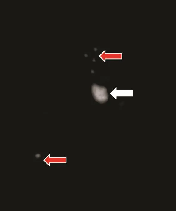
图1 同时重建出弹簧圈缠绕团(白色箭头)和支架标记点(红色箭头)Fig.1 Coils bolus (white arrow) and stent marks ( red arrow) reconstructed in the same image
2.2 动脉瘤栓塞程度 完全栓塞的13个动脉瘤瘤体、瘤颈均被灰色的弹簧圈占据,无造影剂渗入。有3个瘤体可见红色的造影剂占位,瘤体未完全填塞。有4个瘤颈处有红色造影剂占位,瘤颈未完全栓塞(图2)。
2.3 术前术后瘤体长度比较 测量双容积重建影像中瘤颈、瘤体的长度,并与术前双容积重建图像进行比较。对于未完全栓塞的动脉瘤,以弹簧圈和造影剂的共同长度作为瘤体、瘤颈的长度(图3)。结果表明,栓塞前后瘤颈、瘤体的长度均有变化,但差异无统计学意义(P>0.05,表1)。
2.4 不同重建模式的影像特点 双容积重建图像有4种不同的显示模式:①颅骨、弹簧圈、支架显示模式,该模式下弹簧圈缠绕团能清楚显示,可见17个支架的标记点;②血管显示模式,该模式下可显示1-4级脑血管,分支血管清楚,可见未栓塞的4个小动脉瘤;③弹簧圈和颅内血管融合显示模式,该模式下可见颅骨、弹簧圈、血管共同显示在一个影像上(图4、5),4例瘤颈和3例瘤体有造影剂渗入;④重建颅内血管(B)并将颅内组织(A)做多平面重建的融合显示模式(B+A MPR),该模式下可见颅内组织各层面与颅内血管的相互关系,可观察造影剂和弹簧圈同层面的占位范围,显示颅内组织与血管的毗邻关系(图6)。
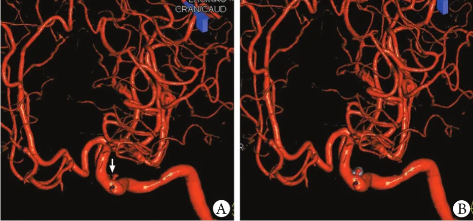
图2 血管和弹簧圈缠绕团的融合图像Fig.2 Fused image of vessel and coils bolusA.Only reconstructing the intracranial vessel, we can discover the contrast medium (white arrow) flow into the aneurysms sac, manifesting the pathway, extent and caliber of inflow tract; B.Fused imaging display the occupation of contrast medium and coils bolus, showing the embolization extent of intracranial aneurysm
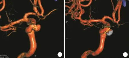
图3 术前和术后动脉瘤瘤颈长度Fig.3 Length of aneurysm neck before and after embolizationA.The length o f aneurysms neck before embolization; B.At the same position, the length of aneurysms neck after embolization measured in the dual volume reconstruction imaging
表1 栓塞前后瘤颈及瘤体长度比较(mm,±s,n=20)Tab.1 The length of aneurysm neck and sac before and after embolization (mm±s, n=20)

表1 栓塞前后瘤颈及瘤体长度比较(mm,±s,n=20)Tab.1 The length of aneurysm neck and sac before and after embolization (mm±s, n=20)
Time point Neck Length of sac Width of sac Before embolization 3.84±2.41 4.90±2.94 2.95±1.73 After embolization 4.74±1.89 5.51±3.01 3.15±1.84
3 讨 论
与二维DSA比较,3D-DSA造影中患者接受的辐射剂量更低[3],其重建图像能提供丰富的影像信息,在动脉瘤的介入诊疗中起重要作用[4]。2008年,西门子公司推出了双容积重建技术,该技术为一次采集两次重建,一次为高密度弹簧圈、支架、颅骨的容积重建,一次为颅内血管重建,血管显示为红色,弹簧圈等显示为灰白色,两种不同密度的组织影像可以单独或融合显示,为动脉瘤提供了新的影像显示平台。
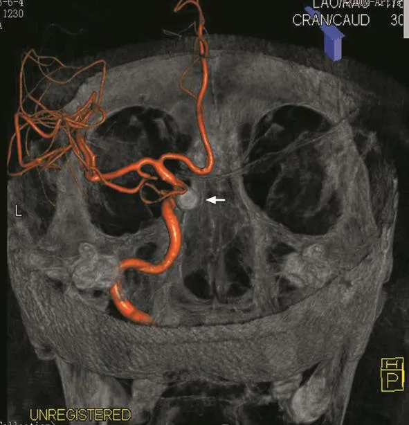
图4 颅内血管、颅骨和弹簧圈缠绕团(白色箭头)的融合图像(用于帮助解剖定位)Fig.4 Fusion image composed by intracranial vessel, skull bone and coils bolus (white arrow, with a great help in anatomy position)

图5 弹簧圈、颅骨和颅内血管的融合图像Fig.5 Fusion image composed by coils bolus, skull bone and intracranial vesselA.Part of coils bolus insert into parent artery, and the distal end of intracranial vessel display clearly; B.Fusion imaging show the skull bone and coils bolus, discovering part of coils inserting into parent artery

图6 B+A MPR图像模式显示动脉瘤多平面重建和血管的融合影像Fig.6 B+A MPR image mode, showing aneurysms fused imaging of multiplanar reconstruction and intracranial vesselA, B.In the oblique transverse and sagittal section, contrast medium occupy the right area of aneurysm, indicating the right area of aneurysm sac without embolization; C, D.In the oblique transverse and coronal section, contrast medium occupy a tiny area in the extrem ity of coli bolus, indicating the distal aneurysms sac without embolization
在动脉瘤栓塞术中,技师常规都会进行三维重建来显示瘤体和瘤颈的详细信息[5-6]。但在容积重建影像上,弹簧圈、颅骨等组织会被减影,很难观察到血管与弹簧圈的相互关系,影响了手术效果的评估和手术方案的设计[7]。双容积重建技术突破了既往重建技术的局限,在本研究中,所有病例均重建出弹簧圈缠绕团,显示了弹簧圈的位置、范围、形状,并可结合血管影像,重建出更多显示动脉瘤栓塞情况的图像。
动脉瘤栓塞不完全可增加术后复发的概率,并可能引发术后再出血[8-10]。有研究发现,完全栓塞后动脉瘤再出血的概率仅为0.28%,而不完全栓塞的再出血概率为7.41%[11],因此如何准确评估动脉瘤的栓塞程度是临床关注的重点,目前可采用二维造影或三维造影判断动脉瘤致密栓塞程度[12],或通过动脉瘤栓塞容积闭塞率[13]、阻塞体积百分比[14]以及特殊的软件程序[15-16]对填塞程度进行评估,但这些方法对弹簧圈在瘤体内的成篮形状和占位情况判断不够直观和准确,不能直接评估动脉瘤的栓塞情况。
在动脉瘤栓塞术前,首先应准确测量出瘤颈、瘤体的长度,以此作为第一枚弹簧圈的选择依据[17]。双容积重建影像可融合显示弹簧圈缠绕团及颅内血管,实现对术中动脉瘤的测量,可以得到新形态下瘤体和瘤颈的长度。本文对术前、术后数据比较,瘤颈和瘤体的长度均有所变化,术后瘤颈长度增大的有13例,瘤体长径增大的有14例,瘤体短径增大的有16例,但术前术后的瘤颈、瘤体长度差异并无统计学意义,与向伟楚等[18]的研究相似。动脉瘤填塞后,由于弹簧圈在瘤体内占位的影响或血流动力学改变等原因,动脉瘤瘤颈、瘤体的形态、位置和长度都可能发生变化,甚至每填塞一个弹簧圈后,瘤体都可能发生不同程度的改变,双容积重建影像可以对未完全栓塞的动脉瘤进行测量,得到新形态下动脉瘤的长度数据,为下一步选择弹簧圈和支架提供准确的数据参考。
在动脉瘤栓塞术中,必要时造影重建出动脉瘤的双容积图像,区分出瘤体内已栓塞区域和未栓塞区域,测量不同区域的长度,以精确的数据来评估栓塞程度,使造影影像数据化,有助于提高栓塞的安全性和有效性。双容积重建图像可以帮助医生和技师精确观察分析瘤颈、瘤体的栓塞程度[19],再结合患者的血管情况,设计或调整手术方案,选择更精准的治疗方法[20],以直观的医学影像评价栓塞的效果,且采用该技术无需各种动脉瘤栓塞率软件,避免了栓塞率计算公式的繁琐和动脉瘤容积换算的模型化,为选择弹簧圈、支架的型号,评估栓塞程度等提供了量化指标。双容积重建技术进行了两次不同阈值的重建,可以组合成不同的影像模式,满足临床需求。如果重点观察弹簧圈、血管、颅骨的相互关系,可以重建出三者的融合图像,分析解剖定位信息。如果要观察血管、动脉瘤、弹簧圈之间的相互关系,可以选择B+A MPR的显示模式,理清动脉瘤内部弹簧圈和血管的毗邻位置,每个层面的栓塞程度和区域,显示造影剂流入动脉瘤腔内的路径。技师可以结合软件的测量功能,为评估动脉瘤栓塞率提供准确的数据,为优化手术方案、选择恰当的弹簧圈型号、定位最佳的手术体位、调整微导管位置等提供帮助,以提高手术的安全性和栓塞效果。
在目前常用的各类血管瘤影像检查方法中,头颅CT血管成像对靠近颅骨的动脉瘤显示不够清楚,易出现伪影,头部的磁共振血管成像不容易发现直径≤3mm的小动脉瘤[21-22],三维超声只能诊断直径≥6mm的较大动脉瘤,都存在一定局限。DSA能检查出直径3mm以下的微小动脉瘤,且能提供非常重要的血流动力学影像,因此在动脉瘤的诊疗中仍然是不可替代的金标准[23]。双容积重建技术作为新的图像后处理手段,在动脉瘤的介入诊治中具有巨大的应用价值,其潜力仍待影像技师们深入研究和探索。
[1] Yoon W.Current update on the randomized controlled trials of in-tracranial aneurysms[J].Neurointervention, 2011, 6(1): 1-5.
[2] Anxionnat R, Bracard S, Ducrocq X, et al.Intracranial aneurysms: clinical value of 3D digital subtraction angiography in the therapeutic decision and endovascu lar treatment[J].Radiology, 2001, 218(3): 799-808.
[3] Schueler BA, Kallmes DF, Cloft HJ.3D cerebral angiography: rad iation dose com parison w ith d igital sub traction angiography[J].AJNR Am J Neuroradiol, 2005, 26(8): 1898-1901.
[4] Piotin M, Gailloud P, Bidaut L, et al.CT angiography, MR angiography and rotational digital subtraction angiography for vo lum etric assessm en t o f intracranial aneurysms.An experimental study[J].Neuroradiology, 2003, 45(6): 404-409.
[5] Loh Y, M cArthur DL, Tateshima S, et al.Safety of intracranial endovascular aneurysm therapy using 3-dimensional rotational angiography: a single center experience[J].Surg Neurol, 2008, 69(2): 158 -163.
[6] Wang HS, Wang H, Xu XW, et al.Diagnostic value of 64-slice spiral CT angiography in the diagnosis of multiple intracranial aneurysms: a report of 25 cases[J].Med J Chin PLA, 2013, 38(2): 147-150.[王洪生, 王辉, 徐新文, 等.64排螺旋CT血管造影对颅内多发动脉瘤的诊治价值:附25例报告[J].解放军医学杂志, 2013, 38(2): 147-150.]
[7] Zhou B, Li MH, Wang W, et a l.The clin ical value o f 3-dimensional volume-rendering technique in the follow-up checkups with DSA for intracranial aneurysms after embolization treatment[J].J Intervent Radiol, 2010, 19(10): 762-766.[周兵,李明华, 王武, 等.三维容积重建技术在栓塞后颅内动脉瘤DSA随访中的价值探讨[J].介入放射学杂志, 2010, 19(10): 762-766.]
[8] Hayakawa M, Murayama Y, Duckwiler GR, et al.Natural history of the neck remnant of a cerebral aneurysm treated with the Guglielm i detachable coil system[J].J Neuro Surg, 2000, 93(4): 561-568.
[9] Huang HD, Gu JW, Zhao K, et al.Preliminary clinical experience of one-stage endovascular embolization for multiple intracranial aneurysms[J].Med J Chin PLA, 2011, 36(3): 287-288.[黄海东,顾建文, 赵凯, 等.一期血管内栓塞治疗颅内多发动脉瘤的临床研究[J].解放军医学杂志, 2011, 36(3): 287-288.]
[10] Wang J, Li BM, Li S, et al.Logistic regression analysis on the risk factors of hemorrhagic cerebral arteriovenous malformation[J].Med J Chin PLA, 2011, 36(12): 1342-1344.[王君, 李宝民, 李生, 等.颅内微小动脉瘤的经血管内栓塞治疗(附23例报告) [J].解放军医学杂志, 2011, 36(12): 1342-1344.]
[11] CARAT invertigator.Rate of delayed rebleeding from intracranial aneurysms are low after surgical and endovascular treatment[J].Stroke, 2006, 37(6): 1437-1442.
[12] Wang DM, Ling F, Li M, et al.An occlusive evaluation proposal to intra-saccular embilization of intracranial aneurysm[J].Chin J Surg, 2000, 38(11): 844-846.[王大明, 凌峰, 李萌, 等.颅内动脉瘤囊内栓塞结果影像学判断标准的探讨[J].中华外科杂志, 2000, 38(11): 844-846.]
[13] Zhang XL, Ling F, Shen TZ, et al.Three dimensional digital subtraction angiography in volum e embo lization ratio measurement of densely packing experimental aneurysms[J].J Neuro Surg, 2002, 40(6): 430-433.[张晓龙, 凌锋, 沈天真, 等.经旋转3D-DSA测量实验动脉瘤弹簧圈致密栓塞的栓塞容积比率[J].中华外科杂志, 2002, 40(6): 430-433.]
[14] Gaba RC, Ansari SA, Roy SS, et al.Embolization of intracranial aneurysms w ith hydrogel-coated coils versus inert platinum coils: effects on packing density, coil length and quantity, procedure performance, cost, length of hospital stay, and durability of therapy[J].Stroke, 2006, 37(6): 1443-1450.
[15] Wu YF, Shen J, Huang QH, et al.Im pact of different coil packing densities on intra-aneurysmal hemodynam ics during embolization of intracranial aneurysm: a numerical simulative study[J].Acad J Second M il Med Univ, 2012, 33(2): 195-199.[吴永发, 沈洁, 黄清海, 等.不同栓塞程度对颅内动脉瘤介入治疗后囊内血流动力学影响的数值模拟研究[J].第二军医大学学报, 2012, 33(2): 195-199.]
[16] Xu Q, Hong Y, Hu Y, et al.Real-time monitoring the packing density of intracranial aneuryms during embolization procedure by VB6 program[J].Chin Comput Med Imag, 2012, 18(4): 347-350.[徐强, 洪泳, 胡宇, 等.利用VB6编制程序对动脉瘤栓塞手术进行实时质量控制[J].中国医学计算机成像杂志, 2012, 18(4): 347-350.]
[17] Zhai ST, Li TX, Zong DW, et al.Clinical value of 3D DSA in the therapeutic decision and endovascular treatment of intracranial aneurysms[J].J Pract Radiol, 2006, 22(8): 982-985.[翟水亭, 李天晓, 宗登伟, 等.3D DSA在颅内动脉瘤介入诊疗中的应用价值[J].实用放射学杂志, 2006, 22(8): 982-985.]
[18] Xiang WC, Li J, Wang Q, et al.Size coparison of aneurysm before and after embolization[J].M il Med J South Chin, 2012, 26(5): 484-487.[向伟楚, 李俊, 王强, 等.动脉瘤栓塞前后大小对比[J].华南国防医学杂志, 2012, 26(5): 484-487.]
[19] Zhao L,Wang J, Xu JR, et al.C linical application of double volume reconstruction technique in evaluating interventional embo lization for cerebral aneurysms[J].J Intervent Radiol, 2012, 21(12): 1020-1022.[赵亮, 王嵇, 许建荣, 等.双容积重建评价脑动脉瘤介入栓塞中的应用价值[J].介入放射学杂志, 2012, 21(12): 1020-1022.]
[20] Sun J, Shi WC, Su ZG, et al.Clinical value o f dual vo lume reconstruction in in terven tional treatm ent o f 16 cases o f intracranial aneurysm[J].Harbin Med J, 2012, 32(5): 337-341.[孙靖, 史万超, 苏治国, 等.16例双容积重建技术在颅内动脉瘤血管内栓塞中的价值分析[J].哈尔滨医药, 2012, 32(5): 337-341.]
[21] Blum MB, Schmook M, Schernthaner R, et al.Quantification and detectability of in-stent stenosis w ith CT angiography and MR angiography in arterial stents in vitro[J].AJR Am J Roentgenol, 2007, 189(5): 1238-1242.
[22] Hiratsuka Y, M iki H, Kiriyama I, et al.Diagnosis of unruptured intracranial aneurysms: 3T MR angiography versus 64-channel multidetector row CT angiography[J].Magn Reson Med, 2008, 7(4): 169-178.
[23] Rooij W J, Sp rengers ME, Gast AN, et al.3D ro tational angiography: the new go ld standard in the detection o f additional intracranial aneurysms[J].AJNR Am J Neuroradiol, 2008, 29(5): 976-979.
Application of dual volume reconstruction technique in embolization of intracranial aneurysms
ZHANG Xiang-hai, CHEN Jin-hua*, WANG Xiao-feng, ZHOU Lin, YAN Hong-ye
Department of Radiology, Institute of Surgery Research, Daping Hospital, Third Military Medical University, Chongqing 400042, China
*
, E-mail: jhchenmri@163.com
ObjectiveTo explore the value of dual volume reconstruction technique in Guglielm i detachable coil (GDC) embolization of intracranial aneurysms.MethodsThree-dimensional imaging data of 20 patients received GDC embolization of intracranial aneurysms from Jun.2012 to Apr.2013 were analyzed for dual volume reconstruction.The value of application of dual volume reconstruction was evaluated by the detection rate of coils bolus, degree of aneurysm occlusion, the length of aneurysm sac and aneurysm neck before and after embolization, and the characteristics and clinical value of the reconstructed images.ResultsA total of 20 coil boluses were detected by dual volume reconstruction images, and the detection rate was 100%.Among all of 20 patients, no visualization of contrast medium in the aneurysm was found in 13 patients, while contrast agent was found in the aneurysm sac in 3 patients and in the aneurysm neck in 4 patients.The length of aneurysm neck and sac was somewhat changed before and after embolization with no statistically significant difference (P>0.05).The dual volume reconstruction could reveal coil bolus, vessels, cranium and fusion images, and the aneurysms could be shown by different imaging modes according to the clinical requirement.ConclusionDual volume reconstruction technique can display the location of coil bolus, degree of occlusion and aneurysm size, and evaluate the embolization effect by multifarious imaging modes, providing a great deal of information for the evaluation of GDC embolization of intracranial aneurysm.
intracranial aneurysm; embolization, therapeutic; image processing, computer-assisted
R651.122
0877-7402(2014)02-0144-05
10.11855/j.issn.0577-7402.2014.02.13
2013-08-11;
2013-10-29)
(责任编辑:李恩江)
张祥海,医学学士,技师。主要从事介入技术工作
400042 重庆 第三军医大学大坪医院野战外科研究所放射科(张祥海、陈金华、王晓峰、周林、闫红野)
陈金华,E-mail:jhchenm ri@163.com
