Persistent port-site sinus in a patient after laparoscopic cholecystectomy: watch out for gallbladder tuberculosis
2011-07-03TariqMansoorSyedAmjadAliRizviandRizwanAhmadKhan
Tariq Mansoor, Syed Amjad Ali Rizvi and Rizwan Ahmad Khan
Aligarh, India
Persistent port-site sinus in a patient after laparoscopic cholecystectomy: watch out for gallbladder tuberculosis
Tariq Mansoor, Syed Amjad Ali Rizvi and Rizwan Ahmad Khan
Aligarh, India
BACKGROUND:The gallbladder is rarely affected bymycobacterium tuberculosis. The diagnosis of gallbladder tuberculosis is often not suspected prior to surgery or biopsy.
METHOD:A young female patient underwent laparoscopic cholecystectomy but presented with a persistently discharging sinus from the port site.
RESULTS:The gallbladder biopsy revealed granulomas typical of chronic granulomatous tuberculosis. The condition of the patient was improved by antitubercular treatment.
CONCLUSIONS:Presentation of gallbladder tuberculosis as a persistent discharging sinus at the port site in a patient who has undergone a laparoscopic cholecystectomy is extremely rare. The diagnosis was reached by histopathology only. The rarity of the presentation prompted us to report the case.
(Hepatobiliary Pancreat Dis Int 2011; 10: 328-329)
port-site sinus; laparoscopic cholecystectomy; gallbladder tuberculosis
Introduction
The rarity of gallbladder tuberculosis has been attributed either to failure of the pathologist to recognize the lesion or to resistance of the gallbladder to the mycobacterium. The gallbladder is usually resistant to tuberculosis due to the presence of concentrated bile acids.[1-3]Despite the presence of the causative microorganism, the disease process is not initiated unless the gallbladder is injured when a patient becomes afflicted with gallstones.[4]Cholelithiasis is associated in more than 70% of cases.[3,5]The usual clinical picture is that of chronic cholecystitis, a gallbladder mass mimicking carcinoma, or nonspecific symptoms. We report a case that should also be considered the possibility of gallbladder tuberculosis in the differential diagnosis when such a presentation occurs.
Case report
A 32-year-old female patient underwent laparoscopic cholecystectomy for multiple gallstones. She had a smooth postoperative recovery and was discharged on the second postoperative day only. She presented again on the seventh postoperative day with persistent discharge from the right port site. Besides the persistence of her initial symptoms of dyspepsia and upper abdominal pain, she complained of poor appetite. Her abdominal examination revealed a discharging sinus at the right port site (Fig. A). It was serous in nature and occasionally yellowish in color. Liver functions were normal. The culture from the port site was sterile but the microbiologic examination revealed occasional cheesy material which was suggestive of a caseation process. Histopathological examination ofthe gallbladder revealed atrophic mucosal epithelium with varying degrees of mononuclear infiltration and fibrosis. The gallbladder showed fibrosis and collection of epitheloid granuloma consisting of giant cells, epitheloid cells, lymphocytes and fibroblasts consistent with gallbladder tuberculosis (Fig. B). She was put on antitubercular treatment, and follow-up showed considerable improvement, i.e., the pain subsided, the sinus showed signs of healing, and her appetite improved.
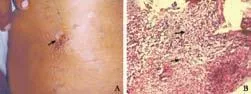
Fig. A: Clinical photograph with arrow depicting the site of persistent port sinus with which the patient presented; B: Photomicrograph demonstrating lymphocytic infiltration and typical granulomas suggestive of tuberculosis.
Discussion
With the prevalence as high as 1.5%,[6]tuberculosis is still rampant in this part of the world. The most common form of extrapulmonary tuberculosis is the involvement of lymph nodes (responsible for 30%-40% of cases).[7]In spite of such a high prevalence, reports of gallbladder tuberculosis from this region have been few. Hepatobiliary tuberculosis is rare and is seen in approximately 1% of all abdominal cases.[4]The normal gallbladder has an unusually high resistance to tubercular infection due to presence of inhibitory factors in the bile.[4,5]The presence of an underlying pathology in the form of cholelithiasis or cystic duct obstruction is said to be essential for the development of gallbladder tuberculosis.[2,4,6]The route of infection can be lymphatic (adjacent caseating lymph nodes), canalicular (ascending directly through an inflamed bile duct) or hematogenous.[1-3,5]More often the clinical features are not vague and non-specific; they include anorexia, fever, weight loss, abdominal pain, diarrhea or jaundice. Features pertaining to chronic cholecystitis due to cholelithiasis like upper gastric discomfort, dyspepsia, vomiting and right upper abdomen pain do not make the life of the clinician easier.[1-3]Various imaging findings have been described, the most common being an abnormally thick-walled gallbladder with underlying cholelithiasis.[4]Besides this, an intraluminal mass simulating a gallbladder carcinoma,[5]acute cholecystitis,[2]intrahepatic biliomas[1]or a multiloculated gallbladder mass have also been reported.[7]The presence of portal, mesenteric and retroperitoneal lymphadenopathy, mesenteric thickening, and ascites support the diagnosis of tuberculosis.[2,4]Although imaging with ultrasound and computed tomography may provide important information, the final diagnosis is usually accomplished by demonstration of caseating granulomas on microscopy and isolation of M. tuberculosis.[8]When detection of the mycobacterium is not possible in difficult cases, employment of the PCR method is a reasonable approach.[8]The treatment when there are gallstones consists of cholecystectomy followed by antitubercular chemotherapy.[6]The diagnosis of gallbladder tuberculosis is often not suspected prior to surgery or biopsy.
In our case, it was unusual as there was no history and her examination was also normal. Ultrasound of the abdomen as a part of the preoperative workup revealed only multiple stones in the gallbladder. Presentation as a persistent port site sinus has not been reported. It was fortunate that the patient presented within the time frame in which the histopathology report usually arrives at our center; that led us to think along the lines of tuberculosis. In our case, there were stones in the gallbladder which may have caused the development of tuberculosis. To our knowledge, such a presentation of gallbladder tuberculosis has not been described. Furthermore, it underlines the role of diligent histopathological examination of resected gallbladder specimens.
Funding: None.
Ethical approval: Not needed.
Contributors: KRA wrote the first draft of this report. All authors contributed to the intellectual context and approved the final version. KRA is the guarantor.
Competing interest:No benefits in any form have been received or will be received from a commercial party related directly or indirectly to the subject of this article.
1 Yu R, Liu Y. Gallbladder tuberculosis: case report. Chin Med J (Engl) 2002;115:1259-1261.
2 Abu-Zidan FM, Zayat I. Gallbladder tuberculosis (case report and review of the literature). Hepatogastroenterology 1999;46:2804-2806.
3 Alvarez SZ. Hepatobiliary tuberculosis. J Gastroenterol Hepatol 1998;13:833-839.
4 Goyal SC, Goyal R, Malhotra V, Kaushik K. Tuberculosis of the gall bladder. Indian J Gastroenterol 1998;17:108.
5 Tanwani R, Sharma D, Chandrakar SK. Tuberculosis of gallbladder without associated gallstones or cystic duct obstruction. Indian J Surg 2005;13:1001-1003.
6 Tauro LF, Martis JJ, Shenoy HD. Tuberculosis of gall bladder presenting as empyema. Saudi J Gastroenterol 2008;14:101.
7 Gulati MS, Seith A, Paul SB. Gallbladder tuberculosis presenting as a multiloculated cystic mass on CT. Indian J Radiol Imaging 2002;12:237-238.
8 Amarapurkar DN, Patel ND, Amarapurkar AD. Hepatobiliary tuberculosis in western India. Indian J Pathol Microbiol 2008; 51:175-181.
Received January 20, 2010
Accepted after revision September 9, 2010
Author Affiliations: Department of Surgery, Jawaharlal Nehru Medical College Hospital, Aligarh Muslim University, Aligarh, India (Mansoor T, Rizvi SAA and Khan RA)
Rizwan Ahmad Khan, MD, 4/817-F, S. S. Nagar, Nagla Road, Aligarh, U.P., India (Tel: +91 9410210281; Email: drrizwanahmadkhan@ yahoo.co.in)
© 2011, Hepatobiliary Pancreat Dis Int. All rights reserved.
杂志排行

Hepatobiliary & Pancreatic Diseases International的其它文章
- Hepatobiliary & Pancreatic Diseases International (HBPD INT)
- Iron overload and HFE gene mutations in Polish patients with liver cirrhosis
- Cytokine and apoptosis gene polymorphisms influence the outcome of hepatitis C virus infection
- Emergency re-routing of anterior sector venous outflow for right lobe living donor liver transplantation including the middle hepatic vein
- Long-term survival of a patient after resection of a gastrointestinal stromal tumor arising from the pancreas
- Predictive value and main determinants of abnormal features of intraoperative cholangiography during cholecystectomy