Tilianin extracted from Xiangqinglan (Herba Dracocephali Moldovicae) inhibits apoptosis induced by mitochondrial pathway and endoplasmic reticulum stress in H9c2 cells after oxygenglucose deprivation/reoxygenation
2023-02-15JIANGWenZHANGWeiZHANGYuxiangYANGHaoPANXiaomeiCHENQiangCHENJunhui
JIANG Wen,ZHANG Wei,ZHANG Yuxiang,YANG Hao,PAN Xiaomei,CHEN Qiang,CHEN Junhui
JIANG Wen,YANG Hao,PAN Xiaomei,CHEN Qiang,CHEN Junhui,Department of Pharmacy,the Sixth Affiliated Hospital of Xinjiang Medical University,Urumqi 830000,China
ZHANG Wei,Department of Anesthesiology,the First Affiliated Hospital of Xinjiang Medical University,Urumqi 830000,China
ZHANG Yuxiang,Department of Laboratory,the Sixth Affiliated Hospital of Xinjiang Medical University,Urumqi 830000,China
Abstract OBJECTIVE: To investigate the efficacy of tilianin extracted from Xiangqinglan (Herba Dracocephali Moldovicae) on apoptosis of H9c2 cell after oxygenglucose deprivation/reoxygenation (OGD/R) and the mechanism.METHODS: Tilianin was obtained from Beijing Inluck Science and Technology Development Co.Ltd.,with purity ≥ 98%.The OGD/R model was established in H9c2 cells.Flow cytometry detected the mitochondrial membrane potential,apoptosis rates,mitochondrial reactive oxygen species (ROS) and calcium ion concentration.Succinate dehydrogenase (SDH) activity,succinate content and levels of tumor necrosis factor-α(TNF-α),interleukin-6 (IL-6) and interleukin-1 β (IL-1β)were detected with enzyme-linked immunosorbent assay.Western blot measured protein levels.RESULTS: Tilianin significantly reduced the apoptotic rates,ROS levels,calcium ion concentration,succinate content,and,levels of TNF-α,IL-6 and IL-1β of OGD/R cells,while significantly increased the membrane potential and SDH activity in mitochondria.Western blot analysis showed that tilianin significantly up-regulated p-Calmodulin-dependent protein kinase Ⅱ and voltagedependent anion selective channel levels in OGD/R cells,while significantly down-regulated p-protein kinase B,Bcl-2-associated X,and dynamin-related protein 1 levels related to apoptosis in the mitochondrial pathway.Moreover,tilianin significantly up-regulated B-cell lymphoma-2 and mitochondrial protein 2 related to the inhibition of apoptosis.Furthermore,tilianin downregulated phosphorylated-apoptosis signal-regulated kinase 1,phosphorylated-p38 and C/EBP homologous protein related to endoplasmic reticulum stress.CONCLUSIONS: Tilianin may inhibit OGD/R-induced H9c2 cell apoptosis mediated by mitochondrial pathway and endoplasmic reticulum stress,thus protecting cardiomyocytes.
Keywords: apoptosis;mitochondria;endoplasmic reticulum stress;tilianin;oxygen-glucose deprivation/reoxygenation
1.INTRODUCTION
Globally,cardiovascular disease is still the most common cause of death,and the myocardial ischemiareperfusion injury is a common risk factor leading to impaired heart function.However,the exact pathogenesis of myocardial ischemia-reperfusion injury is still unclear.It has been shown that the mitochondrial dysfunction and endoplasmic reticulum stress can promote cardiomyocyte apoptosis and aggravate the process of myocardial ischemia-reperfusion injury.1Mitochondrial dysfunction in cardiomyocytes can induce abnormal production of reactive oxygen species (ROS)and intracellular calcium overload,which are important factors leading to myocardial ischemia-reperfusion injury.2Calmodulin-dependent protein kinase II(Calcium/CaMKII) has been considered to be a vicious mediator of cardiac ischemic injury,which participates in the regulation of endogenous injury of myocardial cells during myocardial ischemia-reperfusion.3The apoptosis mediated by the mitochondrial pathway can exacerbate heart diseases.4In addition,endoplasmic reticulum stress has also been involved in cardiomyocyte apoptosis,and myocardial ischemia-reperfusion injury.It has been shown that the inhibiting endoplasmic reticulum stress-induced apoptosis can protect the heart and attenuate the myocardial ischemia-reperfusion injury.5Moreover,the endoplasmic reticulum stress can also activate the Nod-like receptor protein 1 inflammasome by activating nuclear factor kappa-B (NF-κB)pathway and participating in the development of myocardial ischemia-reperfusion injury.6Therefore,regulating mitochondrial function and endoplasmic reticulum stress can inhibit cell apoptosis and help improve myocardial ischemia-reperfusion injury.
The dry aerial part of Xiangqinglan (Herba Dracocephali Moldovicae) has good effects on improving cardiovascular diseases.7The highest content in the total flavonoids of Xiangqinglan (Herba Dracocephali Moldovicae) is tilianin.Studies have shown that tilianin mainly exert anti-inflammatory and antioxidant effects.8,9In a previous study from our lab,talianin has been used to interven myocardial ischemia-reperfusion injuries in rats,which has been shown to be able to mediate the phosphatidylinositol 3 kinase/protein kinase B (PI3K/ AKT) signaling pathway to reduce the protein expression levels of myocardial HtrA2/Omi,Smac/Diablo and BCL2-Associated X (Bax),and to increase the expression levels of B-cell lymphoma-2(Bcl-2) and X-linked inhibitor-of-apoptosis protein in myocardium.10Therefore,we speculate that tilianin may participate in the apoptosis signaling pathway in cardiomyocytes to improve myocardial ischemiareperfusion injury.There are three main apoptosis pathways,including the exogenous pathway (death receptor pathway),endo-plasmic reticulum pathway,and endogenous pathway (mitochondrial pathway).The latter is the most important pathway,which has been called as the executor of the apoptosis process.
Herein,the protective effects of tilianin on injury induced by oxygen-glucose deprivation/reoxygenation (OGD/R)in H9c2 cells were explored.The tilianin was used to interfere with the OGD/R H9c2 cell models,and its effects on mitochondrial function,mitochondrial pathways and endoplasmic reticulum stress,which might lead to cardiomyocyte apoptosis,were analyzed.These findings would help the development of drugs to protect myocardial damages in clinic.
2.METHODS
2.1.Cell model establishment,grouping and treatment
The H9c2 rat cardiomyocytes were provided by the Jiangsu Kaiji Biotechnology Co.,Ltd.,Nanjing,Jiangsu,China.The cellular model was established with the OGD/R induction.Briefly,the H9c2 cells were inoculated onto the 24-well plate,at the density of 1 ×10⁵cells/well,and cultured for 24 h before changing the culture medium.DMEM medium (Gibco,GrandIsland,NY,USA) without serum and glucose was used to culture the cells under hypoxia (95% N2,5% CO2) for 3,6,and 12 h,respectively.For the reoxygenation,fresh DMEM complete medium was used to continue the cell culture for 3 h.The optimal hypoxia duration and drug treatment concentration for model establishment were determined.
The following groups were set up: (a) the control group,in which the cells were normally cultured,without any drug treatment;(b) the OGD/R model group,in which the cells were cultured under normal conditions for 6 h,and under hypoxia in serum-free and glucose-free medium for 6 h,following by culturing under normal reoxygenation for 3 h;and (c) the model+tilianin group,in which the cells were cultured under normal culture condition,with tilianin (1,3,10,20,and 30 µM) (purity≥ 98%;Beijing Inluck Science and Technology Development Co.,Ltd.,Beijing,China) pre-intervention for 6 h,followed by the culture with serum-free and glucose-free culture medium of hypoxia for 6 h(maintaining the drug concentration),and the subsequent normal reoxygenation culture for 3 h (maintaining the drug concentration).
2.2.Observation of cell morphology
The growth conditions of different groups of cells were observed under a fluorescent inverted microscope(Eclipse TS100-F;Nikon,Tokyo,Japan),such as the cellular morphology and density,with 100 ×magnification.Pictures were taken and recorded.
2.3.Cell viability assessment
CCK-8 kit (Transgene Biotech.,Beijing,China) was used to assess the cell viability.The OGD/R-induced H9c2 cells were treated as above described.Totally 100 μL prepared 10% CCK-8 was added for incubation.After 1 h,the OD450 nm was measured on xMarkTM(Bio-Rad,Richmond,CA,USA),and the cell viabilities were calculated accordingly.
2.4.Lactate dehydrogenase (LDH) activity assessment
The assessment of LDH activity was performed in accordance with the instructions of the LDH Assay Kit.Totally 20 μL OGD/R-induced H9c2 cell supernatant was collected from each group for measurement of LDH activity.The absorbance value was measured,and the LDH values were calculated.
2.5.Flow cytometry analysis of apoptosis
After drug intervention,the cells were collected and resuspended with 500 μL 1 × Binding Buffer.Then,5 μL Annexin V-PE (BD,Franklin Lakes,NJ,USA) and 10 μL 7-AAD (BD) were added and incubated at 4 ℃ in dark for 30 min.Cell apoptosis was analyzed on a flow cytometer (LSRFortessa;BD,Franklin Lakes,NJ,USA).
2.6.Detection of mitochondrial membrane potential
After washing,the collected cells were re-suspended and treated with 500 μL Rhodamine 123 (10 μM;Sigma,St.Louis,MO,USA) in a 37 ℃ incubator for 30 min.After washing,the fluorescence intensity was detected with a flow cytometer (LSRFortessa;BD,Franklin Lakes,NJ,USA).
2.7.Mitochondrial ROS detection
After cell collection and washing with PBS,the cells were incubated with 1 mL MitoSOX Red (Thermo Fisher Scientific;St.Walsam,MA,USA),at a final concentration of 5 μM,at 37 ℃ for 10 min.Then,the cells were washed and re-suspended with 500 μL PBS,and the fluorescence intensity was detected with a flow cytometer (LSRFortessa;BD,Franklin Lakes,NJ,USA).
2.8.Calcium ion concentration detection
After cell collection and washing,the cells were incubated with 1 mL Fluo-3/AM (Solarbio,Beijing,China),at a final concentration of 5 μM,in a 37 ℃incubator for 15 min.After washing with pre-cooled PBS,the cells were re-suspended with 500 μL PBS,and the fluorescence intensity was detected with a flow cytometer (LSRFortessa;BD,Franklin Lakes,NJ,USA).
2.9.Enzyme-linked immunosorbent assay (ELISA)
After drug intervention,the mitochondria were extracted.The succinate dehydrogenase (SDH) activity was detected with succinate dehydrogenase activity colorimetric assay kit (Sigma;St.Louis,MO,USA).The cell supernatant was collected,and the succinate concentration was determined with succinate colorimetric assay kit (Sigma;St.Louis,MO,USA).The ELISA kits for tumor necrosis factor α (TNF-α) (Beijing Solarbio Science &Technology Co.,Ltd.,Beijing,China),interleukin-6 (IL-6) (Beijing Solarbio Science &Technology Co.,Ltd.,Beijing,China) and the interleukin-1β (IL-1β) (Elabscience Biotechnology Co.,Ltd.,Wuhan,China) were used to detect the levels of TNF-α,IL-6 and IL-1β in the supernatant,respectively.
2.10.Western blot
Cells were collected and lysed with RIPA (Boster,Wuhan,China) at 4 ℃ for 60 min.After centrifugation,the supernatant was collected.After determining the protein concentrations,the protein sample was separated by SDS-PAGE and electronically transferred onto PVDF membrane (Millipore;St.Boston,MA,USA).After blocking,antibody incubation with primary antibodies,including β-actin (1:800),ox-CaMKII (1:500),p-CaMKII (1:1000),protein kinase B (AKT) (1:500),p-AKT (1:500),Bcl-2 (1:500),apoptosis signal-regulated kinase 1 (ASK1) (1:500),p-ASK1 (1:500),p38 (1:800),p-p38 (1:800),C/EBP homologous protein (CHOP)(1:1000),dynein-related protein 1 (DRP1) (1:500),voltage dependent anion channel (VDAC) (1:1000),mitochondrial protein 2 (MFN2) (1:1000),and Bax(1:500) (Abcam,Cambridge,MA,USA),was performed at 4 ℃ overnight.Then,goat anti-rabbit IgG H &L (HRP)(1:5000;Abcam,Cambridge,MA,USA) (except for βactin and Bax) was added and incubated at room temperature for 1 h.For β-actin and Bax,the goat antirabbit IgG H &L (HRP) (1:15000;Abcam,Cambridge,MA,USA) and goat anti-mouse IgG H&L (HRP)(1:5000;Abcam,Cambridge,MA,USA) were used,respectively.The color development was performed with enhanced chemiluminescence (Thermo Fisher,Waltham,MA,USA).The protein bands were observed with the AlphaView SA version 3.4.0.0 software (ProteinSimple,San Jose,CA,USA).The internal reference was β-actin.
2.11.Statistical analysis
Data were expressed as mean ± standard deviation ().Statistical analysis was conducted with the SPSS 19.0 software (IBM Corp.,Armonk,NY,USA).After normal distribution test,the multiple comparisons were conducted with the analysis of variance followed by Student-Newmnan-Keuls post-hoc test.Statistically significant difference was determined atP< 0.05.
3.RESULTS
3.1.Determination of optimal hypoxia duration of H9c2 cell model induced by OGD/R
The H9c2 cells were induced with OGD/R for different periods,and the cell survival rate and LDH activity were detected.Our results showed that,compared with the control group,the survival rate (Table 1) and LDH activity (Table 2) of cells in the model group after 3-h hypoxia/ 3-h reoxygenation were not significantly changed than control group (P> 0.05).However,the conditions of 12-h hypoxia/3-h reoxygenation,as well as the 6-h hypoxia/3-h reoxygenation,significantly changed the cell survival rate and LDH activity (P<0.05).Based on the overall findings,the condition of 6-h hypoxia/3-h reoxygenation was selected as the optimal treatment duration for the OGD/R model of subsequent experiments.
3.2.Cell morphology
The cells of each group were observed under an inverted microscope.As shown in Figure 1A,the cells in the control group were intact,with smooth edges and fibrous extensions.After the OGD/R treatment,the number of cells in model group was sharply reduced,and the cells were slightly shrunk,with debris.Moreover,the number and morphology of cells were obviously improved by 1 and 3 μM tilianin,but not by 10,20 and 30 μM tilianin.
3.3.Determination of optimal dosage of tilianin treatment on OGD/R-induced H9c2 cells
The model cells were intervened with different doses of tilanin,and the cell survival rate and LDH activity were detected.Our results showed that,the model group had significantly lower cell survival rate (Table 3),while significantly higher LDH activity (Table 3) than control group (P< 0.05).The cell survival rates of the cells treated with 1 and 3 μM tilianin were significantly increased,whereas the LDH activities were significantly reduced than model group (P< 0.05).Compared with the 1 μM tilianin group,the cell survival rate of the 3 μM tilianin treatment group did not significantly changed,while the LDH activity increased significantly (P< 0.05).Based on these findings,the 1 μM tilianin was selected as the optimal dosage for the following experiments.
3.4.Effect of tilianin on cellular apoptosis
To investigate effects of tilianin on cellular apoptosis,flow cytometry was performed.Our results showed that,the model group had significantly elevated apoptosis rate than control group (P< 0.05) (Figure 1B).However,the apoptosis rate of the cells treated with 1 μM tilianin was significantly reduced than model group (P< 0.05).
3.5.Effect of tilianin on cell mitochondrial membrane potential,ROS activity and mitochondrial calcium ion concentration
Flow cytometry detected the effects of tilianin on the mitochondrial membrane potential,ROS activity and mitochondrial calcium ion concentration.Our results showed that,model group had significantly lower mitochondrial membrane potential (Figure 2A) and higher ROS activity (Figure 2B) (bothP< 0.05) than control group.However,the cell mitochondrial membrane potential was significantly increased,while the ROS activity was significantly decreased,in the 1-μM tilianin intervention group than model group (P< 0.05).Additionally,model group had significantly higher fluorescence intensity of mitochondrial calcium ion than control group (P< 0.05) (Figure 2C).However,the fluorescence intensity of mitochondrial calcium ion in the 1 μM tilianin intervention group was significantly reduced than model group (P< 0.05) (Figure 2C).
3.6.Effect of tilianin on ox-CaMKII,p-CaMKII,and VDAC protein expression levels
The expression levels of proteins were measured with Western blot analysis.Our results showed that,model group had significantly up-regulated protein levels of p-CaMKII and VDAC than control group (P< 0.05)(Figure 3).However,their levels in the 1 μM tilianin intervention group were significantly down-regulated than model group (P <0.05) (Figure 3).
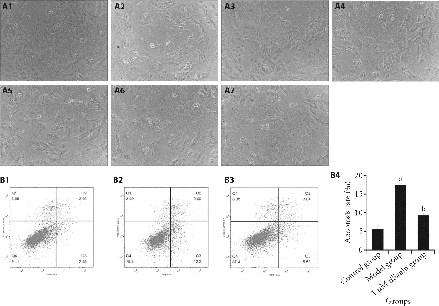
Figure 1 Effect of tilianin on cell morphology and cellular apoptosis
3.7.Effect of tilianin on SDH activity and succinate content in cellular mitochondria
The mitochondrial SDH activity and succinate contents were detected by ELISA.The SDH activity of the modelgroup was significantly lower (Table 4),and the succinate content was significantly increased than control group (Table 4) (P <0.05).However,there was no significant changes in SDH activity between the 1 μM tilianin intervention group and model group,while the succinate content in 1 μM tilianin intervention group was significantly reduced (P< 0.05) (Table 4).
Table 1 Effects of different hypoxia time on the survival of OGD/R-induced H9C2 cell model ()
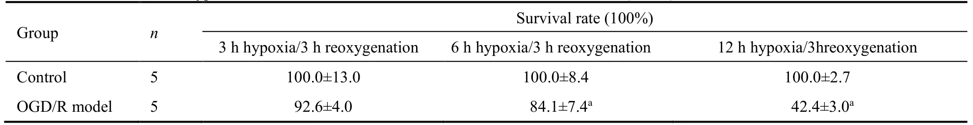
Table 1 Effects of different hypoxia time on the survival of OGD/R-induced H9C2 cell model ()
Notes: control: in which the cells were normally cultured,without any drug treatment;OGD/R model: in which the cells were cultured under normal conditions for 6 h,and under hypoxia in serum-free and glucose-free medium for 6 h,following by culturing under normal reoxygenation for 3 h.OGD/R: oxygen-glucose deprivation/reoxygenation.Compared with control,aP < 0.05.
Table 2 Effects of different hypoxia time on LDH activity of OGD/R-induced H9C2 cell model ()
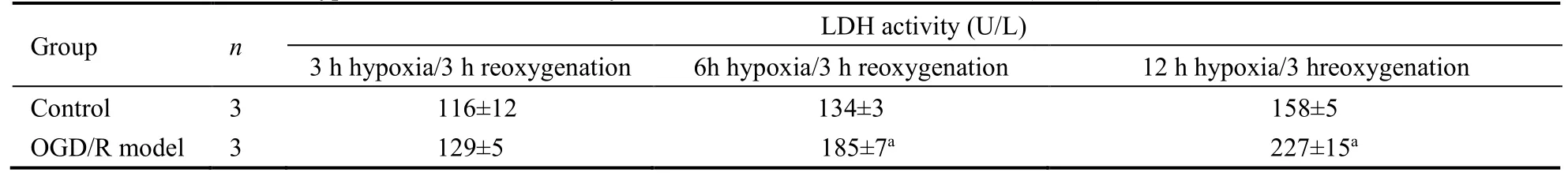
Table 2 Effects of different hypoxia time on LDH activity of OGD/R-induced H9C2 cell model ()
Notes: control: in which the cells were normally cultured,without any drug treatment;OGD/R model: in which the cells were cultured under normal conditions for 6 h,and under hypoxia in serum-free and glucose-free medium for 6 h,following by culturing under normal reoxygenation for 3 h.OGD/R: oxygen-glucose deprivation/reoxygenation.Compared with control,aP < 0.01.
Table 3 Effect of tilianin on the survival and LDH activity in OGD/R-induced H9C2 cell model ()

Table 3 Effect of tilianin on the survival and LDH activity in OGD/R-induced H9C2 cell model ()
Notes: control: in which the cells were normally cultured,without any drug treatment;OGD/R model: in which the cells were cultured under normal conditions for 6 h,and under hypoxia in serum-free and glucose-free medium for 6 h,following by culturing under normal reoxygenation for 3 h.OGD/R: oxygen-glucose deprivation/reoxygenation;1 μM tilianin,3 μM tilianin,10 μM tilianin,20 μM tilianin,30 μM tilianin: in which the cells were cultured under normal culture condition,with tilianin (1,3,10,20,and 30 µM) pre-intervention for 6 h,followed by the culture with serumfree and glucose-free culture medium of hypoxia for 6 h (maintaining the drug concentration),and the subsequent normal reoxygenation culture for 3 h (maintaining the drug concentration).OGD/R: oxygen-glucose deprivation/reoxygenation.Compared with control group,aP < 0.01;compared with OGD/R model group,bP < 0.01;compared with 1 μM tilianin group,cP < 0.05.

Table 4 Analysis of mitochondrial SDH activity and succinate contents
3.8.Analysis of proteins of the mitochondrial mediated apoptosis pathway
The levels of apoptosis-related proteins in the mitochondrial pathway were then detected with Western blot.As shown in Figure 4A,the model group had significantly up-regulated levels of p-AKT,Bax,and DRP1,while significantly down-regulated levels of Bcl-2 and MFN2 than control group (P< 0.05).The protein levels of p-AKT,Bax,and DRP1 in the 1 μM tilianin intervention group were significantly down-regulated,while those of Bcl-2 and MFN2 were significantly upregulated than model group (P< 0.05) (Figure 4A).
3.9.Analysis of endoplasmic reticulum stress-related proteins
Western blot also detected the levels of endoplasmic reticulum stress-related proteins.The protein expression levels of p-ASK1,p-p38,and CHOP in model group were up-regulated significantly than control group (P<0.05) (Figure 4B).However,their levels in the 1 μM tilianin intervention group were significantly downregulated than model group (P< 0.05) (Figure 4B).
3.10.Effect of tilianin on inflammatory factors in culture supernatant
The levels of TNF-α,IL-1β and IL-6 in the culture supernatant were detected with ELISA.Compared with the control group,the levels of TNF-α,IL-1β and IL-6 in the model group were significantly increased (P< 0.05)(Figure 4C).Compared with the model group,1 μM tilianin significantly reduced the levels of TNF-α,IL-1β and IL-6 (P< 0.05).
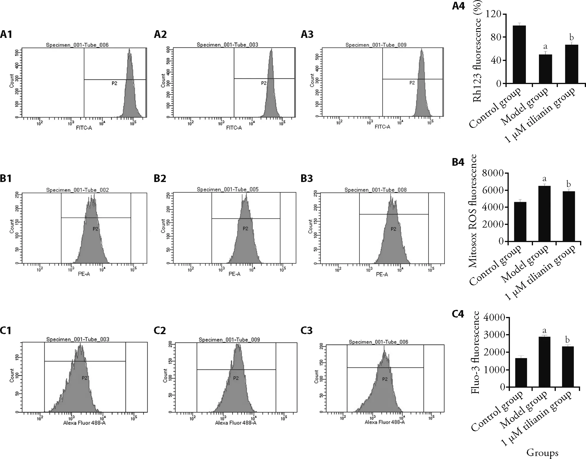
Figure 2 Effect of tilianin on mitochondrial functions
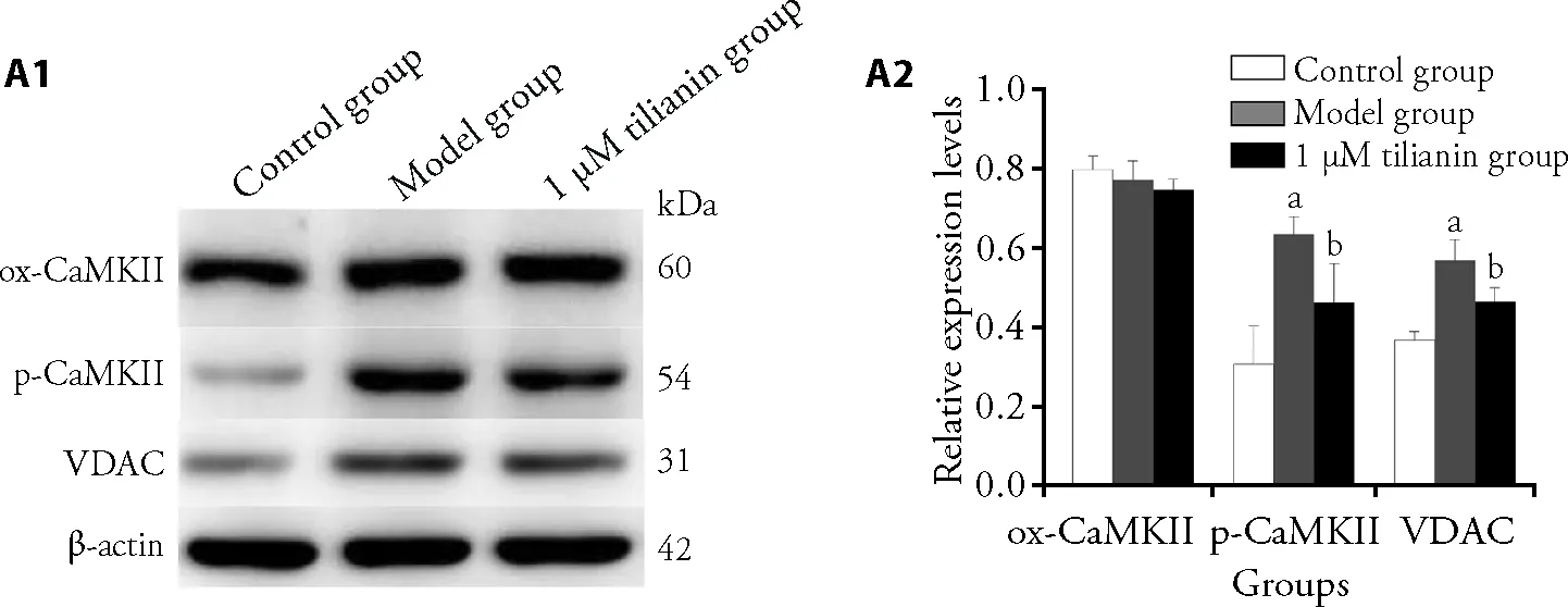
Figure 3 Effects of tilianin on expression levels ox-CaMKII,p-CaMKII,and VDAC

Figure 4 Analysis of expression levels of michondrial pathway-related proteins and endoplasmic reticulum stress-related proteins as well as cytokine levels
4.DISCUSSION
In this study,the H9c2 cell model of OGD/Rin vitrowas established and the effects of tilianin on myocardial ischemia-reperfusion injuries were explored in this cell model.The optimal duration for OGD/R-induced H9c2 cell model was determined as 6-h hypoxia/3-h reoxygenation,and the optimal intervention concentration of tilianin was determined at 1 μM.Our results suggest that tilianin may reduce OGD/R-induced apoptosis of H9c2 cells by targeting mitochondria,thereby protecting cardiomyocytes.
The Ca2+/CaMKII in mitochondria has an extremely important role in Ca2+overload and oxidative stress.11The main factor involved in myocardial ischemiareperfusion injury is the δ subtype CaMKII,which can be activated in two ways: one is the phosphorylationmodified CaMKII generated by autophosphorylation at threonine 287 (p-CaMKII),and the other is the oxidized CaMKII (ox-CaMKII) generated by the reversible oxidation of the CaMKII methionine regulatory domain 281/282.12,13Activated CaMKII can promote the opening of a large number of mitochondrial permeability transition pores,and aggravate the Ca2+overload,and myocardial ischemia-reperfusion injury.14In addition,the Ca2+transport of the outer mitochondrial membrane is mainly achieved through VDAC.As the main permeation pathway of the outer mitochondrial membrane,VDAC participates in the metabolite transport and ion flow.15In addition,VDAC also exxerts important roles various cellular processes,including the ATP generation,Ca2+homeostasis and cellular apoptosis,which therefore is of great significance for maintaining mitochondrial function and cell viability.16In this study,based on the Western blot analysis,our results showed that the tilianin reduced the expression levels of p-CaMKII and VDAC,but did not significantly affect ox-CaMKII.These results demonstrate that tilianin may inhibit mitochondrial ROS levels and calcium ion overload through regulating p-CaMKII and VDAC protein expression levels.
In our previous study with animal models,10our results showed that tilianin may inhibit myocardial cell apoptosis by activating the PI3K/AKT signaling pathway,thus playing a role in protecting against myocardial ischemia-reperfusion injury.The PI3K/AKT signaling pathway can mediate cell survival and apoptosis,which can phosphorylate the glycogen synthase kinase 3β(GSK3β) and other downstream signals to maintain mitochondrial homeostasis and inhibit mitochondrialmediated cellular apoptosis.17The activation of PI3K/AKT/GSK3β pathway can up-regulate the Bcl-2 expression and down-regulate the Bax expression to inhibit cardiomyocyte apoptosis,thereby reducing the area of myocardial infarction.18MFN in the mitochondrial membrane dominates the mitochondrial fusion,while the translocation of DRP1 to the outer mitochondrial membrane would lead to mitochondrial fission.19Myocardial ischemia-reperfusion induces the excessive division and fragmentation of mitochondria,and down-regulates the fusion and mitochondrial phagocytosis,which leads to the release of caspase family proteins and cytochrome C,further resulting in the apoptotic cascade effect.19In the present study,our results showed that OGD/R down-regulated AKT expression in H9c2 cells,leading to up-regulation of Bax and DRP1,as well as down-regulation of Bcl-2 and MFN2,thus promoting cellular apoptosis.Moreover,tilianin inhibited these changes,in line with the findings from flow cytometry.
In addition to endogenous and exogenous pathways,apoptosis could also be induced by the endoplasmic reticulum stress.Endoplasmic reticulum stress reestablishes the endoplasmic reticulum homeostasis through integrated signals called ER-unfolded protein response (UPRER).Prolonged endoplasmic reticulum stress may trigger cell apoptosis by up-regulating CHOP,converting the pro-survival function of UPRER into a lethal signal,which is transmitted to the mitochondria to perform the apoptosis response.20,21In addition,ASK1 can regulate the P38 pathway and is involved in apoptosis induced by endoplasmic reticulum stress.22,23ASK1 can mediate the activation of p38 and exacerbate the development of myocardial ischemia-reperfusion injury.24Our study showed that tilianin inhibited the ASK1/p38 pathway and CHOP expression level,which reduced OGD/R-induced endoplasmic reticulum stress response in H9c2 cells,thereby inhibiting cell apoptosis.Myocardial ischemia-reperfusion can induce the production and release of a variety of pro-inflammatory cytokines and chemokines.25A number of studies have found that the levels of TNF-α,IL-6,IL-1β and MCP-1 are significantly increased in OGD/R model cells.26,27The results suggest that inflammatory factors are involved in myocardial ischemia-reperfusion injury.Tilianin is a representative flavonoid component ofXiangqinglan (Herba Dracocephali Moldovicae)and has anti-inflammatory effects.8,9The anti-inflammatory effect of tilianin on macrophages and VSMCs has been shown to be mainly through down-regulating the TNFα/NF-κB pathway.28Consistently,our results herein showed that tilianin significantly reduced the levels of TNF-α,IL-6 and IL-1βin H9c2 cells induced by OGD/R,indicating that tilianin may protect cardiomyocytes.In conclusion,our results showed that tilianin not only reduced the ROS levels and calcium ions to protect the mitochondria,but also inhibited apoptosis through the mitochondrial and endoplasmic reticulum stress pathways in OGD/R-induced H9c2 cell models.Additionally,tilianin could inhibit the inflammatory responses induced by OGD/R.Our findings suggest that tilianin may serve as a protection factor for myocardial ischemia-reperfusion injuries.Noteworthy,due to lack of appropriate positive control drugs,positive control was not included in this study.The lack of positive control is a limitation of this study.Further studies are warranted to verify the therapeutic effects of tilianin on myocardial ischemia-reperfusion injuries.
杂志排行
Journal of Traditional Chinese Medicine的其它文章
- Effects of the Huangkui capsule (黄葵胶囊) on chronic kidney disease: a systematic review and Meta-analysis
- Effectiveness of moxibustion alone on lumbar disc herniation: a Meta-analysis of randomized controlled trials
- Efficacy of acupuncture therapy for post-stroke fatigue: a systematic review and Meta-analysis
- Efficacy of luteolin on the human gastric cancer cell line MKN45 and underlying mechanism
- Comparing the effects of three decoctions for coronavirus disease 2019 on severe acute respiratory syndrome coronavirus 2-related tolllike receptors-mediated inflammations
- Astragaloside IV ameliorates insulin induced insulin resistance in HepG2 cells through reactive oxygen species mediated c-Jun Nterminal kinase pathway