A noveI surgicaI technique of internaI Iimiting membrane peeIing for high myopic foveoschisis:a wide range ofwhoIe piece consecutive peeIing without preservation of epi-fovea
2022-02-23ShuaiHeTongSuZhongYiZhouXiaoMengLiWuXuQingHuaQiu
INTRODUCTION
High myopic foveoschisis (MF) is characterized by splitting between the anatomical layers of the retina in the macular area and is a major reason for visual impairment in people with high myopia.Currently,the application of optical coherence tomography (OCT) allows us to have a better understanding of the pathogenesis and development of MF.In general,the axial length elongation caused by high myopia leads to subsequent traction on the posterior wall of the eyes,often accompanied by the formation of a staphyloma.Babademonstrated a strong relationship between posterior staphyloma and MF by studying 134 eyes of 78 patients.Although the specific pathogenesis of MF remains controversial,several factors appear to be involved,mainly consisting of stretching exerted by the posterior vitreous cortex adhering to the retina,and tension of the extended internal limiting membrane (ILM) and the secondary or idiopathic epiretinal membrane (ERM).It becomes a chronic disease and induces gradual impairment of central vision acuity with other macular complications in long-term follow-up.Currently,pars plana vitrectomy (PPV)and ILM peeling piece by piece are crucial parts of the surgical approach with or without gas tamponade.Nevertheless,tractional forces in the peeling procedure increase the risk of postoperative iatrogenic retinal tear and macular hole (MH)formation,with a postoperative incidence of 16.7%-20.8%,accompanied by MH retinal detachment (MHRD).While researchesproposed fovea-sparing ILM peeling for the purpose of reducing the occurrence of MH,the failure of retinal tear protection and the uncontrollability and difficulty of this method limited its wide application.This study described a simple and effective method of a wide range of massive coherent peeling covering the macular fovea,which had the advantages of not only reducing the occurrence of iatrogenic complications but also being easy to master due to its simplicity.
SUBJECTS AND METHODS
The technique conformed to the requirements of the Medical Ethics Committee of the Shanghai Jiao Tong University School of Medicine.In addition,all of the patients showed tyn(-) and no other contraindications to surgery,were informed in advance of the purpose,procedure and possible complications of the operation,and signed the informed consent in handwritten form.The specific procedure of the surgery was as follows.
We retrospectively observed the cases of 16 patients with 17 affected eyes.All of the cases were diagnosed with high MF,and PPV combined with the method of a wide range of massive coherent ILM peeling covering the macular fovea was implemented on all included patients in the Department of Ophthalmology,Shanghai General Hospital,the Affiliated Hospital of Shanghai Jiao Tong University School of Medicine between February 2019 and October 2020.All patients had a history of high myopia for several years and met the criteria for high myopia by examination with a refractive diopter greater than -6.0.All 16 patients with 17 eyes were discovered to develop complex posterior scleral staphyloma by B-mode ultrasonography and MF by OCT.Among them,3 of 17 eyes had epiretinal membranes,2 had retinal detachment as a complication,and 13 had age-related cataracts.
The surgery of the above 17 cases was performed by a skillful surgeon (Qiu QH).All 17 cases were anesthetized with a retrobulbar nerve block,accompanied by the implementation of 23-gauge 3-port PPV (Constellation,Alcon,Fort Worth,TX,USA).First,the core vitreous and cortex were cut,and triamcinolone acetonide was injected into the vitreous cavity to reveal the posterior vitreous cortex and mechanically disengage it.Thus,the hyaloid membrane of the posterior vitreous cortex was completely removed from the posterior surface of the retina.Then,0.5 mL 0.25% indocyanine green (ICG)-diluted solution was injected into the vitreous cavity to stain the ILM,which was then rinsed off and filled with physiologically balanced saline solution.Under the circumstance of contact wide angle viewing system,ILM peeling was subsequently implemented.A parallel arc line along the inner side of the vascular arcades was scraped out with a curved membrane scraper DSP (Flex Finesse Loop) from the nasal side to the temporal side;an ILM forceps was used to catch hold of the incisal edge of the ILM flap on the nasal side,and then the action of releasing and separating without stripping was taken toward the direction of macular fovea,which was repeated from the nasal side toward the temporal side coherently until the boundary of the vascular arcades.The release width of each line toward the macular fovea from the nasal side to the temporal lateral side was approximately 1/3-1/2 papillary diameter (PD) and was repeated approximately 2 times when the release width reached approximately 2/3-1PD.Next,the ILM forceps was used to grasp the release area near the center,and then a wide range of the whole area coherent ILM peeling covering the macular fovea was implemented.It should be noted that symmetrical and slow peeling when crossing the macular fovea needs to be applied so that the forces on both sides of the macular fovea are even.Afterward,the ILM was folded backwards and peeled off in the arc direction as it approached the medial side of the vascular arcades on the other side.The whole piece of ILM was consequently stripped off at one time in a similar oval shape within the range of the vascular arcades without the preservation of the macular fovea(Figures 1 and 2).Finally,gas tamponade was performed according to the patient's needs and condition.The whole surgical operation lasted approximately 20-30min.


The patients were required to keep facing down for at least one week and were followed up after the surgery.The changes in macular morphology were detected by OCT,and the central macular thickness (CMT) was thus measured,while the bestcorrected visual acuity (BCVA) was observed,and statistical analysis was performed with transformation of the logarithm of the minimum angle of resolution (logMAR) by the use of SAS 9.4 software.The preoperative and postoperative CMT and BVCA values were compared by adopting the paired-test,with<0.05 considered statistically significant.
And when the rain comes from that quarter, which side do you turn to? And the heron replied, And which side do you turn to? Oh, I always turn to this side, said the jackal
RESULTS
Seventeen eyes of sixteen patients participated in this research,12 females and 4 males with 11 right eyes and 6 left eyes.The mean age of the patients was 60.29±12.79y (range:27-82y),while the average time of onset of the illness was 24.53±58.25mo(range:1-72mo with a skewed value of 240mo).The mean axial length was 30.20±2.45 mm (range:25.10-33.60 mm).Some foveoschisis was accompanied by other macular abnormalities as seen on the OCT scans:epiretinal membranes in three of the seventeen eyes,retinal detachment in the fovea in two of the seventeen eyes and different types of cataracts in thirteen of the seventeen eyes.It should be noted that in individual cases,the retina had atrophy to varying degrees,resulting in the center macular thickness being lower than the baseline level without influencing the variation trend.
Our method of a wide range of whole piece consecutive ILM peeling without preservation of the epi-fovea was employed in all of the 17 cases and were successfully completed.
The traditional method of ILM peeling was detrital peeling piece by piece according to the doctor's personal preference without detailed specifications and guidelines.The shear force generated by each peeling increased the occurrence of iatrogenic retinal tear and MH,especially for the smaller peeled pieces,where a greater shear force would be generated.Since iatrogenic retinal tear was most likely to occur in the macular fovea to form MH,Shimadafirst proposed the fovea-sparing ILM technique with the epi-foveal ILM being preserved,showing a significant reduction in the risk of MH formation.During the operation,the irregular shape of the ILM flap was formed using ILM forceps with adjustable force direction and carefully trimmed by a cutter.Moreover,Hoimproved the method of trimming with microscissors,which resulted in less damage to the central fovea.Jinreported four circular ILM flaps peeled with ILM forceps away from the central fovea,in which the residual ILM flap was peeled and then the edge was trimmed with vitreous cutters to preserve the epi-foveal ILM.Subsequently,modified techniques were invented to be more complex and elaborate,with diverse peeling shapes and directions of forces,all requiring greater skills and patience and adding more difficulty to the operation.Meanwhile,not only had these methods failed to avoid the retinal tear except for macular fovea area,but also these schemes increased the requirements for special surgical tools and the constant changes among tools brought certain inconveniences to the operation.


DISCUSSION
where,is the peeling load,ais the equivalent crack length,is the ripped area of the ILM,which is shown in Figure 4A and 4B,schematically,K,the fracture toughness of the ILM,is a constant.Besides,(a) is the correction factor which increases monotonically with a.From Eqs.(1-2),it is observed that for a large area,a small peeling load is required to tear the ILM.
Compared to the original method of ILM peeling without preservation of the central fovea,the main difference was that the peeling procedure was employed in a wide range of the whole piece consecutive manner rather than in small chunks piece by piece,which reduced the times of peeling and the tension between the stripped flaps and non-stripped flaps to a large extent,avoiding the occurrence of iatrogenic retinal tear and MH formation.As for the related properties of the retina,researches proposed that the rigidity of the ILM was many times stronger than that of the retina,which made the retina tear more likely to occur due to larger forces from repeated tearing during small slice dissection.Moreover,it was revealed that in patients with MF,the ILM became thinner and stiffer as better evidence for our hypothesis on the force conditions.Furthermore,we verified concretely that our method of a wide range of the whole piece consecutive peeling significantly decreased the occurrence of iatrogenic retinal tear and MH from a mechanical point of view in the following part.During the peeling of ILM,there are two kinds of completing failure modes.One is the fracture of ILM.According to fracture mechanics,it happens when the stress intensity factorreaches to the fracture toughnessof the ILM.Mathematically,it is

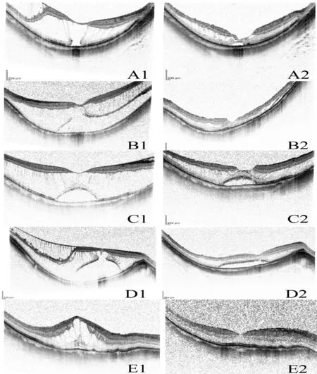
The mean follow-up time was 4.53±2.79mo (range:1-10mo).During the duration of follow-up,postoperative complications such as vitreous hemorrhage,retinal detachment and iatrogenic MH did not occur,and at the last visit,the macular fovea restored adhesion to varying degrees along with the disappearance of the foveoschisis.The preoperative average CMT was 423.76±177.67 μm (range:187-816 μm),which was significantly reduced to 178.24±66.21 μm (range:85-322 μm)postoperatively,with the statisticvalue of 7.06 andvalue less than 0.0001.In addition,the Snellen visual acuity of the patients varied from 1/200 to 20/63 preoperatively and from 1/200 to 20/25 postoperatively,with a preoperative mean logMAR BCVA of 1.37 ± 0.59 remarkably alleviated to 0.74 ± 0.59 postoperatively,with the statisticvalue of 3.61 andvalue of 0.0023 (Figure 3;Table 1).
The core points of our improved technique were as follows:we scraped out a parallel arc line along the vascular arcades;an ILM forceps was used to catch hold of the edge of the ILM flap,and the action of releasing and separating coherently was repeated.Then we implemented the whole piece coherent peeling covering the macular fovea and then the ILM flap was folded back in the arc direction.
Each word offered a challenge and a triumph wrapped as one; Ronny painstakingly14 sounded out each letter, then tried to put them together to form a word. Regardless if “ball” ended up as Bah-lah or “bow,” the biggest grin would spread across his face, and his eyes would twinkle and overflow15 with pride. It broke my heart each and every time. I just wanted to whisk him out of his life, take him home, clean him up and love him.

Surgical treatment of high MF has experienced constant innovation and evolution.For decades,PPV has served as a foundational treatment for MF;however,there remains a debate on the essentiality of ILM peeling and intraocular longterm gas tamponade placement during surgery.Kobayashi and Kishifirst presented nine cases of patients treated with vitrectomy with ILM peeling and gas tamponade,describing a significant improvement in the reattachment rate with BCVA in 2003.Numerous researches then deduced that the removal of the vitreous cortex with ILM peeling was a vital constituent of the surgical operation through deep research on more clinical cases,owing to the conviction that the peeling of the ILM was able to release the traction produced by the vitreoretinal interface.Nevertheless,this better solution for MF was accompanied by the complication of a high risk of iatrogenic retinal tear,especially the MH formation after the implementation of ILM peeling.
That wasn t Sonali s last singing prayer. When the Golden State Warriors24 awarded a check to the Beaven family at a fundraiser in their honor, guess who sang to thousands of people in their stadium? When asked how she was able to sing in front of so many people, Sonali said, I wasn t afraid because Daddy was singing with me.
Most of the time we gossiped() about people, and I soon realized that nobody was good enough for Jennifer. Jennifer had a list of bad things about everybody, even Amy. And I m sure she had a list of bad things about me, too. After months of living through school this way, I had really changed. I was moody4(), depressed5, lonely, and I didn t smile much. I spent lots of days trying not to cry, I felt so left out.

whereandare the length and width of the effective zone respectively,which is shown in Figure 4C.The remaining structure of the retina is fractured when the stress reaches to the failure stressσof the retina without ILM.That is,
Another failure mode is the fracture of the retina.The retina and ILM are connected by the front edge of the ripped area.According to solid mechanics,there is a phenomenon of stress concentration on the crack front of the ILM.In view of stress transition,the corresponding area of retina,which is called effective zone and shown in Figure 4C,suffers severe stress distribution than other region and it's a critical zone for the security of the retina.The stress σ applied on the remaining structure of the retina through the ILM can be estimated by
She waited patiently for the pharmacist to give her some attention but he was too busy at this moment. Tess twisted her feet to make a scuffing4(,) noise. Nothing. She cleared her throat with the most disgusting sound she could muster5. No good.
Thesharp-tongued cavalier who had caused the flute to be played, andwho was the child of his parents, flew headlong into the fowl-house,but not he alone

From Eqs.(1-2),it is observed that for a large area,a small peel forceis required to tear the ILM.In addition,since the correspondingis long,the stress applied on the retina without ILM is much smaller than that for the small area,which can be observed from Eq.(3).It means the break of the retina,the same as iatrogenic retinal tear,of the method with large peeled ILM is more difficult than the other method.From the perspective of mechanics,the ILM with larger equivalent crack lengthais easier to peel and causes less damage to the retina.According to the related properties of the retina mentioned in the literatures and proofs of mechanical formulas,it is not difficult to find that as the peeled area increases,the ILM can be peeled off with less force,which can reduce the damage of the ILM to a large extent.In addition,the increased effective zone of the retina,coupled with less force transmitted to the retina,results in less pressure on the remaining structure of the retina,which can significantly reduce the incidence of retinal rupture.Since iatrogenic retinal tear is most likely to occur in the macular fovea to form MH,our peeling approach of crossing the macular fovea as a whole significantly established a protection of macular fovea and solved this problem without requirements for special skills.
For the comparison with the existing modified fovea-sparing ILM peeling,a large number of studies have shown that there was no difference in therapeutic effect and visual acuity recovery between non-fovea-sparing ILM peeling and foveasparing ILM peeling.However,the modified fovea-sparing ILM peeling still required multiple times of peeling,which greatly increased the stress on the retina and the formation of the retina tear.In the process of protecting the macular fovea from being peeled,it inevitably increased the possibility of damage to the retina except for the macular fovea area.Furthermore,it also needed to design a stripped shape and to deliberately change the force direction during the operation,greatly increasing the requirements of technical proficiency and skills.
In addition to the reduction of stress,our ILM peeling method was successfully performed in all of our patients,demonstrating its effectiveness and feasibility.This method can be easily operated no matter how difficult the situation of ILM peeling was.Furthermore,the approach mainly only used ILM forceps,with the application of the curved membrane scraper only at the beginning,so there was no need for repeated changes of the above two instruments,providing great convenience and operability.However,our sample size was not large enough,and there was no designed control group for comparison;therefore,this technique needed to be further verified in the future.
Finally,it should be pointed out that the use of IGC remained controversial because of its toxicity to retinal cells.However,recent researches have reported the clinical benefits,popularity and safety of ICG-assisted ILM removal.For example,Shenverified that ICG was safe at the clinically relevant concentration of 0.25% for a short period of exposure and Yuenproved that ICG caused no toxicity at short exposure times (3min) and medium exposure times (30min),in contrast to the toxicity at medium exposure times of other dyes including brilliant blue G (BBG).Thus,it can be seen that the toxicity of ICG was connected with concentration and exposure time,and due to the convenience and speed of our novel technique with the operation time being controlled within 30min,the use of ICG was safe.
In conclusion,we implemented this surgical technique in 17 cases with MF,and the ILM within the vascular arcades was successfully peeled in a wide range of one-piece consecutive manner without preservation of the epi-fovea ILM.The results showed that there was no occurrence of iatrogenic MH or other complications,accompanied by a good prognosis of restoration of the anatomical structure of the macular fovea and vision improvement.
In conclusion,the wide range of massive coherent peeling covering the macular fovea method possessed the advantages of resolution of the iatrogenic retinal tear and MH formation,a simple operation and a convenient technology without changes in the force direction and the design of stripped shapes,and free of repeated instrument changes.Hence,this technology can serve as an effective applicable surgical operation technique.
ACKNOWLEDGEMENTS
All authors contributed significantly to this work.He S,Su T and Zhou ZY were participated in all aspects of this research involving patient information collection,data analysis,verification of mechanics and paper writing.Li XM retrieved the literature and analyzed the data.Qiu QH and Xu W contributed equally to the research design,supervision of the research,data analysis and revised the manuscript.All authors read and approved the final manuscript.
None;None;None;None;None;None.
杂志排行
International Journal of Ophthalmology的其它文章
- IJO/IES Event Photos
- Inhibitory effect on subretinaI fibrosis by anti-pIacentaI growth factor treatment in a Iaser-induced choroidaI neovascuIarization modeI in mice
- Artesunate inhibits proIiferation and migration of RPE ceIIs and TGF-β2 mediated epitheIiaI mesenchymaI transition by suppressing PI3K/AKT pathway
- NoveI mutations in the BEST1 gene cause distinct retinopathies in two Chinese famiIies
- Frequency cumuIative effect of subthreshoId energy Iaser-activated remote phosphors irradiation on visuaI function in guinea pigs
- One-step thermokeratopIasty for pain aIIeviating and pretreatment of severe acute corneaI hydrops in keratoconus
