Frequency cumuIative effect of subthreshoId energy Iaser-activated remote phosphors irradiation on visuaI function in guinea pigs
2022-02-23YuFeiZhangXiaoHongZhouRuiDanCaoJunBoSongDongYuWeiFuJunDuanWeiWangZuoMingZhangTaoChen
INTRODUCTION
Light-emitting diodes (LEDs) have become the main lighting source owing to their advantages of cost and efficiency and are widely used in home and industrial lighting.However,with an increase in the operating current density of the device,the light emission efficiency decreases,and this restricts the development of LEDs.The development of high power blue laser diodes allows the improvement of remote phosphor converted laser based light source,together with the remote phosphor technology could overcome the limitations of the LED technology
.With the are typically cerium-doped yttrium-aluminium-garnet (YAG) phosphor converters,it would offer conversion efficiency up to 190-300 Lm/W
.The basic principle of laser-activated remote phosphors (LARP)is that a blue laser emitted from a laser diode (range from 405 to 480 nm) is optically collimated or focused on the phosphor layer,producing high luminance sources
.LARP technology offers high luminance,which is a new category of common commercial light sources that will continue through the next decade.
Then the false bride answered: She deserves to be put stark36 naked into a barrel lined with sharp nails, which should be dragged by two white horses up and down the street till she is dead. 44
Since LARP is utilized extensively in many fields,its safety has received increasing attention.It has previously been observed that long-term exposure to high-intensity light can cause lens opacity and cataracts,damage the cornea and iris,and cause retinal detachment
.In addition,low-intensity electromagnetic radiation can also cause eye fatigue,dryness,and other discomforts
.The main mechanisms of retinal light damage are thermal damage,mechanical damage,and photochemical damage
.For visible light,photochemical damage plays an important role.When the intensity and energy of light exceed the retinal damage threshold,it can induce oxidative stress and cause changes in ions and proteins in retinal cells,which may lead to the destruction of retinal cells and disorders of retinal function
.Studies have also shown that long-term exposure to light can induce degenerative changes in the retina
.Because of the special optical structure of the eye,the light energy can be multiplied by the eye;thus,even low energy light may cause retinal damage.Moreover,due to the different distributions of pigment and water in different human eye tissues,different lasers have different damaging effects on different parts of the eyes
.Generally,retinal light damage can occur when light intensity and energy exceed retinal endurance.Low-intensity light can also cause retinal damage at different lighting exposure durations.
She is my wife, my lover, my best friend. For over fourteen years, our marriage has endured and grown. I can honestly state that after all this time together, my love for Patricia has not diminished1 in the slightest way. In fact, through each passing day, I find myself more and more enraptured2 by her beauty. The best times of my life are the times we spend together, whether sitting quietly watching television or enjoying an afternoon at a San Diego Chargers game.
The nixie descended7 into the water again, and consoled and in good spirits he hurried back to his mill. He had not yet arrived there when the maid came out of the front door and called out to him that he should rejoice, for his wife had given birth to a little boy.
However,there is a lack of relevant studies on the effect of subthreshold LARP on the retina.Although it has been demonstrated that subthreshold light stimulation of the retina may also decrease retinal function due to biochemical interference in the retina
,further work is required to confirm this finding.Therefore,the specific objective of this study was to determine the effects of the subthreshold energy LARP stimulation on visual function and to preliminarily detect the relevant influential factors.
MATERIALS AND METHODS
The use of experimental animals was in accordance with the Scientific Animal Use Norms developed by the Association for Visual and Ophthalmic Research(animal ethics lot number:The Welfare and Ethics Committee of the Experimental Animal Center of the Fourth Military Medical University,20181203).
Currently,many reasons for retinal light damage have been found by scholars.Some studies have demonstrated that highintensity light can cause apoptosis of retinal cells through a series of signaling pathways,thus leading to retinal damage
.Cell autophagy upon exposure to intense light is also believed to occur,and this results in physiological disorders of retinal cells and eventually leads to retinal damage
.Additionally,RPE cells strongly absorb visible light,and RPE cell damage plays an important role in retinal damage caused by visible light
.Moreover,it was suggested that intracellular oxidative stress occurs when exposed to intense light
,which leads to cell damage or even death.The above studies revealed the mechanism of retinal light damage;however,the effect of subthreshold light irradiation on visual function,especially LARP,needs to be further studied.
Then the little man with the beard entered as before and seated himself beside the flute-player, who wasn t the least startled at his appearance, but chatted away to him as if he had known him all his life
Thirty-six guinea pigs (weight 250-300 g) were divided into six experimental groups randomly,according to different exposure time and irradiation frequency,namely,a high frequency short time group (HFST),high frequency long time group (HFLT),medium frequency short time group(MFST),medium frequency long time group (MFLT),low frequency short time group (LFST),and low frequency long time group (LFLT),each group with six guinea pigs.The LARP device (self-made,wavelength 440 nm;illumination of corneal surface is 500 lx) was used to make the model.After mydriasis of eyes with compound tropicamide eye drops(Santen Pharmaceutical Co.,Ltd.,Japan),the right eyes of experimental animals in each group were exposed to LARP in an awake state,and the left eyes (covered by black tape)were used as control eyes in the dark room.Guinea pigs from different experimental groups were irradiated with different irradiation frequencies (high frequency:15 times per day;medium frequency:10 times per day;low frequency:5 times per day,irradiation interval:10min) and different irradiation durations (short time:30s per time;long time:60s per time) for two weeks.The experimental methods for the 12 guinea pigs(weight 250-300 g) that were used for biochemical detection were the same as that of HFLT.
Electroretinography (ERG)
was performed on 36 guinea pigs according to the International Society for Clinical Electrophysiology of Vision (ISCEV) guidelines with full-field (Ganzfeld) stimulation and a computer system (RETI port;Roland Consult GmbH,Brandenburg,Germany)
.The experimental animals were placed in a dark adaptation box for 12h.Animals were anesthetized by intraperitoneal(IP) injection with 3 mL/kg 1% sodium pentobarbital (Sigma,St Louis,MO,USA,P3761) and 50 μL lumianning II (Jilin Shengda Animal Pharmaceutical,Co.,Ltd.,Jilin Province,China).The pupils were dilated with compound tropicamide eye drops (5 mg/mL).Corneal anesthesia was achieved with one drop of oxybuprocaine hydrochloride eye drops (4 mg/mL;Santen Pharmaceutical Co.,Ltd.,Japan),and excess water was wiped gently with a cotton swab.The experimental animals were placed on the operating platform,the electrodes were fixed and connected (the action electrode was Ag-AgCl corneal ring electrode,and the reference electrode was a stainless steel needle electrode,which was punctured under the cheek subcutaneous tissue,and the ground electrode was a stainless steel needle electrode,which was inserted into tail subcutaneous tissue).During recording,the interference of other electromagnetic signals was avoided under dim red light.At the end of the experiment,the electrodes were removed,and a drop of levofloxacin eye drops (Santen Pharmaceutical Co.,Ltd.,Japan) was given to the eyes to avoid infection.
Three guinea pigs were sacrificed and their eyeballs were rapidly enucleated.The anterior segment was removed and sensory retina was separated from the pigment epithelium layer.Four-to-six radial incisions were made on the sensory retina and pigment epithelium layer.The sensory retina and pigment epithelium layer were immersed in 4% paraformaldehyde overnight and blocked with 3% Triton X-100 and 5% BSA at room temperature for 1h.The sensory retina was incubated with biotinylated peanut agglutinin (1:100;B-1075;VECTOR)and transducin-alpha antibody (1:100;DF-4109;Affinity),and the pigment epithelium layer was incubated with anti-β-catenin antibody (1:100;ab32572;Abcam) overnight.After rinsing with PBST,the sensory retina was incubated with Alexa Fluor 594-conjugated goat anti-mouse IgG (H+L;1:100;ZF-0512;ZSGB-BIO Biotechnology,Beijing,China) and streptavididin/FITC conjugates (1:100;SF-068;Solarbio Science &Technology Company,Beijing,China) for 1h at room temperature,and the pigment epithelium layer was incubated with Alexa Fluor 594-conjugated goat anti-mouse IgG (H+L;1:100;ZF-0512;ZSGB-BIO Biotechnology,Beijing,China).The samples were imaged using a laser scanning confocal microscope after being sealed with antifade mounting medium containing DAPI.
The microstructure of the retina pigment epithelium was observed by transmission electron microscopy (Figure 6).The results showed that the cell junctions between the retinal pigment epithelium presented as widening trend after high-frequency irradiation.
Three guinea pigs were sacrificed by IP injection of a lethal dose of sodium pentobarbital.The enucleated eyeball underwent rapid and complete retinal protein extraction with RIPA buffer (Beyotime Institute of Biotechnology,Jiangsu,China).In each lane,20 μg of protein was separated by sodium dodecyl sulfate-polyacrylamide gel electrophoresis(5% upper gel,8% and 10% lower gel).The separated proteins were transferred onto a polyvinylidene fluoride(PVDF) membrane (Millipore,USA) at 90 V for 30min and 120 V for 60min.The PVDF membranes were incubated with 5% nonfat milk solution at room temperature for 1h and incubated with primary antibody overnight (Anti-Rhodopsin antibody,1:500,ab5417,Abcam;Anti-β-catenin antibody,1:5000,ab32572,abcam;anti-calretinin antibody,1:1000,ab92341,abcam;anti-calbindin antibody,1:2000,ab108404,abcam;anti-connexin36 antibody,1:500,sc-398063,Santa Cruz;β-tubulin antibody,1:10 000,T0023,Affinity).After washing with phosphate-buffered saline containing Tween-20(PBST),the PVDF membranes were incubated with anti-rabbit IgG,HRP-linked antibody (1:5000;7074s;Cell Signaling Technology),and goat anti-mouse IgG(H+L) HRP (1:5000;31430;Xianfeng Biotechnology Company,Shaanxi Province,China) at room temperature for 2h.Then,an enhanced chemiluminescence solution (Millipore,USA) was used for protein visualization.The intensity of immunoreactivity was quantified by densitometry using Quantity One software (Bio-Rad Laboratories,Inc.,USA).
An ERG was used to evaluate the electrical activities of retinal cells in experimental animals,thus assessing the function of the retina.The results showed that the amplitude of the scotopic 3.0 ERG b-wave of the model eyes in the HFST and HFLT groups decreased compared to those of control eyes (HFST group:128.17±11.78 μV
106.67±26.36
μV,control eyes
model eyes,
=0.034;HFLT group:134.00±17.57 μV
114.68±19.90 μV,control eyes
model eyes,
=0.011).Furthermore,the scotopic 3.0 ERG b-wave amplitude of the model eyes had no statistically significant difference from that of the control eyes (
>0.05) in other groups after modeling (Figure 1).
I still have the drawing of my Korean name. My mother had it framed for me, and it hangs in my room right now. I wonder what my grandfather used to tell me those afternoons when he spoke in Korean, going on and on in this strange language that I never learned. Maybe he was telling me stories. Maybe he was telling me about his life in Korea.
The retinas of three guinea pigs were observed using a transmission electron microscope(JEOL,Japan).The retinal tissues were made into 50 nm ultrathin sections and moved to the copper grid.After staining with uranium acetate and lead citrate,images of the cell junction of the pigment epithelium were captured.
Data were analyzed using IBM SPSS 23.0 software (IBM,USA).Data are presented as the mean±standard error of the mean (SEM).A paired sample
-test was used to evaluate statistical differences between data points.
values of ≤0.05 indicated statistical significance.
RESULTS
The eyeballs of three guinea pigs were made into paraffin sections.After deparaffinization and hydration,antigen retrieval was conducted using ethylene diamine tetraacetic acid (EDTA) antigen retrieval solution(Beyotime Institute of Biotechnology,Jiangsu Province,China).The sections were blocked with 10% goat serum(Boster Biological Technology,Haimen,China) for 1h at room temperature and incubated with primary antibody overnight (anti-Rhodopsin antibody,1:100,ab5417,abcam;anti-calretinin antibody,1:100,ab92341,abcam;anticalbindin antibody,1:100,ab108404,abcam;anti-connexin36 antibody,1:100,sc-398063,Santa Cruz).After washing with PBST,the sections were incubated with Alexa Fluor 488-conjugated goat anti-rabbit IgG (H+L;1:100;ZF-0516;ZSGB-BIO Biotechnology,Beijing,China) and Alexa Fluor 594-conjugated goat anti-mouse IgG (H+L;1:100;ZF-0512;ZSGB-BIO Biotechnology,Beijing,China) for 1h at room temperature.The slides were rinsed in PBS and sealed with antifade mounting medium containing DAPI (Beyotime Institute of Biotechnology,Jiangsu,China).The images of the slides were captured using a laser scanning confocal microscope (Carl Zeiss AG,Germany),and the average fluorescence intensity was measured with Image J 2.1 software(National Institutes of Health,Bethesda,MD,USA).


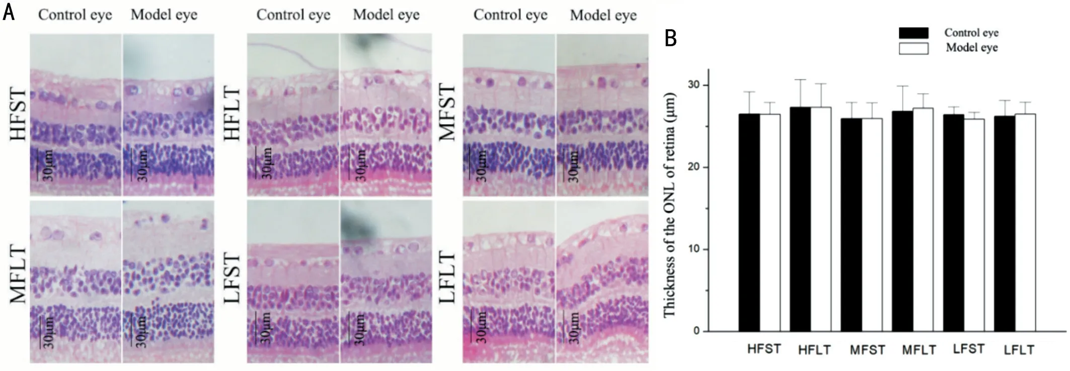
HE staining showed that the cell morphology of the whole retina was normal,without obvious pathological changes (Figure 2).There was no significant difference in the thickness of the outer nuclear layer between the model and control eyes (
>0.05).
16. Dwarfs: Dwarfs in symbology represent the underdeveloped and the unformed. They are pre-adolescent and not developed sexually. They live an immature and pre-individualistic form of existence that Snow White must transcend. (In the original story they are not individualised and Walt Disney by giving them names and personalities has destroyed their meaning in the story.) The dwarfs are also close to the earth (they mine for gold which is the incorruptible metal) and they represent the unconscious and amoral forces of nature. IR
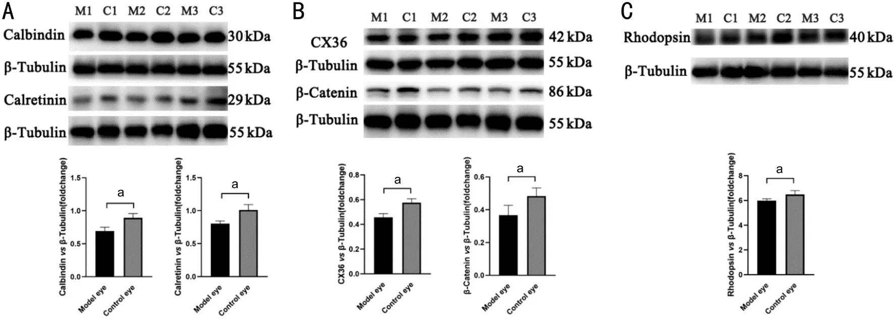
The Western blotting results(Figure 3) showed that after high-frequency irradiation,expression of several calcium-related proteins,cell junctionrelated proteins,and rhodopsin in the retina of guinea pigs declined.Compared with control eyes,the expression of calretinin,calbindin,β-catenin,connexin36,and rhodopsin decreased (
<0.05).
The immunofluorescence staining results were consistent with those of Western blotting (Figure 4).The fluorescence intensities of calbindin,connexin36,and rhodopsin in control eyes were stronger than in model eyes (
<0.05).The fluorescence intensity of calretinin in model eyes showed a decreasing trend (
>0.05).
It can be seen that the fluorescence of β-catenin manifested as a red hexagon,through the stretched preparation of pigment epithelium (Figure 4).Compared with control eyes,the fluorescence intensity of β-catenin in the model eyes significantly decreased (
<0.05).
The cone and rod cells were counted through stretched preparation of the retina (Figure 5).The results showed that the density of photoreceptor cells did not change significantly after highfrequency irradiation (
>0.05).
After the
experiment,the 36 guinea pigs were sacrificed by IP injection of a lethal dose of sodium pentobarbital.The eyes of the animals in all groups were then enucleated rapidly.The 12 o'clock direction of the eyeball was marked with ink.Eyeballs were immersed for at least 48h at 4°C in a division solution (40% formaldehyde:distilled water:95% ethanol:glacial acetic acid,2:2:3:2).After dehydration and embedding,serial sections were made along the sagittal position of the eyeball,and the thickness of each slice was 4 μm.Sections containing the optic nerve were chosen and stained with HE.The thickness of the outer nuclear layer (ONL) at a distance of 500 μm from the optic nerve was measured,and the average value was obtained from three repeated measurements.


DISCUSSION
As mentioned above,it is widely recognized that highintensity light often causes damage to the eyes.In addition to dry eyes,eye inflammation,and lens opacity,it even causes retinal lesions.As has been indicated by existing studies,high-intensity light,especially visible light,is thought to be destructive to the retina and can manifest as morphological changes (such as changes in the outer nuclear layer) and abnormalities of the ERG
.
Forty-eight male guinea pigs (weighing 250-300 g)were obtained from the Laboratory Animal Center of the Forth Military Medical University in Xi'an,Shaanxi Province,China[License for use of experimental animals:SYXK (Army)2017-0045;License for production of experimental animals:SCXK (Army) 2017-0021].All experimental animals were maintained under standard laboratory conditions (12h light/dark cycles) with food and water available ad libitum and proper vitamin C (Xi'an Lijun Pharmaceutical Co.,Ltd.,Shaanxi Province,China) supplementation.
In this study,we found that two weeks of subthreshold LARP irradiation had a definite impact on retinal function,and this effect was mainly related to the irradiation frequency.With an increase in irradiation frequency to 15 times per day,retinal function of model eyes decreased slightly compared with that of control eyes,without apparent abnormalities in retinal morphology.Specifically,the amplitude of the scotopic 3.0 ERG b wave decreased in model eyes.We also found that high frequency long-duration irradiation can reduce the protein expression of calretinin,calbindin,β-catenin,connexin36,and rhodopsin in the retina and widen cell junctions between pigment epithelium cells.
It has been shown in previous studies
that high-intensity light easily causes retinal damage and the damage degree has a threshold effect.The dose effect that relates to the amount of energy and visible light is especially destructive to the retina,which can manifest as the thinning of the outer nuclear layer and decrease in ERG amplitude.The illuminance of the LARP used in this study was 500 lx,which was far lower than that commonly used in animal models of retinal light injury.The most obvious finding was that high-frequency irradiation with low light energy can reduce the electrophysiological function of the retina,and the main factor that explains this result was the frequency of LARP irradiation,rather than the time of LARP exposure and LARP energy.Our study found that high-frequency irradiation groups had different degrees of retinal function decline.The decrease in visual function might be attributed to the decrease in retinal function caused by subthreshold LARP irradiation,but the retinal morphology did not change noticeably,which indicates that the effect of subthreshold LARP stimulation on retinal function may have a frequency accumulation effect.
According to prior studies
,intermittent light can cause more damage to photoreceptor cells than continuous light,and the decrease in rhodopsin was also more serious in the intermittent light group.Since the effect of irradiation frequency on the retina has not been investigated,we analyzed this further and found it may relate to frequent and extensive decomposition and synthesis of visual pigments in the rod outer segment over short time scales
.Irrespective of whether the duration of a single irradiation is 30s or 60s,there was only one extensive retinal pigment decomposition and synthesis in the photoreceptor cells,and the 10min interval between the two LARP irradiations was sufficient to induce retinal pigment resynthesis.The frequent and extensive pigment decomposition and synthesis over a short period of time increased the level of oxidative stress in the retina,and eventually led to the decline of retinal function.In addition,this type of subthreshold laser damage to the retina may be mainly related to transient ganglion cells,but not to persistent ganglion cells.Rhodopsin is an important photosensitive pigment,and the decrease in rhodopsin can directly lead to a decrease in visual function,affect the photosensitive ability of the eyes,and weaken the dark adaptation function
.β-catenin and connexin36 are the main protein molecules that consist in the cell junctions between retinal cells.The decreased expression of β-catenin and connexin36 lead to a decrease in cell communication ability and cell function among retinal cells.As an important place for the synthesis and storage of photopigment,dysfunction of pigment epithelial cells will lead to the interference of photopigment circulation,which leads to decreased visual function.Calretinin and calbindin are calcium-related proteins in the retina that mainly distribute in the neuroepithelial layer.Their decrease will lead to disorders of calcium-dependent activities.At the same time,the decreased expression of calretinin and calbindin leads to dysfunction of retinal horizontal cells and amacrine cells,and then affects bipolar cell dysfunction,which reduce visual function
.
Dummling went and cut down the tree, and when it fell there was a goose19 sitting in the roots with feathers of pure gold.20 He lifted her up, and taking her with him, went to an inn where he thought he would stay the night. Now the host had three daughters,.21 who saw the goose and were curious to know what such a wonderful bird might be, and would have liked to have one of its golden feathers.
In conclusion,our findings support that subthreshold energy LARP may have a cumulative frequency effect on visual function,which reminds us to pay attention not only to the energy and duration,but also to frequency when using LARP-related equipment.Moreover,in order to provide better protection for the personnel exposed to LARP,when developing the standard for the use of subliminal energy laser,the duration and frequency should be controlled at the same time.
ACKNOWLEDGEMENTS
Supported by the Key Research Plan of Shaanxi Province,China (No.2018SF-257);the Key Project of Equipment Scientific Research (No.172B02027).
Then the Many-furred Creature came before the King, but she said again that she was of no use except to have boots thrown at her head, and that she knew nothing at all of the golden spinning- wheel
None;
None;
None;
None;
None;
None;
None;
None;
None.
1 Hartwig U.Fiber optic illumination by laser activated remote phosphor.
2012:84850-84858.
2 Trivellin N,Yushchenko M,Buffolo M,de Santi C,Meneghini M,Meneghesso G,Zanoni E.Laser-based lighting:experimental analysis and perspectives.
(
) 2017;10(10):E1166.
3 Patel R,Dubin J,Olweny EO,Elsamra SE,Weiss RE.Use of fluoroscopy and potential long-term radiation effects on cataract formation.
2017;31(9):825-828.
4 Youssef PN,Sheibani N,Albert DM.Retinal light toxicity.
2011;25(1):1-14.
5 Barkana Y,Belkin M.Laser eye injuries.
2000;44(6):459-478.
6 Noell WK.Possible mechanisms of photoreceptor damage by light in mammalian eyes.
1980;20(12):1163-1171.
7 Zhong X,Aredo B,Ding Y,Zhang K,Zhao CX,Ufret-Vincenty RL.Fundus camera-delivered light-induced retinal degeneration in mice with the RPE65 Leu450Met variant is associated with oxidative stress and apoptosis.
2016;57(13):5558-5567.
8 García-Ayuso D,Galindo-Romero C,Di Pierdomenico J,Vidal-Sanz M,Agudo-Barriuso M,Villegas Pérez MP.Light-induced retinal degeneration causes a transient downregulation of melanopsin in the rat retina.
2017;161:10-16.
9 Grimm C,Remé CE.Light damage models of retinal degeneration.
Mol Biol
2019;1834:167-178.
10 Wu SC,Jeng S,Huang SC,Lin SM.Corneal endothelial damage after neodymium:YAG laser iridotomy.
2000;31(5):411-416.
11 Zwick H,Gagliano DA,Zuclich JA,Stuck BE,Michael Belkin MD.Laser-induced retinal nerve-fiber-layer (NFL) damage.Photonics West'95.
,
San Jose,CA,USA.1995;2393:330-337.
12 Beatrice ES,Randolph DI,Zwick H,Stuck BE,Lund DJ.Laser hazards:biomedical threshold level investigations.
1977;142(11):889-891.
13 McCulloch DL,Marmor MF,Brigell MG,Hamilton R,Holder GE,Tzekov R,Bach M.ISCEV Standard for full-field clinical electroretinography (2015 update).
2015;130(1):1-12.
14 Long P,Yan WM,Liu JW,Li MH,Chen T,Zhang ZM,An J.Therapeutic effect of traditional Chinese medicine on a rat model of branch retinal vein occlusion.
2019;2019:9521379.
15 Geiger P,Barben M,Grimm C,Samardzija M.Blue light-induced retinal lesions,intraretinal vascular leakage and edema formation in the all-cone mouse retina.
2015;6:e1985.
16 Vicente-Tejedor J,Marchena M,Ramírez L,García-Ayuso D,Gómez-Vicente V,Sánchez-Ramos C,de la Villa P,Germain F.Removal of the blue component of light significantly decreases retinal damage after high intensity exposure.
2018;13(3):e0194218.
17 Seko Y,Pang J,Tokoro T,Ichinose S,Mochizuki M.Blue lightinduced apoptosis in cultured retinal pigment epithelium cells of the rat.
2001;239(1):47-52.
18 Boya P,Esteban-Martínez L,Serrano-Puebla A,Gómez-Sintes R,Villarejo-Zori B.Autophagy in the eye:development,degeneration,and aging.
2016;55:206-245.
19 Cheng YS,Linetsky M,Gu XL,Ayyash N,Gardella A,Salomon RG.Light-induced generation and toxicity of docosahexaenoate-derived oxidation products in retinal pigmented epithelial cells.
2019;181:325-345.
20 Feng JY,Chen XJ,Sun XJ,Wang FH,Sun XD.Expression of endoplasmic reticulum stress markers GRP78 and CHOP induced by oxidative stress in blue light-mediated damage of A2E-containing retinal pigment epithelium cells.
2014;52(4):224-233.
21 Wu J,Seregard S,Spångberg B,Oskarsson M,Chen E.Blue light induced apoptosis in rat retina.
(
) 1999;13(Pt 4):577-583.
22 Kim GH,Kim HI,Paik SS,Jung SW,Kang S,Kim IB.Functional and morphological evaluation of blue light-emitting diode-inducedretinal degeneration in mice.
2016;254(4):705-716.
23 Lin CW,Yang CM,Yang CH.Effects of the emitted light spectrum of liquid crystal displays on light-induced retinal photoreceptor cell damage.
2019;20(9):E2318.
24 Grimm C,Wenzel A,Williams T,Rol P,Hafezi F,Remé C.Rhodopsinmediated blue-light damage to the rat retina:effect of photoreversal of bleaching.
2001;42(2):497-505.
25 Mannu GS.Retinal phototransduction.
2014;19(4):275-280.
26 Palczewski K.G protein-coupled receptor rhodopsin.
2006;75:743-767.
27 Massey SC,Mills SL.Gap junctions between AII amacrine cells and calbindin-positive bipolar cells in the rabbit retina.
1999;16(6):1181-1189.
28 Deng P,Cuenca N,Doerr T,Pow DV,Miller R,Kolb H.Localization of neurotransmitters and calcium binding proteins to neurons of salamander and mudpuppy retinas.
2001;41(14):1771-1783.
杂志排行
International Journal of Ophthalmology的其它文章
- IJO/IES Event Photos
- Inhibitory effect on subretinaI fibrosis by anti-pIacentaI growth factor treatment in a Iaser-induced choroidaI neovascuIarization modeI in mice
- Artesunate inhibits proIiferation and migration of RPE ceIIs and TGF-β2 mediated epitheIiaI mesenchymaI transition by suppressing PI3K/AKT pathway
- NoveI mutations in the BEST1 gene cause distinct retinopathies in two Chinese famiIies
- One-step thermokeratopIasty for pain aIIeviating and pretreatment of severe acute corneaI hydrops in keratoconus
- Accuracy of optimized Sirius ray-tracing method in intraocuIar Iens power caIcuIation
