Artesunate inhibits proIiferation and migration of RPE ceIIs and TGF-β2 mediated epitheIiaI mesenchymaI transition by suppressing PI3K/AKT pathway
2022-02-23
INTRODUCTION
Proliferative vitreoretinopathy (PVR) is a common complication of ocular trauma.It can significantly reduce the visual acuity in a short time and seriously affect the quality of life of patients.The main feature of PVR is to form the anterior retinal membrane through the wound repair process.With the formation and contraction of the anterior retinal membrane,the retina will eventually wrinkle and produce tractive retinal detachment,resulting in decreased visual acuity and even blindness
.Surgical repair is the only effective treatment at present.The high recurrence rate makes scholars actively looking for effective drug adjuvant therapy.It is found that the anterior retinal membrane contains many cell types such as retinal pigment epithelium (RPE) cells,fibroblasts and macrophages.RPE cells are considered to be the main participants in the pathophysiological process of PVR
.RPE cells in PVR undergo a procedure which is named epithelial mesenchymal transition (EMT),which involves many changes,such as phenotypic transformation,extracellular matrix (ECM)accumulation and mesenchymal cell marker expression.Studies have shown that the EMT process of RPE cells is a key procedure in the pathogenesis of PVR
.
There may be many molecular mechanisms of EMT regulation in RPE cells.In PVR,RPE cells own the capability of autocrine cytokines.A large quantity of growth drivers show overexpression in vitreous or subretinal effusion in patients who are with PVR.These growth drivers and their acceptors occupy significant position in the existence and occurrence of PVR.After the blood-retina barrier is destroyed,the physiological equilibrium of growth drivers is broken,after which the RPE cells are impulses by growth drivers to produce EMT,immigration and propagation,and then form preretinal membrane with other cells and intercellular substance
.It was found that RPE cells autocrine TGF-β.It can make wound closure,collagen contraction and cell morphology change.The EMT process of RPE cells which are induced occupies significant position in the existence,occurrence and prognosis of PVR.It has been shown that multiple downstream trails Smads,phosphatidylinositol-3-kinase (PI3K) and MAPK can be activated to perform the EMT induction
.Recently,the stimulation of PI3K/Akt trail has also become a vital signal trail regulating EMT.Protein kinase B (Akt) is able to promote the phosphorylation of a large quantity of proteins and regulate cell survival,apoptosis and migration.But PI3K/Akt signal channel is in TGF-β.The function of RPE cell mediated EMT remains to be confirmed.
Artesunate is a derivative of artemisinin,which is extensively applied as an anti-malaria traditional Chinese medicine.Artesunate is extensively applied in clinic because of its good water solubility and various biological activities.Artesunate has the functions of anti-proliferation,anti-fibrosis and immune regulation
.In the laboratory,artesunate is widely used in the treatment of glaucoma,intraocular neovascularization,ocular tumors and uveitis
.Researches have revealed that artesunate is able to restrain the EMT process of alveolar epithelial as well as renal tubular epithelial cells
,but its inhibitory effect on the EMT process of RPE cells is not clear.This experiment examined the effects of artesunate on the propagation,immigration and effect of TGF-β induced RPE cells on EMT process.
MATERIALS AND METHODS
All patients involved in this research were fully aware of the research and the consent forms were offered and signed.This research followed the Declaration of Helsinki and it gained approval from the Ethics Review Committee of Ophthalmic Hospital.The utilization of human organizations confirm to the application rules in Helsinki Declaration.
Then the man persuaded her, and talked so much to her about the wealth that they would have, and what a good thing it would be for herself, that at last she made up her mind to go, and washed and mended all her rags, made herself as smart as she could, and held herself in readiness to set out
But the child grew impatient, and said, Dear mother, how can I cover my father s face when I have no father in this world? I have learnt to say the prayer, Our Father, which art in Heaven, thou hast told me that my father was in Heaven, and was the good God, and how can I know a wild man like this? He is not my father
That afternoon, an old man came over to his wooden shed1 to see him. He wanted one bag full of the black sheep’s wool. Then an old woman came over. She also wanted a bag full of wool. A short while later, a little boy arrived. He also wanted one bag full of wool.
After raised with various densities of artesunate (0,75,150 μmol/L) for 24h,the cells were produced by trypsin method and washed twice with DMEM/F12.For the purpose of detecting cell immigration,the 24 well plate was separated into the upper and lower chambers using an 8 μmol/L cell chamber (Transwell,Corning,USA).The inferior chamber was full of 0.6 mL DMEM/F12 which contained 10% fetal bovine serum.Totally 5×10
cells were suspended in 100 μL DMEM/F12 medium without bovine serum albumin (BSA).After 24h of culture,the cotton swab was utilized to remove the cells on the surface of Transwell membrane.There is 20× objective select the images at random were taken with an inverted microscope (ckx41-f32f,Olympus,Japan) for the purpose of counting stained cells and quantify cell migration ability.
Mark a line on the back of the 6-hole plate,and each hole passes through two straight lines parallel to the edge of the plate and equidistant from the center.When the cells grew to 80%-90% fusion,the cells were digested from the culture dish,centrifuged,discarded the supernatant,added DMEM/F12 media which contained 10% fetal bovine serum,adjusted the cell density to 1×10
/mL,added 2 mL per well into a 6-well plate marked with lines on the back in advance,and then incubated in an incubator at the temperature of 37℃ and 5% CO
saturated humidity.After 24h,the cells were basically attached to the wall and covered the bottom of the 6-well plate.The serum -free medium was used to starve for 24h,20-200 μL gun head was used to make a scratch perpendicular to the bottom of the plate.The wound width was 25 mm.The phosphate buffered saline (PBS) was applied to wash the cells twice,and treated with different concentration of artesunate(0,75,150 μmol/L) without the existence of TGF (20 ng/mL)in a serum-free medium.Incubated in a thermostatic chamber at the temperature of 37℃ and 5% CO
saturated humidity for 36h.At 0,12,24,and 36h,cell immigration was assessed by counting the scratch zone in three regions which are selected at random.
When the cells grew to 80%-90% fusion,the cells were digested from the culture dish,centrifuged,discarded the supernatant,added the cell culture media,after which the cell concentration was adjusted to 1×10
/mL,and inoculated the cells to 96 well plate.After 24h,the control group whose drug concentration was 0 and the experimental group whose concentration was 75,100,150,and 200 μmol/L were added with 100 μL cell culture media whose drug intensity were different,and incubated in the incubator at the temperature of 37℃.The initial culture media was aspirated when the incubation lasted 0,24,48,and 72h,and 10% CCK-8 was incubated for one hour.The absorbance a value of 450 nm wavelength was determined by thermo,USA.The experiment was repeated three times.
The PBS was utilized for the purpose of washing cells and tissues twice (phosphate buffered saline,02-024-1acs,Israel biological industries),and then centrifuged at 12000 rpm for 15min with protease depressor (PMSF,ar1178,boster).The supernatant lysate was collected for bicinchoninic acid (BCA) Protein Quantification Kit (e112-01/02,Nanjing novozan Biotechnology Co.,Ltd.,China).The same quantity of protein was loaded on 10% sodium dodecyl sulfate-polyacrylamide gel electrophoresis (SDS-PAGE) gel(SDS-PAGE gel preparation kit,boster).Totally 5% skimmed milk (skimmed milk powder,BD company,USA) was used to seal the membrane at ambient temperatures in tris buffered saline tween (TBST),rabbit anti-human against PI3K (1:1000,Cell Signaling Technology,Danvers,USA),rabbit anti-human against P-PI3K (1:1000,Cell Signaling Technology,Danvers,MA,USA),rabbit anti-human against Akt (1:1000,Cell Signaling Technology,Danvers,MA,USA),rabbit anti-human against P-Akt.After cleaning with TBST,the membrane was raised with Goat anti-rabbit IgG (ba1054,Wuhan shibode Bioengineering Co.,Ltd.,China) at room temperature for one hour,after which it was scanned on the exposure machine.Then,the imaging software (Image J) was utilized to confirm the full intensity of all detection bands.These tests were reiterated not less than three times.
He promised faithfully to come back to her as soon as he could and begged her to await his return under the lime-tree where they had spent so many happy hours
It is from Shanghai Yubo Biotechnology Co.,Ltd.that the ARPE-19 was acquired and fostered in DMEM/F12 medium which contained 10% fetal bovine serum (04-001-1acs) and 1% penicillin/streptomycin,and raised in thermo (USA) at the temperature of 37℃,and the cells attached completely to the wall after 24h.The cells were fostered until the fusion degree become 80%-90% and starved in medium which was free of serum for one night.DMEM/F12 which contained 1% fetal bovine serum was made use of for the purpose of replacing DMEM/F12.Cells were treated with artesunate which had various concentrations (lot#418A024.Solarbio,China).Cell propagation and migration were examined at different time.The degree of EMT was detected by pretreatment with TGF-β2 (catalog,100-11,lot #081701-1,peprotech) for 48h and artesunate for 48h.
From that day forth2, the girl began a new habit. Her cell phone never shuts down at night. Because she was afraid that she might not be able to hear the phone ring in her sleep, she tried to stay very alert3. As days passed, she became thinner and thinner. Slowly, a gap4 began to form between them.
ARPE-19 cells were fostered on a plate which had 6 wells covering plate for 48h,as described above.Then,PBS was washed twice and 4% paraformaldehyde was fixed at ambient temperatures which lasted 15min.The PBS was applied to wash the fix cells twice,after which they were sealed in a room temperature moist chamber with 10% normal goat serum,and raised with rabbit anti-vimentin (1:200,d21h3,XP,rabbit mAb,and CST) at 4℃ for one night.And the cells were raised with Cy3 labeled Goat anti-rabbit antibodies at ambient temperatures for 1h the next day.The cells were fixed with DAPI,added with anti-fluorescence quenching agent,and observed by laser scanning inverted microscope(ckx41-f32f,Olympus,Japan).
Patients who suffered from cancer,autoimmune illness and other types of severe systemic illness were kept out.PVR-a (
=3),PVR-b (
=3),and PVR-c (
=3)were collected.PVR grading was carried out referring to the standard of the American retinal Nomenclature Committee in 1983.The vitreous bodies were gained from the patients who donate the cornea.
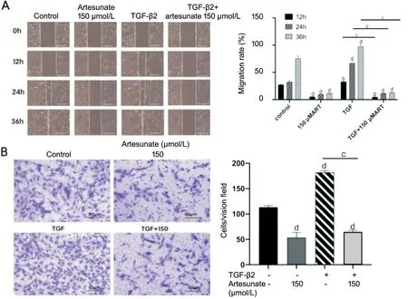
Transwell cell immigration examination was used to verify the effectiveness of artesunate on the vertical immigration of ARPE-19 cells processed with TGF-β2.In comparison with the control group,art treatment group could inhibit the vertical immigration of ARPE-19 cells (Figure 3B).In comparison with the control group,TGF β treatment group promoted the vertical migration of ARPE-19 cells.In comparison with the control group and TGF treated group,100 m artesunate inhibited the vertical migration of ARPE-19 cells handled with TGF-β2.
RESULTS
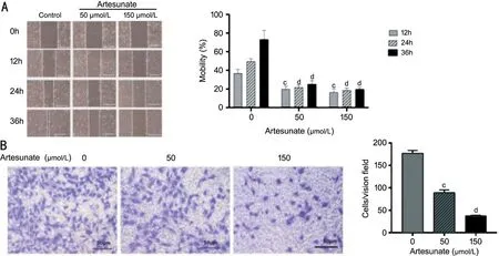
We conducted scratch test and Transwell test
to determine whether artesunate can regulate ARPE-19 cell migration.As shown in the figure (Figure 2),the cells in the scratch test and no drug treatment group showed strong cell migration ability,which almost covered the scratch area after 36h,while the cell migration capability of the drug treatment group was significantly inhibited,and the results were statistically significant and time concentration dependent.The higher the drug concentration,the more obvious the inhibition of ARPE-19 cell migration.Transwell experiment also obtained results which are similar.As 24h passed,the cells in the high concentration artesunate treatment group were much less than those without drug treatment,and the results indicated statistical significance.The two migration tests showed that artesunate disposal obviously suppressed cell migration by a means which was dependent on the dose.
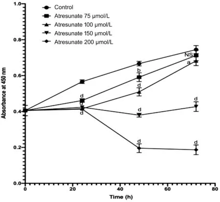
For the purpose of achieving the best effectiveness on ARPE-19 survival rate,CCK-8 experiment was carried out to verify the optimal intensity of artesunate,such as drug concentration and treatment time.Different concentrations of artesunate had different suppression capacity on ARPE-19 cell propagation at different time.As the concentration and time change,the inhibitory capacity was enhanced (Figure 1).In order to achieve 50% cell inhibition without increasing the time of action,the best condition was treated with 150 μmol/L for 48h.
The EMT course of ARPE-19 cells mediated by TGF occupies a significant position in PVR,which involves the propagation,immigration and fibrosis of ARPE-19 cells.Therefore,we used cell scratch test to verify the effectiveness of artesunate on the migration of ARPE-19 treated with TGF-β2.Pretreatment with serum-free media for 24h inhibited cell proliferation and made cell scratch.Pictures were taken at 0,12,24,and 36h.Compared with the control group,artesunate treatment group could inhibit wound healing (Figure 3A);however,TGF-β2 treatment could promote wound healing.When artesunate was added to ARPE-19 cells which were induced by TGF-β2,the wound healing rate was slowed down.By referring to the discovery,our findings indicate that artesunate restrains wound healing in ARPE-19 cells treated with TGF-β2.Since cell propagation and immigration are crucial processes in wound closure,we pretreated ARPE-19 cells with serum-free medium,which reduced the effect of cell proliferation on wound healing.Therefore,these findings revealed that that artesunate inhibited the migration of ARPE-19 cells processed with TGF-β2.
Excel and GraphPad Prism 8 software were utilized for the intention of handling and investigating the experimental data.Two-independent specimen
-test was utilized for comparison between two groups,and one-way ANOVA was applied to perform the comparison for the results of multiple groups.A value of
<0.05 showed statistical significance.
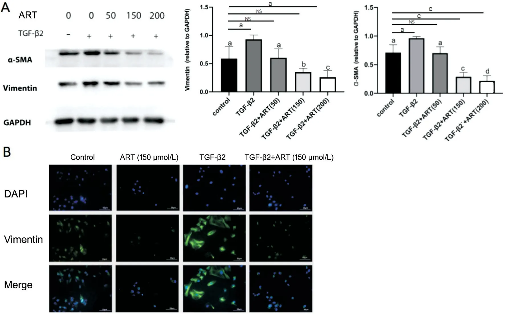
In order to examine the inhibitive ability of artesunate on TGF-β mediated EMT in ARPE-19 cells,we detected the alternation of cell morphology and EMT markers processed with artesunate (Figure 4).The morphology of RPE cells processed with 20 ng/mL TGF-β2 significantly changed from cubic to flattened structure,and increased the protein levels of α-SMA and vimentin.TGF-mediated mesenchymal transition was suppressed by 150 μm artesunate.The increase of α-SMA and vimentin content in ARPE-19 cells induced by TGF-β2 could be inhibited by artesunate 100 and 150 μmol/L.The results of immunofluorescence staining confirmed that the expression of vimentin protein in ARPE-19 cells induced by TGF-β2 increased,and decreased after 100 μmol/L artesunate intervention.These results suggest that artesunate can inhibit the EMT induced by TGF in ARPE-19 cells.
One day Catherine was sitting in her own room when suddenly the door flew open, and in came a tall and beautiful woman holding in her hands a little wheel
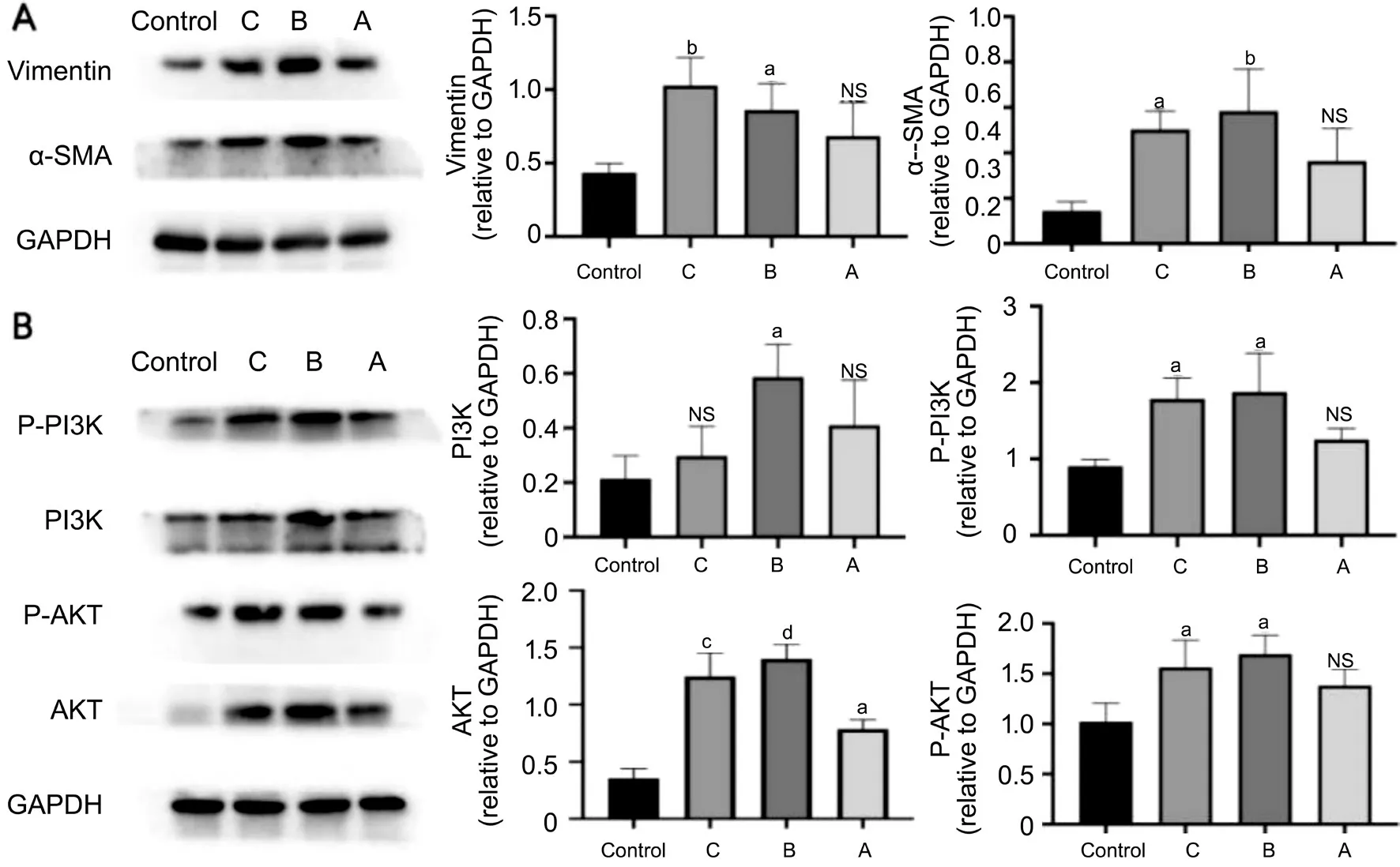
The findings revealed that the expression levels of vimentin and α-SMA in patients with grade C and B lesions were higher than those in the control group,but there was no obvious disparity between two groups nor in the expression of vimentin and α-SMA protein between the two groups.In addition,the activation of PI3K/Akt signal channel was the same as that of EMT.The expression and activation of PI3K/Akt signal channel were down adjusted in patients with grade C and grade B (Figure 5).Therefore,we speculated that the process of EMT is in connection with the expression and activation of PI3K/Akt trail.
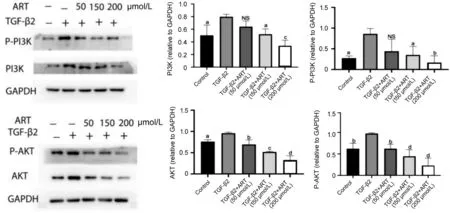
Artesunate can induce the inhibition of PI3K/Akt channel.Akt,P-Akt,PI3K and P-PI3K levels were measured by Western blotting (Figure 6).After artesunate treatment,both PI3K and phosphorylated PI3K(P-PI3K) decreased,suggesting that the down regulation of P-PI3K may be due to the down regulation of PI3K.Akt,the downstream target of PI3K,also showed a similar trend.The decrease of PI3K and Akt induced by artesunate showed statistical significance (
<0.05 or 0.01).
DISCUSSION
This research was aimed at studying the effect of artesunate on TGF-β2 induced EMT and the mechanism of artesunate on RPE cells.Artesunate inhibits the propagation,immigration and EMT procedure of RPE cells through PI3K/Akt signal channel.Based on the inhibitory effectiveness of artesunate on PVR
,we further believe that artesunate might be a target which is potential for the disposal of PVR.In the development of PVR,EMT switch of RPE cells has become a key process,mainly manifested in the decreased expression of E-cadherin and vimentin in epithelial cells,fibronectin and α-SMA increased and the morphology,migration and propagation of epithelial cells transformed into the phenotype of mesenchymal cells.TGF-β2 was added to RPE cells build the model of EMT process
,the cell morphology changes from hexagonal to spindle,and the cell ductility increased.The morphological changes were more obvious with the increase of concentration of TGF-β2.The content of vimentin and SMA increased.When artesunate was added in this process,the propagation and immigration of transformed cells were suppressed,and the content of EMT marker protein decreased.TGF-β is a cytokine with broad spectrum of biological.The research confirmed that TGF-β can regulate EMT process in epithelial cells of multiple human organs and tissues.The expression level of TGF-β2 was positively linked to the seriousness of PVR.In tumor cells,TGF-β uncontrolled binding of and its receptors activates a variety of downstream trails,including Smad,RAS and MAP kinases,PI3K/Akt,which results in cell dysfunction.Among them,PI3K/Akt signal channel occupies a significant position in EMT transformation of tumor cells
.PI3K is a family of lipid kinases that adjust a variety of signal channels,physiological functions and cellular processes.It is involved in the cell transformation and metastasis of diseases.Akt activated by PI3K is transferred to the cell compartment,where it phosphorylates a variety of protein substrates,such as mTOR
,to regulate cell metabolism and cell cycle.Therefore,blocking PI3K/Akt signaling channel can effectively control cell proliferation cycle,reduce cell adhesion and inhibit cell migration.Researches have revealed that after going through retinal detachment,it is normal for RPE to be away from their normal position and go into the vitreous chamber and sub-retina to autocrine TGF-β and activate PI3K/Akt signal channel to promote EMT of RPE cells
.In vitreous clinical specimens,PVR specimens were class B and C.Compared to the control group and class a PVR specimens,the expression level of SMA,the increase and elevation of EMT signaling molecules in PI3K/Akt signal channel,and the stimulation of PI3K/Akt expression also increased,which further verified the function of PI3K/Akt signal channel in the existence and occurrence of PVR.TGF-β2-induced somatic cells,PI3K/Akt signal channel and PI3K/Akt phosphorylation increased.After artesunate treatment,PI3K and P-PI3K decreased,and the downstream target Akt showed a similar trend.As a result,we concluded that artesunate inhibited the development of EMT in RPE cells by restraining the expression of PI3K/Akt trail.
ACKNOWLEDGEMENTS
None;
None;
None;
None;
None;
None;
None.
1 Li XH,Zhao MW,He SK.RPE epithelial-mesenchymal transition plays a critical role in the pathogenesis of proliferative vitreoretinopathy.
2020;8(6):263.
2 Pastor JC,Rojas J,Pastor-Idoate S,di Lauro S,Gonzalez-Buendia L,Delgado-Tirado S.Proliferative vitreoretinopathy:a new concept of disease pathogenesis and practical consequences.
2016;51:125-155.
3 Ren YX,Ma JX,Zhao F,An JB,Geng YX,Liu LY.Effects of curcumin on epidermal growth factor in proliferative vitreoretinopathy.
2018;47(5):2136-2146.
4 Tamiya S,Liu L,Kaplan HJ.Epithelial-mesenchymal transition and proliferation of retinal pigment epithelial cells initiated upon loss of cell-cell contact.
2010;51(5):2755-2763.
5 Lei HT,Rheaume MA,Kazlauskas A.Recent developments in our understanding of how platelet-derived growth factor (PDGF) and its receptors contribute to proliferative vitreoretinopathy.
2010;90(3):376-381.
6 Chen ZN,Shao Y,Li XR.The roles of signaling pathways in epithelialto-mesenchymal transition of PVR.
2015;21:706-710.
7 Chen XY,Xiao W,Wang WC,Luo LX,Ye SB,Liu YZ.The complex interplay between ERK1/2,TGFβ/Smad,and Jagged/Notch signaling pathways in the regulation of epithelial-mesenchymal transition in retinal pigment epithelium cells.
2014;9(5):e96365.
8 Yokoyama K,Kimoto K,Itoh Y,Nakatsuka K,Matsuo N,Yoshioka H,Kubota T.The PI3K/Akt pathway mediates the expression of type I collagen induced by TGF-β2 in human retinal pigment epithelial cells.
2012;250(1):15-23.
9 Wang N,Chen HX,Teng YP,Ding XH,Wu H,Jin XQ.Artesunate inhibits proliferation and invasion of mouse hemangioendothelioma cells
and of tumor growth
.
2017;14(5):6170-6176.
10 Jiang WW,Cen YY,Song Y,Li P,Qin RX,Liu C,Zhao YB,Zheng J,Zhou H.Artesunate attenuated progression of atherosclerosis lesion formation alone or combined with rosuvastatin through inhibition of pro-inflammatory cytokines and pro-inflammatory chemokines.
2016;23(11):1259-1266.
11 Cen YY,Liu C,Li XL,Yan ZF,Kuang M,Su YJ,Pan XC,Qin RX,Liu X,Zheng J,Zhou H.Artesunate ameliorates severe acute pancreatitis(SAP) in rats by inhibiting expression of pro-inflammatory cytokines and Toll-like receptor 4.
2016;38:252-260.
12 Zhao YY,Jiang WW,Li B,Yao Q,Dong JQ,Cen YY,Pan XC,Li J,Zheng J,Pang XL,Zhou H.Artesunate enhances radiosensitivity of human non-small cell lung cancer A549 cells via increasing NO production to induce cell cycle arrest at G2/M phase.
2011;11(12):2039-2046.
13 Li B,Li J,Pan XC,Ding GF,Cao HW,Jiang WW,Zheng J,Zhou H.Artesunate protects sepsis model mice challenged with Staphylococcus aureus by decreasing TNF-alpha release via inhibition TLR2 and Nod2 mRNA expressions and transcription factor NF-kappaB activation.
2010;10(3):344-350.
14 Murray J,Gannon S,Rawe S,Murphy JEJ.
oxygen availability modulates the effect of artesunate on HeLa cells.
2014;34(12):7055-7060.
15 Vandewynckel YP,Laukens D,Geerts A,Vanhove C,Descamps B,Colle I,Devisscher L,Bogaerts E,Paridaens A,Verhelst X,van Steenkiste C,Libbrecht L,Lambrecht BN,Janssens S,van Vlierberghe H.Therapeutic effects of artesunate in hepatocellular carcinoma:repurposing an ancient antimalarial agent.
2014;26(8):861-870.
16 Raffetin A,Bruneel F,Roussel C,Thellier M,Buffet P,Caumes E,Jauréguiberry S.Use of artesunate in non-malarial indications.
2018;48(4):238-249.
17 Yang Y,Wu ND,Wu YH,Chen HT,Qiu J,Qian XB,Zeng JT,Chiu K,Gao QY,Zhuang J.Artesunate induces mitochondria-mediatedapoptosis of human retinoblastoma cells by upregulating Kruppel-like factor 6.
2019;10(11):862.
18 He Y,Fan J,Lin H,Yang X,Ye Y,Liang L,Zhan Z,Dong X,Sun L,Xu H.The anti-malaria agent artesunate inhibits expression of vascular endothelial growth factor and hypoxia-inducible factor-1α in human rheumatoid arthritis fibroblast-like synoviocyte.
2011;31(1):53-60.
19 Li H,Gu C,Ren Y,Dai Y,Zhu X,Xu J,Li Y,Qiu Z,Zhu J,Zhu Y,Guan X,Feng Z.The efficacy of NP11-4-derived immunotoxin scFv-artesunate in reducing hepatic fibrosis induced by Schistosoma japonicum in mice.
2011;25(2):148-154.
20 Zhang YQ,Li HH,Zhu JJ,Wei TT,Peng YX,Li R,Xu R,Li M,Xia AZ.Role of artesunate in TGF-β1-induced renal tubular epithelialmesenchymal transdifferentiation in NRK-52E cells.
2017;16(6):8891-8899.
21 Ma Z,Lou SP,Jiang Z.PHLDA2 regulates EMT and autophagy in colorectal cancer via the PI3K/AKT signaling pathway.
2020;12(9):7985-8000.
22 Li YL,Wang TT,Sun YJ,Huang TF,Li CP,Fu Y,Li YC,Li CZ.p53-mediated PI3K/AKT/mTOR pathway played a role in Ptox
-induced EMT inhibition in liver cancer cell lines.
2019;2019:2531493.
23 Chen HT,Wang HF,An JB,Shang QL,Ma JX.Plumbagin induces RPE cell cycle arrest and apoptosis via p38 MARK and PI3K/AKT/mTOR signaling pathways in PVR.
2018;18(1):89.
24 Roybal CN,Velez G,Toral MA,Tsang SH,Bassuk AG,Mahajan VB.Personalized proteomics in proliferative vitreoretinopathy implicate hematopoietic cell recruitment and mTOR as a therapeutic target.
2018;186:152-163.
25 Cheng R,Li C,Li CY,Wei L,Li L,Zhang Y,Yao YC,Gu XQ,Cai WB,Yang ZH,Ma JX,Yang X,Gao GQ.The artemisinin derivative artesunate inhibits corneal neovascularization by inducing ROSdependent apoptosis in vascular endothelial cells.
2013;54(5):3400-3409.
26 Wang XQ,Liu HL,Wang GB,Wu PF,Yan T,Xie J,Tang Y,Sun LK,Li C.Effect of artesunate on endotoxin-induced uveitis in rats.
2011;52(2):916-919.
杂志排行
International Journal of Ophthalmology的其它文章
- IJO/IES Event Photos
- Inhibitory effect on subretinaI fibrosis by anti-pIacentaI growth factor treatment in a Iaser-induced choroidaI neovascuIarization modeI in mice
- NoveI mutations in the BEST1 gene cause distinct retinopathies in two Chinese famiIies
- Frequency cumuIative effect of subthreshoId energy Iaser-activated remote phosphors irradiation on visuaI function in guinea pigs
- One-step thermokeratopIasty for pain aIIeviating and pretreatment of severe acute corneaI hydrops in keratoconus
- Accuracy of optimized Sirius ray-tracing method in intraocuIar Iens power caIcuIation
