The ‘Unsigned highway’ :An alternative route for portal vein anastomosis for non-malignant portal vein thrombosis during pediatric re-transplantation
2021-05-19AlertChiYnChnWingChiuDiPtrickHoYuChungWongHoiSheSuiLingSin
Alert Chi Yn Chn , ,Wing Chiu Di ,Ptrick Ho Yu Chung ,Wong Hoi She ,Sui Ling Sin
a Division of Liver Transplantation, Department of Surgery, The University of Hong Kong, Hong Kong, China
b Division of Paediatric Surgery, Department of Surgery, The University of Hong Kong, Hong Kong, China
TotheEditor:
Non-malignant portal vein thrombosis (PVT) remains an important issue in liver transplantation due to the technical challenges in re-establishing the portal flow to the liver graft.The prevalence of complex PVT (i.e.Yerdel grade 4) was reported to be around 2.0% [1].In the early history of liver transplantation,PVT was regarded as a contraindication.As knowledge and techniques improved over the years,different operative strategies have been derived,including direct endovenous thrombectomy [2],jump grafting to superior mesenteric vein (SMV) [3],and renoportal [4]or portocaval anastomosis [3].However,the majority of these reports focused on flow restoration in adult recipients in whom cirrhosis with portal hypertension was the most common etiology.The wider caliber of mesenteric veins or the availability of shunts rendered such techniques feasible for portal flow restoration in adults.Nonetheless,this may not be possible in pediatric patients,given the size of the vessels,or the lack of spontaneous shunt formations [5].We herein describe a novel,frequently unnoticed route for portal flow restoration.
A 7-year-old girl with a history of living donor liver transplantation using a left lateral section graft for biliary atresia 6 years ago developed chronic graft dysfunction and recurrent variceal bleeding due to main PVT.Computed tomography showed complete PVT down to the SMV,complicated by cavernous transformations.The distal segment of the SMV and its jejunal branches were patent.The spleen was enlarged with dilated splenic vein.She had been wait-listed for two years before a liver graft from a deceased donor of 14 years old became available.On review of the scan,the SMV with a diameter of 6 mm was considered too short for any landing zone for distal vascular control.On the contrary,the central segment of the splenic vein with a diameter of 9 mm was shifted caudally by huge splenomegaly beyond the inferior border of the pancreas before branching off (Fig.1 A).Intraoperatively,this segment of the splenic vein was identified for anastomosis (Fig.1 B).The graft portal vein was brought down over the anterior surface of the pancreas to reach the splenic vein via the mesenteric window.A side-to-end splenoportal anastomosis (Fig.2 A–C) below the body of pancreas was performed using 6–0 Prolene,resulting in a Sshaped configuration (Fig.3 A).Doppler ultrasound confirmed portal flow at 50 0–60 0 mL/min.Convergence of SMV flow and distal splenic flow into the splenoportal anastomosis reconstituted physiological hepatopedal flow (Fig.3 B).Graft function at 6 months remained well with normalization of liver enzymes.
In summary,the caudal shift of the splenic vein out of the posterior aspect of the pancreas caused by splenomegaly provided a direct route for splenoportal anastomosis.Appreciation of the anatomical situation on preoperative imaging is of paramount importance to operative strategic planning.We reported an alternative novel approach for PVT during difficult pediatric liver transplantation.
Acknowledgments
None.CRediT authorship contribution statement
Albert Chi Yan Chan:Conceptualization,Supervision,Writing-original draft,Writing -review &editing.Wing Chiu Dai:Data curation,Writing -review &editing.Patrick Ho Yu Chung:Data curation,Writing -review &editing.Wong Hoi She:Data curation,Writing -review &editing.Sui Ling Sin:Data curation,Writing -review &editing.
Funding
None.

Fig.1.A:Coronal view on the portal circulation in computed tomography.Note the abrupt cutoff of portal vein due to portal vein thrombosis above the uncinate process of pancreas (U) resulting in dilated jejunal veins (red arrow),superior mesenteric vein and splenic vein (SV).Short superior mesenteric vein was not favorable for anastomosis(white double arrowhead).On the contrary,the curved mid-segment of portal vein was shifted caudally (double white arrow) by massive splenomegaly and was used for splenoportal anastomosis.B:Coronal view on the splenoportal anastomosis (red arrow) inferior to the body of pancreas (P).
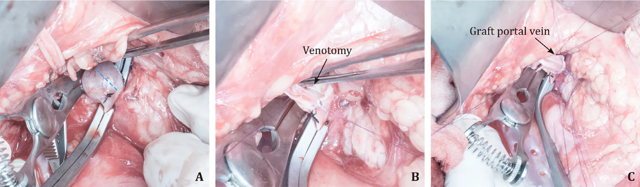
Fig.2.Intraoperative exposure of the native portal vein (A),venotomy on native portal vein for anastomosis (B),and completion of graft portal vein anastomosis (C).
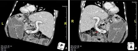
Fig.3.A:An oblique posterior coronal view on graft portal vein (GV).It ran its course from inferior border of pancreas (P) and curved cranially over the anterior surface of pancreas to the liver graft.B:Coronal view on the graft portal vein from splenic vein to the liver graft.
Ethical approval
Not needed.
Competing interest
No benefits in any form have been received or will be received from a commercial party related directly or indirectly to the subject of this article.
杂志排行

Hepatobiliary & Pancreatic Diseases International的其它文章
- Practice of precision surgery in primary liver cancer
- Reporting of longitudinal pancreatojejunostomy with partial pancreatic head resection (the Frey procedure) for chronic pancreatitis:A systematic review
- Hepatobiliary&Pancreatic Diseases International
- Presentation and surgical management of xanthogranulomatous cholecystitis
- Transjugular intrahepatic portosystemic shunt is effective in patients with chronic portal vein thrombosis and variceal bleeding
- Long-term follow-up of HCV patients with sustained virological response after treatment with pegylated interferon plus ribavirin