基于喷射效应的太赫兹高分辨成像研究与进展
2020-06-02马晓茗姜在超屈庆山张振伟杨玉平
马晓茗,姜在超,屈庆山,崔 彬,张振伟,杨玉平*
基于喷射效应的太赫兹高分辨成像研究与进展
马晓茗1,姜在超1,屈庆山1,崔 彬1,张振伟2,杨玉平1*
1中央民族大学理学院,北京 100081;2首都师范大学物理系;北京市成像技术高精尖中心;北京市太赫兹波谱与成像重点实验室;太赫兹光电子学教育部重点实验室,北京 100048
太赫兹(THz)成像技术,因其具有能量低、透射率高、波谱范围宽等独特的分析能力,已经在生物医学、安全检查、航空航天等领域展现出巨大的优势及潜在的应用价值,但是较低的空间分辨率制约了太赫兹成像技术的进一步应用。太赫兹波通过具有适当折射率的介质结构产生的“太喷射”效应调控亚波长尺寸太赫兹光场,突破衍射极限对显微系统空间分辨率的限制,同时不损失光场能量和光谱信息,实现高通量、超宽谱的远场太赫兹高分辨成像。本文首先介绍基于纳米喷射的微球透镜显微技术,接着介绍基于太喷射的太赫兹显微技术,最后对基于喷射效应的太赫兹高分辨成像技术的前景做了展望。
太赫兹成像;太喷射;分辨率
1 引 言
太赫兹(Terahertz, THz)成像技术,因其具有能量低、穿透性强、波谱范围宽等独特的分析能力,已经在生物医学、安全检查、航空航天等领域展现出巨大的优势及潜在的应用价值[1-2]。然而,由于太赫兹波波长较长,受衍射极限的限制,常规远场THz成像技术的分辨率较低,难以满足当前日益提高的应用需求。为了提高分辨率,在过去的几十年里,各种THz超分辨率成像技术相继被提出,包括THz近场成像[3-4]、THz共焦扫描成像[5-6]、THz图像复原处理[7-9]等,使分辨率达到了微米或纳米尺度。其中,近场探测技术通过产生和探测含有超精细信息的倏逝波,突破衍射极限,获得亚波长分辨率,是太赫兹成像领域研究的热点之一。目前,在THz频率范围已经开展的近场显微方法中,大致有四类:分别为利用亚波长物理孔径(或虚拟孔径)的方法[10-13]、亚波长针尖局域(或增强)的方法[14-17]、激光聚焦后产生的亚波长尺寸THz辐射源显微术[18-22]、微纳结构材料局域或增透的THz超透镜[23-26]等。但是以上方法在提高分辨率的同时,严重地限光作用和频率选择特性导致光通量急剧下降,同时损耗了大量的高频分量,不但降低了测量的灵敏度和信噪比,也损失了THz成像的宽谱分析能力,削弱了THz成像的技术优势。
近年来,在可见光领域,研究人员发现纳米、微米尺寸介质球的光子纳米喷射效应(photonic nanojet)具有超分辨能力[27]。类比于可见光波段的纳米喷射(nanojet)效应,在太赫兹波段,基于介观尺寸介质结构的太喷射(terajet)效应的THz显微技术也相继应用在连续和脉冲THz成像技术中,有效提高目前常规的THz远场成像分辨率[28-29]。相对于其它超分辨成像技术,基于太喷射的THz高分辨成像有以下独特的优点:1) 非倏逝波:超分辨的太赫兹喷射光束持续至少2的长度,并能传输到远场,使得对具有一定厚度样品的高分辨成像成为可能;2) 高通量特性:光谱宽度和光谱能量几乎没有损失,适合宽光谱成像,且成像样品不受限制;3) 设备成本低、操作简便:介质超透镜的尺寸在1 mm~6 mm范围内,操作简便,可以进行大面积成像。本文将对基于喷射效应的THz高分辨成像技术进行详细的介绍。第二部分介绍基于纳米喷射的微球透镜显微技术,第三部分介绍基于太喷射的连续波显微技术,第四部分介绍我们提出的几种实现高通量、脉冲太赫兹波高分辨成像的方案,第五部分是总结和展望。
2 基于纳米喷射的微球透镜显微技术
2011年,在可见光领域,英国曼彻斯特大学的Wang等[27]将光学透明微球(直径在2mm到9mm之间的二氧化硅微球)与传统的光学显微镜结合实现微球超显微技术:将直径几微米至几十微米的微球直接置于样品表面,球体收集近场信息,能够成倍提高传统光学显微镜的成像能力,分辨率达到50 nm,如图1所示。
为了进一步改进微球超分辨显微技术,不同性能的微透镜(形状、尺寸、材料以及折射率等)、不同浸没条件(不同液体、浸没、半浸没等)等方案相继被提出,成像分辨率和对比度获得一定的增强效果[30-38]。比如,浙江大学刘旭研究组的郝翔等[30]将介质微球浸入到乙醇溶液内,在透射模式和反射模式下分别进行成像,发现如果把微球半浸没于液体时,成像对比度会大幅增强;但将微球完全浸没于液体时,并没有获得明显的增强效果。由于乙醇具有挥发性,他们还通过视频记录了成像的动态过程。当微球慢慢露出乙醇时,暗条纹出现在微球内,并且逐渐变得清晰,边缘也变得更加尖锐;当乙醇完全挥发后图像变得再次模糊。2013年,曼彻斯特大学的Li等[33]把直径100mm的BaTiO3微球和光学显微镜的物镜同时浸没在水中,利用微球的近场放大效应,用光学显微镜观察到75 nm的腺病毒(adenoviruses)。
以上所发现的介质微球超分辨能力既无法用传统几何光学解释,也不能简单归类在近场光学中。2004年,美国西北大学的Chen等[39]通过理论计算认为微球超分辨成像是微球的光子学纳米喷射(photonic nanojet)效应引起的。微球通过光子纳米喷射效应可以收集、传递倏逝波,减慢倏逝波衰减,从而可以在远场看到超分辨成像,如图2(a)所示。2014年,圣路易斯华盛顿大学Shen等[40]研究了两层电介质微球,发现采用常规材料制成的双层介质微球比单个微球具有更长的纳米喷射能力,如图2(b)所示。当光进入到球体内时,其能量流向前传播的过程中是收敛的,而对于两层微球来说,能量流在球体内交替发生了收敛和发散,其中发散能够起到平衡和减缓收敛的作用,通过适当选择双层介质球的尺寸和折射率参数,可以有效地使焦点处的发散角最小,传输距离延长。2017年,以色列理工学院的Yacob[41]提出了产生纳米喷射的无衍射贝塞尔光束理论,如图2(c)所示,平面波垂直照射到样品上,透射波经微球汇聚到焦平面上。具体来说,通过接触点附近的倏逝波携带样品的超精细结构信息,并通过接触点传输到微球中,转换成传输波;而远离接触点的入射波以入射角in在介质球表面发生折射(折射角为T);最终,将球差叠加在所有几何光线上,得到产生纳米喷射流的贝塞尔光束。由于纳米喷射流受倏逝波的调制,通过将点附近的行波场传输到共焦显微镜中获得物体的精细结构。光子纳米喷射具有优异的光学性能,如非交联性、较强的局部电场以及尖锐的焦点,目前已经应用于纳米颗粒检测、光学纳米光刻和超分辨率成像等领域。
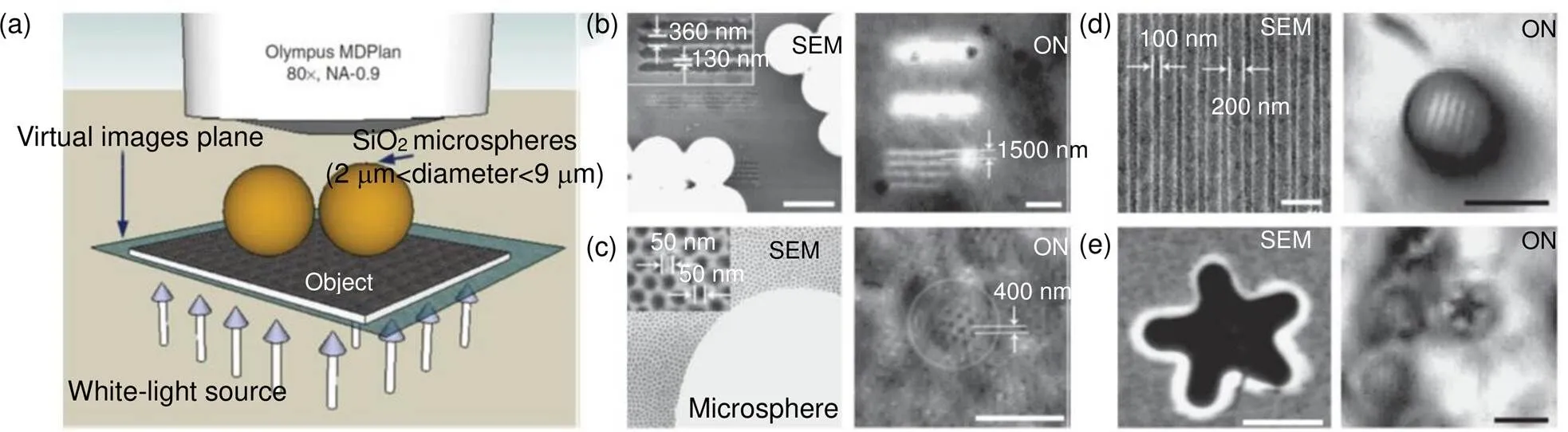
图1 (a) 微球透镜与传统光学显微镜结合显微镜示意图;(b), (c) 电镜图(左)与透射模式下微球超透镜的显微成像图(右)对比:(b) 宽度为360 nm、间隔为130 nm的线条;(c) 渔网状的镀金AAO模板;(d), (e) 电镜图(左)与反射模式下微球超透镜的显微成像图(右)对比:(d) 商业蓝光DVD光盘;(e) 星状GeSbTe薄膜 [27]
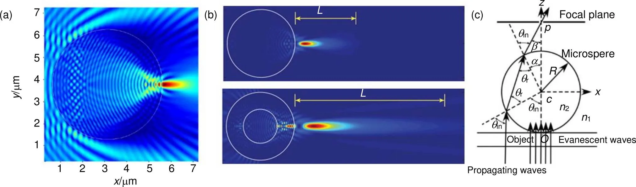
图2 (a) 微球纳米喷射示意图[39];(b) 单层同质微球和双层异质微球的纳米喷射对比图[40];(c) 产生纳米喷射的无衍射贝塞尔光束[41]
3 基于太喷射的连续波显微技术
与可见光波段已经广泛研究的微球显微成像技术相比,基于太喷射效应的THz波远场聚焦特性和高分辨成像研究仅处于起步阶段。2014年,西班牙Pacheco-Pena等人和俄罗斯Minin等[28, 42-45]将200 GHz的平面波垂直照射在无损耗的长方体介质上,在平面波的照射下产生的“太喷射”具有一定的汇聚能力。模拟结果显示,其主要特性在于太喷射波具有亚波长的束腰半径,并且在传播几个波长的距离之后光束发散很小,这表明聚焦光斑焦点附近的半高宽会比较小,可在远场实现亚波长分辨率。另外,通过改变介质长方体的折射率,当折射率从2减小到1.4时,太喷射波束的焦点位置沿着光轴从立方体内部向表面外移,如图3(a)所示[28]。
2017年,日本大阪大学Pham等[45]将折射率为1.46的聚四氟乙烯(Teflon)立方体放置在反射式太赫兹成像系统中(如图3(b)示),对具有0.63沟槽的铝板进行成像,成像结果显示:使用了介质立方体的图像可以清楚地分辨铝板上的空气沟槽,而没有使用介质立方体的图像由于分辨率不足而模糊不清。在此基础上,他们使用耦合长方体阵列的全介质周期性太喷射波导,使THz波束进一步聚焦,光束半高宽达到/2,如图4(a)所示[46]。2018年,又在长方体介质透镜的前表面放置金属掩膜,通过减小喷射光束的束腰半径进一步增加空间分辨率和聚焦光强,如图4(b)所示[47-48]。
另外,研究人员们又设计一种基于具有高金属填充因子和穿孔电介质的金属光子晶体超材料,可以产生太赫兹喷射,如图5(a)所示[49]。仿真结果表明,不同结构和填充因子影响材料的有效介电常数以及太喷射波束的光场分布[50]。Cruz等[51]通过仿真模拟研究了非常规几何结构(如图5(b)~5(e)所示)介电材料在0.1 THz频率下的太赫兹喷射效应,并与球形结构做对比,发现三角椎形介电体对入射波的功率增强了32倍,分辨率高达0.396。
在实际应用中,由于样品具有一定的厚度,喷射波束的作用长度也是提高成像分辨率和对比度的一个重要指标。最近,华中科技大学的王可嘉课题组采用3D打印技术打印的锥透镜,将平行的太赫兹高斯光束变换成无衍射的贝塞尔光束,该光束持续的长度可达50 mm,并应用于太赫兹反射成像中[52]。为了进一步增加无衍射THz波束的作用长度,他们又联合首都师范大学张振伟等人设计了一个透镜组系统,如图6(a)所示[53]。发散的高斯光束经过准直透镜后成为准直的高斯光束,再经过锥透镜(A1)变换为无衍射光束;该无衍射光束进一步经过柱透镜(C1)和锥透镜(A2),使得其作用长度达到250 mm,如图6(c)所示。实验证明,这种光束可以用来扩展太赫兹传输成像系统的景深,其成像效果如图6(b)所示。
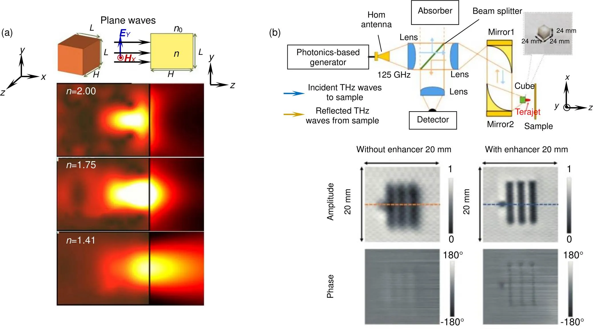
图3 (a) 不同折射率介质长方体产生的太喷射波束示意图[28];(b) 反射模式下介质长方体耦合THz成像系统及其对铝板样品的振幅和相位图像[45]
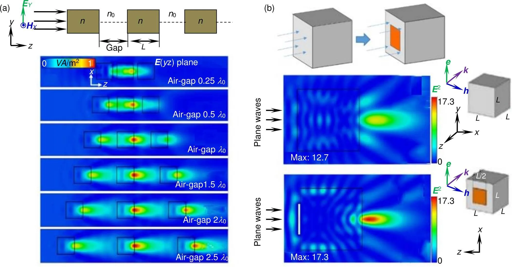
图4 (a) 长方体阵列的太喷射波导侧面示意图及其太喷射波束在不同空隙情况下的光场分布[46];(b) 耦合金属掩膜的介质立方体的示意图及其太喷射波束的光场分布[48]

图5 不同几何形状介质结构产生的喷射波束的光场分布。(a) 金属棒阵列填充长方体[49];(b) 球体[51];(c) 三角椎体[51];(d) 梯形椎体[51];(e) 扇形椎体[51]
4 基于太喷射的脉冲波显微技术
宽谱分析是太赫兹成像技术的重要优势之一,虽然基于连续毫米波的太喷射效应初步提高了成像分辨率,但成像信息少(单频,缺少相位信息),并且无法进行光谱分析。2018年,中央民族大学杨玉平课题组提出并开发了基于介电小球的太赫兹喷射显微技术,即在常规远场8太赫兹脉冲成像系统上进行改进,在两个抛物面镜的中间放置直径为3 mm的聚四氟乙烯介质小球,入射THz波经过二次聚焦和调制,实现亚波长太喷射波束,将样品放置在太喷射光束的合适位置,对透射(或反射)THz波进行测试和成像,如图7(a)所示。利用刀口法测量了常规太赫兹成像系统的分辨率及加入介质球后的分辨率,结果显示,加入介质球后能够显著提高系统的分辨率,最大提高了66.7%,且在一定范围内,介质球与刀片的距离越远,成像分辨率越好,如图7(b)所示。对宽度为110mm的硅基介质光栅进行成像,结果显示:在时域显示模式下,介质球辅助THz最大值成像和最小值成像均能够明显地分辨出介质光栅条纹,而不放介质球直接成像时则不能将两者分辨。在频域显示模式下,高频处的成像效果比低频成像效果更好,这是因为频率越高,太喷射光束的尺寸越小,能量密度越高,分辨率和对比度越高;而不放介质球直接成像时,无论在低频还是高频,均无法辨别出光栅条纹,如图7(c)所示[29]。
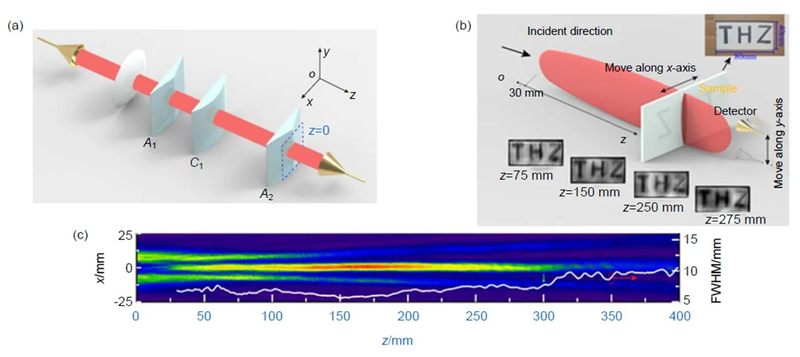
图6 (a) 锥透镜组系统示意图;(b) 基于锥透镜组的太赫兹透射成像;(c) 经锥透镜组系统透射的太赫兹波束光场分布[53]

图7 (a) 太喷射效应示意图;(b) 成像分辨率;(c) 成像效果对比图[29]
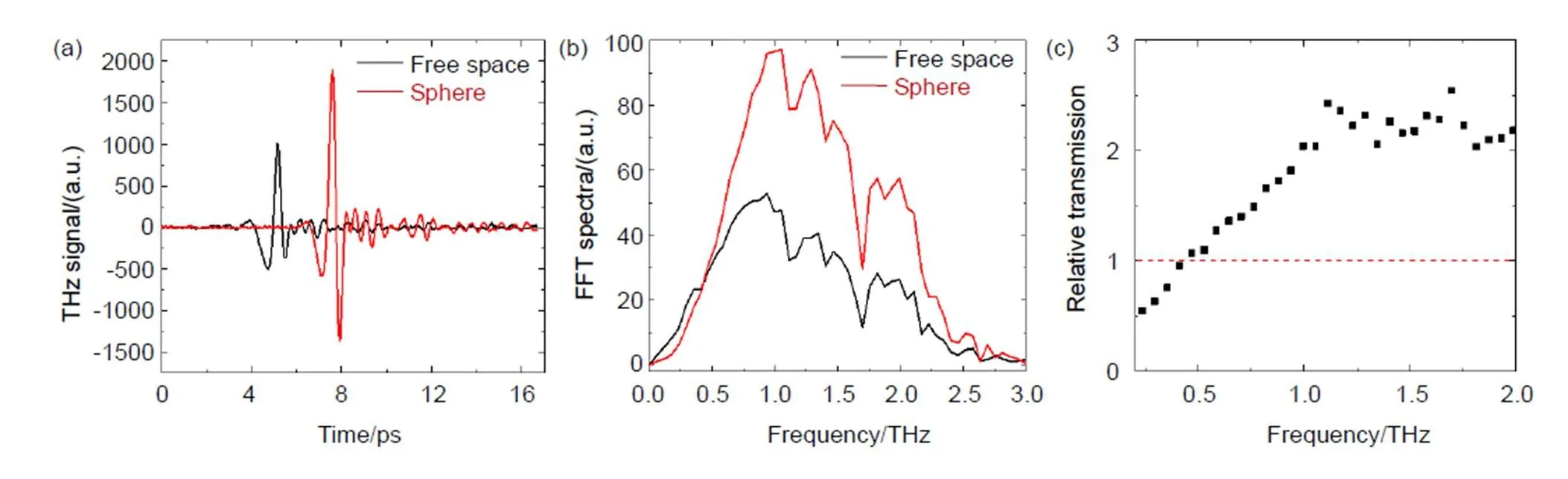
图8 (a) 有无介质球情况下的THz时域信号;(b) 频域信号;(c) 相对振幅透过率[54]

图9 (a) 3 mm直径介质球在不同频率下x-z平面上的空间能量密度分布;(b) 3 mm直径介质球在1.5 THz频率下x-z平面上的空间能量密度分布随折射率的演变[29]
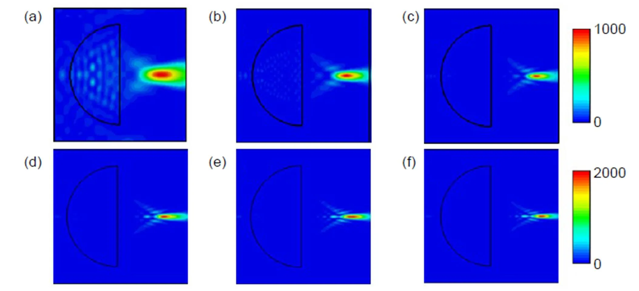
图10 3 mm直径介质半球球在不同频率下x-z平面上的空间能量密度分布。(a) 0.5 THz;(b) 1.0 THz;(c) 1.5 THz;(d) 2.0 THz;(e) 2.5 THz;(f) 3.0 THz[54]
太喷射现象不但具有优异的调控THz波束的能力,在远场实现超分辨THz成像,并且具有高通量和超宽带等优点。我们比较有无介质球的“太喷射”效应下,系统在信号通量和光谱带宽方面的性能,如图8所示。实验发现:由于太喷射对THz波束的调制与优化,和自由空间的THz信号相比,经过介质超透镜的THz脉冲除有一定的时间延迟外,信号还有较大的提高,如图8(a)所示;其对应的FFT频谱如图8(b)所示,在高于0.5 THz的范围内THz信号具有明显的提高;两者的相对振幅透过率如图8(c)所示,在1 THz以上的高频区,加介质球的振幅信号提高了一倍以上,对应的能量(振幅的平方)变为4倍以上,表明介质球耦合THz成像技术具有高通量和超宽带的优点[54]。
模拟结果表明,入射波的频率分量、介质球的尺寸、折射率等因素对太喷射波束具有不同程度的调节作用,极大地影响THz成像的超分辨能力。当介质球尺寸(=3 mm)一定时,仿真结果表明:随着频率的增大,聚焦光斑的尺寸逐渐减小,同时焦点距介质球的距离增大,如图9(a)所示。当介质球尺寸(=3 mm)和频率(=1.5 THz)都固定时,随着折射率的增加,聚焦光束位置发生变化。当折射率从1.4增加到1.6的过程中,喷射光束沿着轴向球体表面移动;折射率从1.6增大到1.8的过程中,喷射状的光斑的位置开始向球体内部移动,而且焦点束腰处的半高宽由0.48减小到0.35。当折射率从1.8继续增大到2.0的过程中,喷射状光斑的位置继续沿着球体半径方向向球中心移动,即距离小于半径,喷射光束束腰从0.33减小到0.30,如图9(b)所示。
不难看出,介质小球产生的太喷射光束都位于球体表面附近,使得实验时小球要紧靠样品表面,可能会污染样品。我们进一步提出了一种介质半球耦合太赫兹高分辨率成像的方法,来提高THz波经过介质后的喷射焦距和喷射光束的工作距离[54-56]。模拟结果显示,介质半球产生的太喷射波束受入射波的频率分量、介质球的尺寸、折射率等因素的调制作用呈现出与介质小球类似的规律。随着频率的增加,THz波通过介质半球后所产生的聚焦光束更加收敛,即“太喷射”的光斑越来越细长,焦点开始外移,且焦点处能量密度越来越大,如图10所示。值得注意的是,焦点的位置(最佳成像分辨率的位置)距离介质半球的范围为1.1 mm~1.5 mm,实际操作空间大,避免了对样品的直接接触。
5 结论和展望
太喷射效应对入射太赫兹波具有较好的调节能力,在提高分辨率的同时频率分量和信号通量也有所增加。结合我们的实验结果和模拟结果可知,与传统的远场太赫兹成像相比,基于太喷射的THz成像技术分辨率提高了一个数量级。此外,该方法有效地避免了强度和频谱分量的损失;且方法简单,易操作;对测试样品没有任何限制(介质与金属都可),为今后提高太赫兹成像分辨率提供了一个新的思路,对高分辨的太赫兹无损检测应用具有重要的实用价值。
然而相对于传统的近场成像,分辨率还有待进一步提高;且太赫兹波经过介质结构后所产生的光场形状、大小是否与仿真结果一致还有待进一步实验验证;携带样品高频信息的倏逝波传向远场的机理也还尚不明确;以及超精细结构信息的增强与探测、超分辨信息的提取等也有待进一步的研究与探讨。
[1] Mittleman D M. Twenty years of terahertz imaging[Invited][J]., 2018, 26(8): 9417–9431.
[2] Tonouchi M. Cutting-edge terahertz technology[J]., 2007, 1(2): 97–105.
[3] Adam A J L. Review of Near-Field Terahertz measurement methods and their applications[J]., 2011, 32(8–9): 976–1019.
[4] Siday T, Natrella M, Wu J,. Resonant terahertz probes for near-field scattering microscopy[J]., 2017, 25(22): 27874–27885.
[5] Zinov’ev N N, Andrianov A V, Gallant A J,. Contrast and resolution enhancement in a confocal terahertz video system[J]., 2008, 88(8): 492–495.
[6] Llombart N, Cooper K B, Dengler R J,. Confocal ellipsoidal reflector system for a mechanically scanned active terahertz imager[J]., 2010, 58(6): 1834–1841.
[7] Balbekin N S, Kulya M S, Belashov A V,. Increasing the resolution of the reconstructed image in terahertz pulse time-domain holography[J]., 2019, 9: 180.
[8] Liu T, Pi Y M, Yang X. Wide-angle CSAR imaging based on the adaptive subaperture partition method in the terahertz band[J]., 2018, 8(2): 165–173.
[9] Ding S H, Li Q, Yao R,. High-resolution terahertz reflective imaging and image restoration[J]., 2010, 49(36): 6834–6839.
[10] Hunsche S, Koch M, Brener I,. THz near-field imaging[J]., 1997, 150(1–6): 22–26.
[11] Mitrofanov O, Brener I, Wanke M C,. Near-field microscope probe for far infrared time domain measurements[J]., 2000, 77(4): 591–593.
[12] Chen Q, Zhang X C. Semiconductor dynamic aperture for near-field terahertz wave imaging[J]., 2001, 7(4): 608–614.
[13] Mitrofanov O, Brener I, Harel R,. Terahertz near-field microscopy based on a collection mode detector[J]., 2000, 77(22): 3496–3498.
[14] Chen H T, Kersting R, Cho G C. Terahertz imaging with nanometer resolution[J]., 2003, 83(15): 3009–3011.
[15] van der Valk N C J, Planken P C M. Electro-optic detection of subwavelength terahertz spot sizes in the near field of a metal tip[J]., 2002, 81(9): 1558–1560.
[16] Moon K, Park H, Kim J,. Subsurface nanoimaging by broadband terahertz pulse near-field microscopy[J]., 2015, 15(1): 549–552.
[17] Klarskov P, Kim H, Colvin V L,. Nanoscale laser terahertz emission microscopy[J]., 2017, 4(11): 2676–2680.
[18] Kiwa T, Tonouchi M, Yamashita M,. Laser terahertz-emission microscope for inspecting electrical faults in integrated circuits[J]., 2003, 28(21): 2058–2060.
[19] Yamashita M, Kawase K, Otani C,. Imaging of large-scale integrated circuits using laser terahertz emission microscopy[J]., 2005, 13(1): 115–120.
[20] Yang Y P, Yan W, Li W. A reflected terahertz-emission microscopy[J]., 2007, 24(1): 169–171.
[21] Yang Y P, Shi Y L, Yan W,. A new microscopy for THz radiation[J]., 2005, 54(9): 4079–4083.
杨玉平, 施宇蕾, 严伟,等.一种新型THz显微探测技术[J],物理学报, 2005, 54(9): 4079–4083.
[22] Zhao J Y, Chu W, Guo L J,. Terahertz imaging with sub-wavelength resolution by femtosecond laser filament in air[J]., 2015, 4: 3880.
[23] Ishihara K, Ohashi K, Ikari T,. Terahertz-wave near-field imaging with subwavelength resolution using surface-wave-assisted bow-tie aperture[J]., 2006, 89(20): 201120.
[24] Chen H, Ma S H, Wu X M,. Diagnose human colonic tissues by terahertz near-field imaging[J]., 2015, 20(3): 036017.
[25] Xu Y H, Zhang X Q, Tian Z,. Mapping the near-field propagation of surface plasmons on terahertz metasurfaces[J]., 2015, 107(2): 021105.
[26] Chen S C, Du L H, Meng K,. Terahertz wave near-field compressive imaging with a spatial resolution of over λ/100[J]., 2019, 44(1): 21–24.
[27] Wang Z B, Guo W, Li L,. Optical virtual imaging at 50 nm lateral resolution with a white-light nanoscope[J]., 2011, 2: 218.
[28] Pacheco-Peña V, Beruete M, Minin I V,. Terajets produced by dielectric cuboids[J]., 2014, 105(8): 084102.
[29] Yang Y P, Liu H L, Yang M H,. Dielectric sphere-coupled THz super-resolution imaging[J]., 2018, 113(3): 031105.
[30] Hao X, Kuang C F, Liu X,. Microsphere based microscope with optical super-resolution capability[J]., 2011, 99(20): 203102.
[31] Darafsheh A, Walsh G F, Negro L D,. Optical super-resolution by high-index liquid-immersed microspheres[J]., 2012, 101(14): 141128.
[32] Lee S, Li L, Ben-Aryeh Y,. Overcoming the diffraction limit induced by microsphere optical nanoscopy[J]., 2013, 15(12): 125710.
[33] Li L, Guo W, Yan Y Z,. Label-free super-resolution imaging of adenoviruses by submerged microsphere optical nanoscopy[J]., 2013, 2(9): e104.
[34] Yang H, Moullan N, Auwerx J,. Fluorescence imaging: super-resolution biological microscopy using virtual imaging by a microsphere nanoscope [J]., 2014, 10(9): 1876.
[35] Wang F, Yang S, Ma H,. Microsphere-assisted super-resolution imaging with enlarged numerical aperture by semi-immersion [J]., 2018, 112:023101.
[36] Yan Y Z, Li L, Feng C,. Microsphere-coupled scanning laser confocal nanoscope for sub-diffraction-limited imaging at 25 nm lateral resolution in the visible spectrum[J]., 2014, 8(2): 1809–1816.
[37] Li P Y, Tsao Y, Liu Y J,. Unusual imaging properties of superresolution microspheres[J]., 2016, 24(15): 16479–16486.
[38] Yang H, Trouillon R, Huszka G,. Super-resolution imaging of a dielectric microsphere is governed by the waist of its photonic nanojet[J]., 2016, 16(8): 4862–4870.
[39] Chen Z G, Taflove A, Backman V. Photonic nanojet enhancement of backscattering of light by nanoparticles: a potential novel visible-light ultramicroscopy technique[J]., 2004, 12(7): 1214–1220.
[40] Shen Y C, Wang L V, Shen J T. Ultralong photonic nanojet formed by a two-layer dielectric microsphere[J]., 2014, 39(14): 4120–4123.
[41] Ben-Aryeh Y. Nano-jet related to Bessel beams and to super-resolutions in microsphere optical experiments[J]., 2017, 4: 3.
[42] Pacheco-Peña V, Beruete M, Minin I V,. Multifrequency focusing and wide angular scanning of terajets[J]., 2015, 40(2): 245–248.
[43] Pham H H N, Hisatake S, Minin I V,. Three-dimensional direct observation of Gouy phase shift in a terajet produced by a dielectric cuboid[J]., 2016, 108(19): 191102.
[44] Pham H H N, Hisatake S, Minin O V,. Asymmetric phase anomaly of terajet generated from dielectric cube under oblique illumination[J]., 2017, 110(20): 201105.
[45] Pham H H N, Hisatake S, Minin O V,. Enhancement of spatial resolution of terahertz imaging systems based on terajet generation by dielectric cube[J]., 2017, 2(5): 056106.
[46] Minin I V, Minin O V, Pacheco-Peña V,. All-dielectric periodic terajet waveguide using an array of coupled cuboids[J]., 2015, 106(25): 254102.
[47] Minin I V, Minin O V, Pacheco-Peña V,. Localized photonic jets from flat, three-dimensional dielectric cuboids in the reflection mode[J]., 2015, 40(10): 2329–2332.
[48] Yue L Y, Yan B, Monks J N,. A millimetre-wave cuboid solid immersion lens with intensity-enhanced amplitude mask apodization[J]., 2018, 39(6): 546–552.
[49] Minin I V, Minin O V. Terajet from 3D anisotropic artificial metamaterial[C]//, 2016: 142–144.
[50] Minin I V, Minin O V. Terahertz artificial dielectric cuboid lens on substrate for super-resolution images[J]., 2017, 49(10): 326.
[51] Cruz A L S, Cordeiro C M B, Franco M A R. Enhanced Terahertz transmission through 3D non-spherical terajets[J]., 2015, 9634: 963412.
[52] Niu L T, Wang K J, Yang Y Q,. Diffractive elements for zero-order Bessel beam generation with application in the terahertz reflection imaging[J]., 2019, 11(1): 5900212.
[53] Zhang Z W, Zhang H Y, Wang K J. Diffraction-free THz sheet and its application on THz imaging system[J]., 2019, 9(5): 471–475.
[54] Yang Z C, Qu Q S, Yang M H,. Propagation characteristics of high-throughput terajet beam and its super Resolution THz imaging[C]//, 2019: 1–2.
[55] Qu Q S, Liu H L, Zhu D,. Terajet effect of dielectric sphere and THz imaging[J]., 2018, 10826: 1082606.
[56] Chernomyrdin N V, Frolov M E, Lebedev S P,. Wide-aperture aspherical lens for high-resolution terahertz imaging[J]., 2017, 88(1): 014703.
Research advances of high-resolution THz imaging based on terajet effect
Ma Xiaoming1, Jiang Zaichao1, Qu Qingshan1, Cui Bin1, Zhang Zhenwei2, Yang Yuping1*
1School of Science, Minzu University of China, Beijing 100081, China;2Department of Physics, Capital Normal University; Beijing Advanced Innovation Center for Imaging Technology; Beijing Key Laboratory for Terahertz Spectroscopy and Imaging; Key Laboratory of Terahertz Optoelectronics, Ministry of Education, Beijing 100048, China
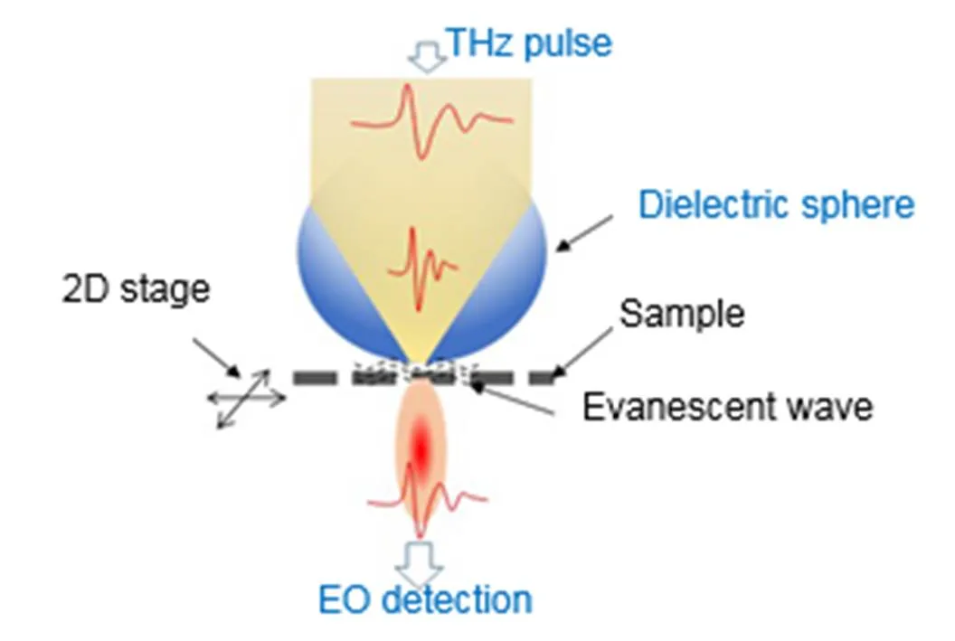
Experimental configuration of dielectric sphere-coupled THz microscopy
Overview: In the past decades, great advancements have been made to achieve super-resolution imaging, including near-field THz microscopy, metamaterial superlens, fluorescence microscopy and so on, pushing the resolution tomm or nm scale. Unfortunately, applications of these methods have been limited in part due to their complication in access and operation, loss of energy and spectral bandwidth, difficulty in information extraction or limited choices of samples. Thus, it is highly desired to develop innovative super-resolution THz imaging modality that is easily accessible and low-cost. Fortunately, at visible frequencies, a unique easy-access super-resolution imaging where dielectric microsphere with appropriate refractive index has been presented and delivered a remarkable 50 nm resolution with white lights in 2010. Furthermore, super-resolution imaging was also presented with a large field-of-view using large polystyrene microspheres (above 30 μm). A strong continuing interest in the technique has led to numerous progress in visible light. In these works, the super-resolution capability of microspheres is determined by the “photonic nanojet” and coupling with evanescent waves. More recently, a straightforward THz imaging method based on terajet effect, analogous to microsphere optical nanoscope, is proposed and developed with spatial resolution beyond the diffraction limit by using either continuous or pulsed THz wave. The terajet beam can break through the restriction of the diffraction limit on the spatial resolution of the microscopic system without losing the energy and spectral bandwidth of the THz field, i.e., a high-resolution, high-throughput and broad-bandwidth THz imaging method. Also, with the extensively longer wavelength of the THz wave, the size of the dielectric spheres is much larger (on the order of millimeters), the spheres are easier to fabricate, simple to manipulate, and capable of handling energy and bandwidth losses. In addition, as unique spectroscopic technique, THz imaging reveals much richer subwavelength structural information, including frequency-dependent amplitude and phase, as well as time-dependent delay and thickness. In this review, firstly, a white-light nanoscopy based on photonic nanojet produced by microspheres is introduced, then the terahertz microscopy based on terajet effect produced by mesoscopic dielectric structures is reviewed. Finally, the prospect of terahertz high resolution imaging technology based on terajet effect is prospected.
Citation: Ma X M, Jiang Z C, Qu Q S,. Research advances of high-resolution THz imaging based on terajet effect[J]., 2020,47(5): 190590
Research advances of high-resolution THz imaging based on terajet effect
Ma Xiaoming1, Jiang Zaichao1, Qu Qingshan1, Cui Bin1, Zhang Zhenwei2, Yang Yuping1*
1School of Science, Minzu University of China, Beijing 100081, China;2Department of Physics, Capital Normal University; Beijing Advanced Innovation Center for Imaging Technology; Beijing Key Laboratory for Terahertz Spectroscopy and Imaging; Key Laboratory of Terahertz Optoelectronics, Ministry of Education, Beijing 100048, China
Terahertz (THz) imaging technology has shown great advantages and potential applications in the fields of biomedicine, security, and aerospace, due to its low energy, high transmittance, wide bandwidth, and unique analysis abilities; while low spatial resolution restricts its further applications. Recently, a high-resolution, high-throughput, and broad-bandwidth THz imaging method has been proposed based on the terajet effect produced by dielectric structures with appropriate refractive index. The terajet beam can break through the restriction of the diffraction limit on the spatial resolution of the microscopic system without losing the energy and spectral bandwidth of the THz field. In this paper, firstly, a white-light nanoscopy based on photonic nanojet produced by microspheres is introduced, then the THz microscopy based on terajet effect produced by mesoscopic dielectric structures is reviewed. Finally, the prospect of THz high resolution imaging technology based on terajet effect is prospected.
terahertz imaging; terajet; spatial resolution
National Natural Science Foundation of China (11574408), the National Key R&D Program of China (2017YFB0405400), the Young-talent Plan of State Affairs Commission (2016-3-02), and the Undergraduate Innovative Test Program funded by Minzu University of China (URTP2019110002)
* E-mail: ypyang@muc.edu.cn
TH742;O439
A
马晓茗,姜在超,屈庆山,等. 基于喷射效应的太赫兹高分辨成像研究与进展[J]. 光电工程,2020,47(5): 190590
10.12086/oee.2020.190590
: Ma X M, Jiang Z C, Qu Q S,Research advances of high-resolution THz imaging based on terajet effect[J]., 2020, 47(5): 190590
2019-09-30;
2020-01-06
国家自然科学基金资助项目(11574408);国家重点研发计划(2017YFB00405400);国家民委“中青年英才”培养计划(2016-03-02);中央民族大学大学生创新性实验计划(URTP2019110002)
马晓茗(1999-),女,主要从事太赫兹光谱和成像的研究。E-mail:2321823700@qq.com
杨玉平(1976-),女,博士,教授,主要从事太赫兹光谱和成像的研究。E-mail:ypyang@muc.edu.cn
版权所有©2020中国科学院光电技术研究所