Lumen-apposing metal stent deployment for walled-off necrosis using semi-free hand technique from the intestine
2020-05-10TakeshiOguraNobuNishiokaKazuhideHiguchi
Takeshi Ogura , Nobu Nishioka, Kazuhide Higuchi
2nd Department of Internal Medicine, Osaka Medical College, 2-7 Daigakuchou, Takatsukishi, Osaka 569-8686, Japan
Lumen-apposing metal stent (LAMS) deployment under endoscopic ultrasonography (EUS) guidance has been used for walled-off necrosis (WON) [1] . Recently, LAMS with electrocautery enhanced delivery system has been developed. One of the advantages of this stent is that the stent can be delivered without any dilation devices. However, to prevent stent migration into the intestinal lumen, it is important that stent delivery system should be inserted within the WON. When the diameter of WON is not large enough to insert stent delivery system, contralateral wall of WON may be injured. To prevent this adverse event, guidewire insertion may be useful after puncturing. In addition, according to the recent reports [2-4] , EUS-guided LAMS placement for WON drainage has been performed. We herein described a case of LAMS deployment from the second part of the duodenum using semi-free hand technique.
A 56-year-old man was admitted to hospital due to acute pancreatitis. After conservative treatment, WON, which was 65 mm diameter in pancreatic head, was diagnosed. The patient was firstly treated with drainage under endoscopic retrograde cholangiopancreatography (ERCP) guidance. Dudenoscope (JF 260V; Olympus Optical, Tokyo, Japan) was inserted into the duodenum, and cannulation using ERCP catheter (MTW Endoskopie, Düsseldorf, Germany) for pancreatic duct was performed. Pancreatography showed that the pancreatic head duct was obstructed by WON and the connection between pancreatic duct and WON was not observed( Fig. 1 (A)). Although pancreatic duct stent deployment was performed, the drainage was not satisfactory ( Fig. 1 (B)). Therefore,EUS-guided access was attempted. First, A convex EUS (GF-UCT240 or 260; Olympus Optical) connected to an ultrasound device(SSD5500; Aloka, Tokyo, Japan), was initially advanced into the second part of the duodenum, and WON was identified ( Fig. 1 (C)).Then, WON was punctured using electrocautery-enhanced LAMS(Hot AXIOS, Boston Scientific). Because diameter of WON was relatively small to completely insert LAMS delivery system, we insert the 0.025-inch VisiGlide guidewire (Olympus Medical Systems, Tokyo, Japan) to prevent injuring the contralateral wall of WON (semi-free hand technique) ( Fig. 2 (A)). Next, stent was released completely within the EUS scope to prevent stent migration( Fig. 2 (B)). Finally, we inserted 7-Fr double pig tail plastic stent within the LAMS ( Fig. 2 (C)), and WON was completely resolved after 7 days ( Fig. 2 (D)). LAMS was removed after 14 days, and the patient was discharged without any adverse events.
In the duodenum, compared with the stomach, the space of the lumen is limited. If conventional metal stent deployment is performed, the contralateral duodenum lumen may be injured by stents itself. Therefore, LAMS might be useful as drainage device compared with conventional metal stent. In addition, if the target is not large enough to insert the LAMS delivery system, semi-free hand technique may be safe.
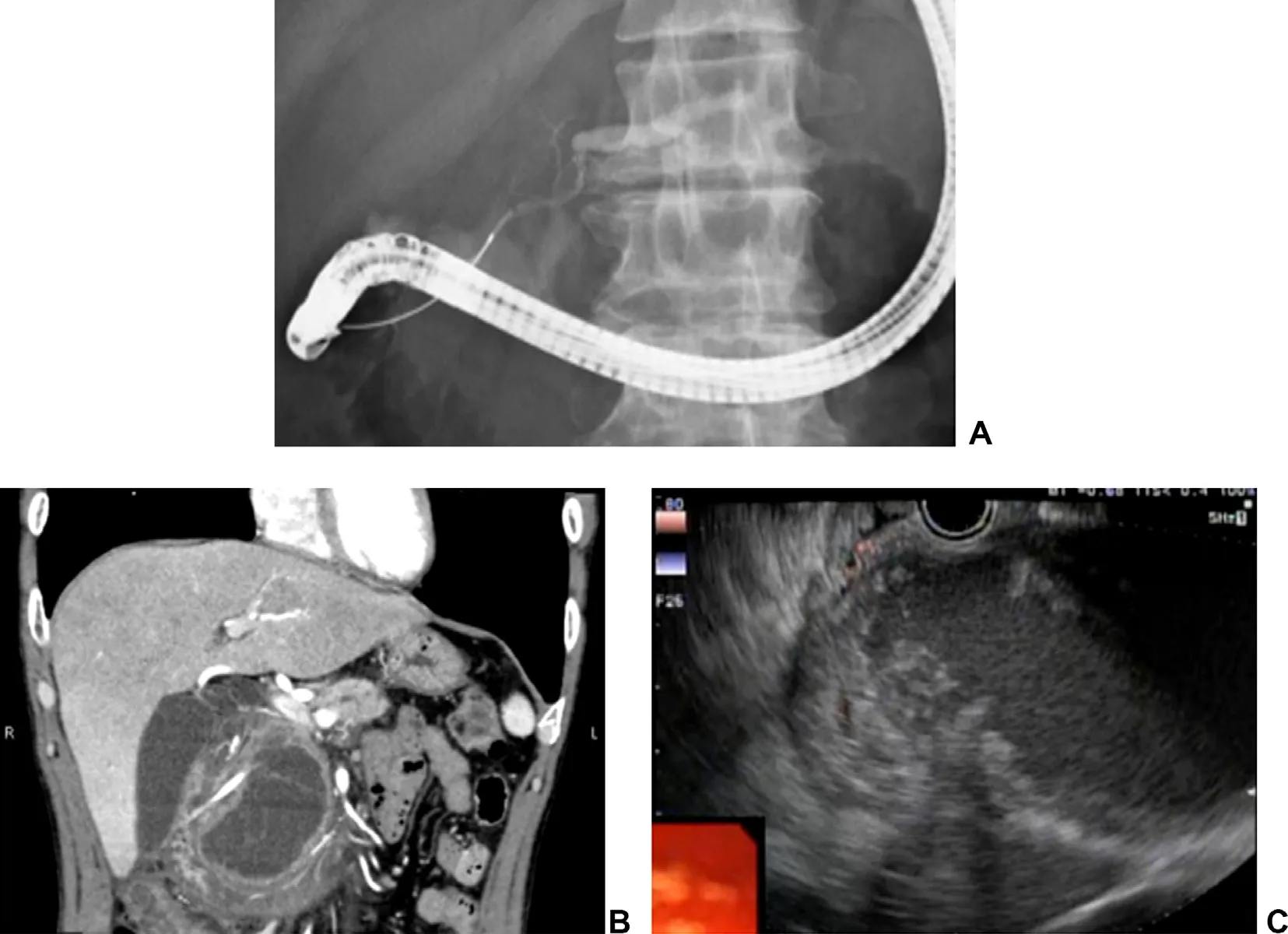
Fig. 1. ( A) Pancreatography shows no connection between pancreatic duct and walled-off necrosis. ( B) Walled-off necrosis is seen in pancreatic head. ( C) EUS imaging of walled-off necrosis.
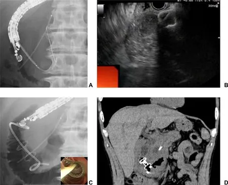
Fig. 2. ( A) Walled-off necrosis was punctured using electrocautery-enhanced LAMS. The guidewire was inserted to prevent the wall injuring by inserting LAMS delivery system. ( B) Stent release was completely performed within the EUS scope to prevent stent migration. ( C) LAMS deployment was successfully performed, and double pig tail plastic stent was also deployed within the LAMS. ( D ) Walled-off necrosis was successfully resolved.
CRediT authorship contribution statement
Takeshi Ogura:Writing - original draft.Nobu Nishioka:Data curation.Kazuhide Higuchi:Data curation.
Funding
None.
Ethical approval
Patient informed content was obtained.
Competing interest
No benefits in any form have been received or will be received from a commercial party related directly or indirectly to the subject of this article.
杂志排行
Hepatobiliary & Pancreatic Diseases International的其它文章
- Neoadjuvant chemoradiotherapy before resection of perihilar cholangiocarcinoma: A systematic review
- Hepatobiliary&Pancreatic Diseases International
- Human microbiome is a diagnostic biomarker in hepatocellular carcinoma
- Current practice of anticoagulant in the treatment of splanchnic vein thrombosis secondary to acute pancreatitis
- Enhanced recovery after surgery program in the patients undergoing hepatectomy for benign liver lesions
- Assessment of biological functions for C3A cells interacting with adverse environments of liver failure plasma