基于薄层标本断面建立可视性三维颅底模型*
2020-05-08孙基栋朱荣江刘树伟
孙基栋 刘 军 朱荣江 刘树伟
(1 中国医科大学航空总医院神经外科,北京 100012;2 湛江中心人民医院神经外科,湛江 524045;3 山东大学断层与影像研究中心,济南 250012 )
The central skull base includes the sellar region,cavernous sinus,and clivus region.It is necessary to understand this complex anatomy for clinical diagnosis and surgical planning in the region[1-2].Several computerbased three-dimensional(3D)surgical models have been developed from computed tomography(CT),magnetic resonance imaging(MRI),and anatomical specimens[3-7].These models have been used to train residents and attend physicians preoperatively,and to practice virtual surgery on a computer screen.The limited image resolution and internal distortion of imaging sections,however,means that some surgical models based on imaging data can’t delineate the minor structures clearly,such as the cranial nerves and vessels[8].These models are inadequate for training.The computerized freezing milling technique,developed for the virtual human project,has become an optimal method to obtain thin histological slices[9-10].These thin slices have enough details to be sufficient for 3D reconstruction.Using thin sections,we created a highresolution 3D model of the central skull base for doctors to master the regional anatomy.
1 Materials and methods
1.1 Acquisition of thin specimen sections
Two adult cadaveric specimens resulting from accidental death were obtained,and voluntary consents were provided by the families of the deceased for medical research.This study was approved by the ethical committee of Shandong university,school of medicine,Shandong,People’s republic of China.The arteries were injected with red-colored latex through common carotid arteries.Veins were injected with bluecolored latex.
After being embedded with 5% gelatin and frozen to -40℃for 1 week,one specimen was sliced into 0.05-mmthick continuous sections in the coronal plane perpendicular to the canthomeatal line,from anterior to posterior,with an SKC500 computerized freezing milling machine(accuracy of 0.001 mm)in a -15 ℃ laboratory.The other specimen was sliced into 0.05 mm continuous sections in the transverse plane along the anterior commissure-posterior commissure line from inferior to superior.The serial sections were photographed with a high-resolution digital camera and imported into an animation computer.Each picture was numbered and placed in an alignment frame[11].
1.2 Segmentation and extracting organs from specimen sections
The two sets of sections were preprocessed for color adjustment and registration.The 780 coronal sections were segmented into 518 432 pixel images of the central skull base,and the 413 transverse sections were segmented into 532 437 pixel images,using Photoshop 7.0(Adobe,San Jose,CA,USA).Some structures(internal carotid artery,ICA;pituitary gland;cavernous sinus;central skull base;Ⅶ-Ⅷ cranial nerve complex)were manually traced in each coronal section throughout the series until the structures could no longer be visualized.Each image was enlarged by 200% and the salient structures were outlined with the magnetic lasso tool.These structures were then copied to a new layer and were filled with a different grey value.Other structures(cranial nervesⅢ,Ⅴ,Ⅵ;Ⅸ-Ⅹ-Ⅺ cranial nerve complex;Ⅻ and vertebral-basilar arteries)were tracked and segmented in transverse sections.All pictures were stored in BMP format(Fig 1,see inside back cover).The entire dataset was transferred and processed using Amira 4.1 software(TGS Company,France).The related structures were separately reconstructed and displayed.A smoothing function was applied to more clearly distinguish the segmented organs,and the structures were color coded.The background was assigned to be light blue,the arteries in red,and the nerves in yellow.With this information,the software reconstructed the 3D structures.
1.3 3D modeling of the central skull base
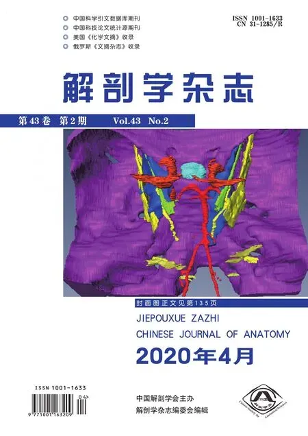
The reconstructed organs from both head specimens were loaded,rotated,translated,and fused together in a single 3D model(using Amiro’s Animation/Demo function).The surfaces of the volume were smoothed appropriately(Fig 2,see inside back cover).
Photographs of one of the cadaver head specimens were taken.The anterolateral and posterosuperior views were studied to evaluate the anatomical accuracy and interactivity of the 3D model.
2 Results
The 3D images of the central skull base,ICA,vertebral-basilar artery,cavernous sinus,pituitary gland,the cranial Ⅲ,Ⅴ,Ⅵ,Ⅶ-Ⅷ cranial nerve complex,Ⅸ-Ⅹ-Ⅺ cranial nerve complex,and Ⅻ nerves were obtained separately.The structures in the 780 thin coronal sections and 413 transverse sections of the base region in the central skull were all traceable.The 3D model of the central skull base was successfully produced.All reconstructed structures could be displayed individually or jointly,rotated in any plane,and reduced or enlarged using the zoom function.The structures in the model could be displayed in any color(Fig 3,see inside back cover),and the 3D model could be cut at defined X,Y,and Z clipping planes.Compared with cadaver specimen photographs,the 3D model could show mutual spatial relationships of anatomical structures(Fig 4-5,see inside back cover).There were some non-anatomical anomalies in the model because it was reconstructed with organs from two head specimens.
3 Discussion
The knowledge of fine anatomical details is crucial during complex skull base surgery[12-14].Traditionally,surgeons study 2D diagrams,photographs,and atlases derived from CT,MRI,and cadaveric dissections[15-16].It is nontrivial,however,to visualize the anatomy of the skull base in 3D from these 2D images[17].Cadaveric dissections are thus the best means of learning skull base anatomy.Because of legal and ethical problems,obtaining cadaveric material can prove difficult in some countries and areas.Cadaveric heads are also associated with the transmission of infection to the students who dissect them.Furthermore,cadaver dissection courses are expensive and time-consuming[18].Several artificial models have been used in skull base anatomical training in recent years.Nonetheless,the anatomic accuracy of these models is limited,which restricts their virtual surgical manipulation[19-20].
The advances in 3D imaging,image processing hardware and software,and virtual reality technology have led to computer-generated models being used for training and rehearsal in skull base surgery[21-22].Those 3D models were derived from 2D data,such as CT,MRI,and specimen sections.Some fine anatomical structures,such as small nerves and vessels in the skull base,however,couldn’t be acquired by imaging devices that had limited signal-to-noise ratio and section depth.Furthermore,the reconstruction from 2D slices makes it difficult to establish the continuity of some structures[23].Compared with low-resolution imaging data,our color photographs of 0.05 mm freezing milling sections were of sufficient resolution for accurate 3D reconstruction.Using high-speed drilling and without decalcification,it had no section depth and maintained normal structures that were easily deformed.In the skull base,some nerves and vessels are not discernable from the background tissues.There are variations in the intensity,contrast,shape,and direction of nerves and vessels,making their segmentation challenging[24-25].From our two head specimens,we got thin coronal and transverse sections,across which we were able to observe and track structures.Cranial nerves and vessels were delineated and preserved continuity with an edge-preserving-smoothing function.On the coronal sections,we delineated some structures aligned medial to lateral(pituitary gland,vertebral-basilar artery,Ⅶ-Ⅷ cranial nerve complex)from the surrounding anatomical structures.On the transverse sections,some nerves running from anterior to posterior(cranial nerves Ⅲ,Ⅴ,Ⅵ;Ⅸ-Ⅹ-Ⅺ cranial nerve complex,Ⅻ,)were able to be tracked.The cranial nerves and vessels were then reconstructed and displayed separately using surface rendering.
The model produced in this study has sufficiently high resolution to adequately and accuratelly show the relevant structures.The 3D data can be made into video animations that continuously and dynamically display anatomic structures in 3D space from any viewpoint.We submit that our model will benefit preoperative surgical rehearsal for the skull base[26].However,there were some anatomical inaccuracies because the model was reconstructed from two specimens.
A 3D model of the central skull base that displays fine anatomical structures is established.We propose that this model be used as part of a preoperative surgical training program and to increase anatomical knowledge of the skull base.
Explanation of figures(See inside back cover)

Fig1 Transverse sections with tracked and labeled Ⅵ and V nerves(dark grey).1:Abducent nerve;2:Trigeminal nerve.

Fig2 Amira editor window showing the cranial V and VI nerves in the skull base model.1:Trigeminal nerve;2 :Abducent nerve.

Fig3 Anatomic relationships of structures of central skull base in the model.A,B :Anatomical relationships between the internal carotid artery,pituitary gland,and cavernous sinuses ;B :The trigeminal nerve was also added.C :Anterior view;D:Superior-posterior view;E:Superior view;F:Anterior-lateral view.This model demonstrated the ability to include or exclude any structures of interest,and use custom color palettes.The transparency effect of the virtual bone in C and F,revealed the important spatial relationships of the model.

Fig4 Posterosuperior view of the specimen and the model.A :Specimen;B:Model.1 :Pituitary gland;2 :Oculomotor nerve;3 :Trigeminal nerve;4 :Abducent nerve;5:Ⅶ-Ⅷ cranial nerve complex;6 :The Ⅸ- Ⅹ- Ⅺ cranial nerve complex;7 :Sublingual nerve;8 :Vertebral-basilar artery.

Fig5 Anterolateral view of the specimen and the model.A :Specimen ;B :Model.1 :Anterior clinoid process ;2 :Trigeminal nerve;3:Cavernous sinus;4:Trochlear nerve;5:Pituitary gland.
