CDK13基因对缺氧性脑损伤小鼠细胞凋亡的影响
2019-02-20王荣跃鲁文洁邱海凡戴芬
王荣跃 鲁文洁 邱海凡 戴芬


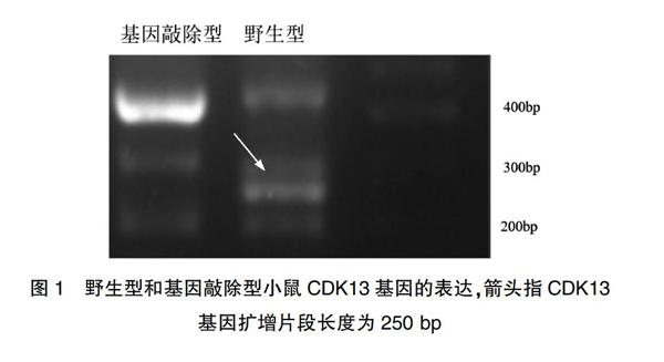
[摘要] 目的 研究CDK13基因对缺氧性脑损伤小鼠细胞凋亡的影响。 方法 将野生型和CDK13基因敲除型小鼠分别分为野生型假手术组(SWT)、野生型模型组(MWT)、基因敲除假手术组(SKO)及基因敲除模型组(MKO)。采用PCR反应法检测CDK13基因表达、TTC染色法检测脑梗死程度、免疫组化法检测脑组织中活化型半胱天冬酶-3(CC3)表达、TUNEL法检测细胞凋亡情况,四组小鼠进行相关指标比较。 结果 野生型模型组(MWT)小鼠脑组织梗死程度较基因敲除模型组(MKO)小鼠明显减轻(P<0.05),且脑组织凋亡阳性细胞数明显减少,凋亡指数降低,凋亡蛋白CC3表达减少(P<0.05)。 结论 敲除CDK13基因具有加剧小鼠缺氧性脑损伤的作用。
[关键词] CDK13基因;基因敲除;细胞凋亡;模型
[中图分类号] R722.1 [文献标识码] A [文章编号] 1673-9701(2019)34-0039-04
Effect of CDK13 gene on apoptosis in mice with hypoxic brain damage
WANG Rongyue1 LU Wenjie2 QIU Haifan1 DAI Fen1
1.Department of Obstetrics and Gynecology, the Second Affiliated Hospital of Wenzhou Medical University, Wenzhou 325000, China; 2.Outpatient Department, the Second Affiliated Hospital of Wenzhou Medical University, Wenzhou 325000, China
[Abstract] Objective To study the effect of CDK13 gene on apoptosis in mice with hypoxic brain injury. Methods Wild type and CDK13 knockout mice were divided into wild type sham operation group (SWT), wild type model group (MWT), gene knockout sham operation group (SKO) and gene knockout model group (MKO). The expression of CDK13 gene was detected by PCR reaction. The degree of cerebral infarction was detected by TTC staining. The expression of activated caspase-3 (CC3) in brain tissue was detected by immunohistochemistry. The apoptosis was detected by TUNEL method. Four groups of mice were compared. Results Compared with the MKO group, the degree of cerebral infarction in the MWT group was significantly reduced (P<0.05), and the number of apoptotic positive cells, apoptotic index and CC3 expression were significantly decreased(P<0.05). Conclusion Knockout of CDK13 gene aggravates hypoxic brain damage in mice.
[Key words] CDK13 gene; Gene knockout; Apoptosis; Model
智力障礙(mental retardation,MR)是指出现在18周岁之前以认知障碍和社会适应能力缺陷为主要表现的一种常见的出生缺陷疾病,在世界范围内患病率约为1%~3%[1,2]。智力障碍严重危害儿童及青少年的身心健康,MR具有终生发病和不可治愈的特点,目前研究认为细胞坏死及凋亡是神经元细胞死亡的主要方式,同时研究发现脑缺氧神经元损伤有多种死亡机制参与[3,4],但细胞凋亡发生的具体机制仍不清楚。
CDK13(cyclin-dependent kinases 13,CDK13)是最晚被发现的细胞周期依赖性激酶家族成员,该家族蛋白C端有一段20个ATP依赖的丝氨酸-苏氨酸蛋白激酶结构,通过整合细胞内外的信号,实现细胞循环和基因转录过程的调控功能。CDK12和CDK13具有较多的同源序列,最近发现其可能在转录和加工RNA过程中发挥重要作用[3,4]。人类CDK13蛋白分子量较大,为165 kDa,已知CDK13与周期蛋白Cyclin K结合形成蛋白复合物可发挥生物学功能[6,7]。CDK13通过磷酸化丝氨酸蛋白酶Omi/HtrA2促进细胞凋亡[8],在前期研究中发现CDK13基因缺失可导致智力障碍[9],推测CDK13可能参与脑缺氧过程引起细胞凋亡。因此本研究以缺氧性脑损伤作为研究区域,采用线栓法建立新生鼠(MCAO)模型,探讨CDK13基因敲除对小鼠缺氧性脑损伤细胞凋亡的影响。
2017年,Bostwic BL等[18]对一组CDK13新发突变患儿的临床表型进行深入分析,发现CDK13变异可致患儿发生先天性心脏病、面容异常及智力发育障碍,但具体分子机制仍不详。本研究发现,两组假手术组小鼠之间神经元凋亡数目无差异,而CDK13基因敲除后凋亡明显增加,提示CDK13基因敲除后可能通过激活其他路径上的蛋白加重脑损伤,如JNK[20,21]、p53[22]、NF-κB[23,24]等,其在细胞凋亡中可能起重要作用,对深入阐明CDK13敲除引起小鼠脑组织损伤的作用机制具有重要意义。CDK13缺失可能导致小鼠突触前谷氨酸等兴奋性递质的过度释放,激活p38MARK,通过p21等表达影响细胞凋亡,降低缺氧小鼠脑神经细胞损伤。本研究同时观察到敲除CDK13后能降低脑神经细胞凋亡的发生,与Wu HJ等[25]的报道基本符合。本研究发现CDK13基因敲除后小鼠脑组织凋亡程度较重,且凋亡蛋白表达明显增加,提示神经细胞损伤增加,但这一路径的中间过程仍有待下一步深入研究。
综上所述,敲除CDK13基因具有加剧小鼠缺氧性脑损伤的作用,为明确智力障碍的原因提供了新思路,但具体分子机制需进一步研究阐明。
[参考文献]
[1] Roehr B. American psychiatric association explains DSM-5[J]. BMJ,2013,346:f3591.
[2] Moeschler JB,Shevell M. Comprehensive evaluation of the child with intellectual disability or global developmental delays[J]. Pediatrics,2014,134(3):e903-e918.
[3] Parikh P,Juul SE. Neuroprotective strategies in neonatal brain injury[J]. J Pediatr,2018,192:22-32.
[4] Wang JY,Xia Q,Chu KT,et al. Severe global cerebral ischemia-induced programmed necrosis of hippocampal CA1 neurons in rat is prevented by 3-methyladenine: A widely used inhibitor of autophagy[J]. J Neuropathol Exp Neurol,2011,70(4):314-322.
[5] Malumbres M,Harlow E,Hunt T,et al. Cyclin-dependent kinases: A family portrait[J]. Nat Cell Biol,2009,11(11):1275-1276.
[6] Chen HH,Wong YH,Geneviere AM,et al. CDK13/CDC2L5 interacts with L-type cyclins and regulates alternative splicing[J]. Biochem Biophys Res Commun,2007,354(3):735-740.
[7] Greifenberg AK,Honig D,Pilarova K,et al. Structural and functional analysis of the Cdk13/Cyclin K complex[J].Cell Rep,2016,14(2):320-331.
[8] Niemi NM,MacKeigan JP. Mitochondrial phosphorylation in apoptosis: Flipping the death switch[J]. Antioxid Redox Signal,2013,19(6):572-582.
[9] 王榮跃,雷婷缨,符芳,等. 染色体微阵列分析技术在489例生长发育迟缓/智力低下患儿中的应用[J]. 中华医学遗传学杂志,2017,34(4):528-533.
[10] Cho SE,Kim YM,Jeong JS,et al. The effect of ultrasound for increasing neural differentiation in hBM-MSCs and inducing neurogenesis in ischemic stroke model[J]. Life Sci,2016,165:35-42.
[11] Zhu J,Qu Y,Lin Z,et al. Loss of PINK1 inhibits apoptosis by upregulating alpha-synuclein in inflammation-sensitized hypoxic-ischemic injury in the immature brains[J]. Brain Res,2016,1653:14-22.
[12] Minami A,Nakanishi A,Matsuda S,et al. Function of alpha-synuclein and PINK1 in Lewy body dementia(Review)[J]. Int J Mol Med,2015,35(1):3-9.
[13] Zhou Z,Fu XD. Regulation of splicing by SR proteins and SR protein-specific kinases[J]. Chromosoma,2013, 122(3):191-207.
[14] Dixon BJ,Reis C,Ho WM,et al. Neuroprotective strategies after neonatal hypoxic ischemic encephalopathy[J]. Int J Mol Sci,2015,16(9):22368-22401.
[15] Trinh J,Kandaswamy KK,Werber M,et al. Novel pathogenic variants and multiple molecular diagnoses in neurodevelopmental disorders[J]. J Neurodev Disord,2019, 11(1):11.
[16] Lipp JJ,Marvin MC,Shokat KM,et al. SR protein kinases promote splicing of nonconsensus introns[J]. Nat Struct Mol Biol,2015,22(8):611-617.
[17] Liang K,Gao X,Gilmore JM,et al. Characterization of human Cyclin-Dependent Kinase 12(CDK12) and CDK13 Complexes in C-Terminal domain phosphorylation, gene transcription,and RNA processing[J]. Molecular and Cellular Biology,2015,35(6):928-938.
[18] Bostwick BL,McLean S,Posey JE,et al. Phenotypic and molecular characterisation of CDK13-related congenital heart defects, dysmorphic facial features and intellectual developmental disorders[J]. Genome Med,2017,9(1):73.
[19] Hamilton MJ,Caswell RC,Canham N,et al. Heterozygous mutations affecting the protein kinase domain of CDK13 cause a syndromic form of developmental delay and intellectual disability[J]. J Med Genet,2018,55(1):28-38.
[20] 韓琳,王洪新,鲁美丽. 黄芪多糖通过抑制NF-κB和JNK信号通路减轻LPS诱导的小鼠心肌细胞凋亡[J]. 中国药理学通报,2018,34(2):243-249.
[21] Wang Q,Gan X,Li F,et al. PM2.5 exposure induces more serious apoptosis of cardiomyocytes mediated by caspase3 through JNK/P53 pathway in hyperlipidemic rats[J]. Int J Biol Sci,2019,15(1):24-33.
[22] Tang R,Mei X,Wang YC,et al. LncRNA GAS5 regulates vascular smooth muscle cell cycle arrest and apoptosis via p53 pathway[J]. Biochim Biophys Acta Mol Basis Dis,2019,1865(9):2516-2525.
[23] 丁丹,焦丽华, 王雪臣,等. 灯盏花素对大鼠心肌缺血再灌注损伤心肌细胞凋亡及NF-κB通路信号分子α7nAChR、p65、IkB-α的影响[J]. 中国循证心血管医学杂志,2018,10(12):1480-1483.
[24] Raish M,Ahmad A,Ansari MA,et al. Momordica charantia polysaccharides ameliorate oxidative stress, inflammation, and apoptosis in ethanol-induced gastritis in mucosa through NF-κB signaling pathway inhibition[J]. Int J Biol Macromol,2018,111:193-199.
[25] Wu HJ,Pu JL,Krafft PR,et al. The molecular mechanisms between autophagy and apoptosis: Potential role in central nervous system disorders[J]. Cell Mol Neurobiol,2015,35(1):85-99.
(收稿日期:2019-06-17)
