Lithium promotes recovery of neurological function after spinal cord injury by inducing autophagy
2018-10-22DuoZhangFangWangXuZhaiXiaoHuiLiXiJingHe
Duo Zhang , Fang Wang, Xu Zhai, Xiao-Hui Li, Xi-Jing He,
1 Department of Orthopedics, Beijing Tiantan Hospital, Capital Medical University, Beijing, China
2 Department of Orthopedics, Second Affiliated Hospital of Xi’an Jiaotong University, Xi’an, Shaanxi Province, China
3 Department of Emergency, Second Affiliated Hospital of Xi’an Jiaotong University, Xi’an, Shaanxi Province, China
4 Department of Radiology, Second Affiliated Hospital of Xi’an Jiaotong University, Xi’an, Shaanxi Province, China
Abstract
Key Words: nerve regeneration; spinal cord injury; lithium; secondary injury; autophagy, diffusion tensor imaging; neuroprotection; functional recovery; immunohistochemistry; Beclin-1; light-chain 3B; neural regeneration
Introduction
Spinal cord injury (SCI) is the direct or indirect damage to any part of the spinal cord that results in permanent impairment in strength, sensation or other body function below the injury site (Wyndaele and Wyndaele, 2006; Krause et al.,2017). Secondary injury mechanisms play important roles in the acute, sub-acute and chronic phases and lead to vasospasm, ischemia, inflammation, and free radical production(Oyinbo, 2011). These events result in neuronal loss, which is the key cause of permanent neurological dysfunction(Kanno et al., 2009; Tang et al., 2014; Li et al., 2015a; Kwan et al., 2017).
Cell autophagy or type II programmed cell death is an intracellular catabolic mechanism for recycling damaged organelles and senescent proteins and plays a very important role in cell survival, differentiation and homeostasis (Erlich et al., 2007; Mizushima et al., 2010). It was reported that enhancing autophagy promotes the recovery of neurological functions by inhibiting apoptosis (Sekiguchi et al., 2012; Liu et al., 2015; Colón and Miranda, 2016; Dai et al., 2017). Sev-eral studies have reported that autophagy is involved in SCI.Lysosomal dysfunction and the disruption of autophagy, as well as increased neuronal apoptosis, have been observed in SCI, suggesting that autophagy is involved in SCI (Silva et al., 2008; Liu et al., 2015). Accordingly, an increasing number of studies have focused on the therapeutic effect of modulating autophagy in SCI.
Lithium is the first line drug for treating bipolar disorder, and provides neuroprotection in multiple neurologic diseases (Young, 2009; Huo et al., 2012). Accumulating evidence suggests that lithium has numerous actions, including neuroprotection, inflammation inhibition, induction of neurotrophic factor secretion, and the enhancement of neurogenesis (Son et al., 2003; Senatorov et al., 2004; Su et al., 2007; Yasuda et al., 2009; Yuskaitis and Jope, 2009; Chi-Tso and Chuang, 2011; De Meyer et al., 2011; Li et al., 2011;Lauterbach, 2016). However, it remains unclear whether autophagy plays a positive or negative role in SCI (O’Donovan et al., 2015; Del Grosso et al., 2016; Fabrizi et al., 2017).
Multiple signaling pathways are involved in autophagy,including the PI3K/Akt/mTOR, AMPK/TSC/mTOR and eIF2α/Atg12 pathways (Periyasamy-Thandavan et al., 2009).It was reported that lithium affects several signaling pathways, including the PI3K/Akt and IP3 pathways, which are involved in autophagy. Therefore, it is reasonable to speculate that lithium promotes functional recovery by inducing autophagy in rat models of SCI, which has not yet been studied (Periyasamy-Thandavan et al., 2009; Chi-Tso and Chuang, 2011; Li et al., 2015b).
In the present study, we investigate the neuroprotective effect of lithium and the role of autophagy in SCI using the autophagy inhibitor 3-methyladenine (3-MA). To objectively and accurately evaluate recovery following SCI, we performed diffusion tensor imaging (DTI), which is an effective method of assessing neurological recovery, in addition to the Basso, Beattie, Bresnahan (BBB) locomotor rating scale(Zhang et al., 2015).
Materials and Methods
Animal care and groups
A total of 72 specific-pathogen-free adult healthy male Sprague-Dawley rats weighing 230–270 g were provided by the Experimental Animal Center of Xi’an Jiaotong University of China (production license No. SCXK (Shaan) 2007-001; user license No. SYXK (Shaan) 2007-003). All rats were housed under a 12-hour light/dark cycle. All experimental procedures were performed in accordance with the Guidelines for the Care and Use of Laboratory Animals published by the US National Institutes of Health. The protocols were approved by the Animal Ethics Committee of Xi’an Jiaotong University of China. All efforts were made to minimize distress to the rats.
The rats were randomly separated into sham, SCI, lithium and 3-MA groups (n = 18 per group). Six rats from each group were randomly selected for BBB scoring and DTI examination, and three rats from each group were sacrificed for immunohistochemical staining at each time point.
SCI model
Rats in the SCI, lithium and 3-MA groups received SCI operation as previously described (Basso et al., 1995). Briefly, the rat was anesthetized with intraperitoneal injection of chloral hydrate (300 mg/kg) and placed in the prone position with a heating pad to maintain body temperature. After shaving and aseptic preparation, the spinal cord was exposed. Dorsal laminectomy was performed at T9–11. The T10 segment of the spinal cord was impacted with an NYU weight-drop impactor (10 g rod dropped from a height of 25 mm; Rutgers University, USA), which led to hemorrhage and edema at the site of impact, wagging tail reflex and lower limb spasm. All manifestations indicated the success of the injury model. The rats in the sham group underwent laminectomy alone. The tissue was sutured layer by layer, with a piece of fat sutured under the skin at the T10 level. After SCI surgery, manual bladder massage was performed three times, and intraperitoneal injection of penicillin 20 U/kg was given once daily until bladder function was reestablished.
Lithium and 3-MA treatments
Lithium chloride (LiCl; Kemiou, Tianjin, China) and 3-MA(Sigma-Aldrich, St. Louis, MO, USA) were dissolved in 0.9%NaCl. Rats in the 3-MA group were given intraperitoneal injection of 1 mL 3-MA (3 mg/kg) 2 hours before SCI (Chen et al., 2013; Tang et al., 2014). Rats in the lithium and 3-MA groups were administered 1 mL lithium by intraperitoneal injection (LiCl, 30 mg/kg) 6 hours after SCI and then once daily until sacrifice (Yick et al., 2004; Zakeri et al., 2014). The sham and SCI groups received 1 mL 0.9% NaCl via intraperitoneal injection.
Neurological function assessment
The BBB locomotor rating scale was used to assess neurological function after SCI (Zhang et al., 2015). The BBB score ranges from 0 (complete paralysis) to 21 (normal), based on the range and extent of motion, weight loading, coordination of the forelimbs and hindlimbs, and motion of the forepaw, hindpaw and tail. Three independent examiners blindly assessed the BBB score before operation, and at 6 hours and 1, 2, 3 and 4 weeks after SCI. The average score was taken as thefinal score for each rat at each time point.
DTI examination
A DTI scan was performed 24 hours before SCI and at 6 hours and 1, 2, 3 and 4 weeks after SCI using a 3.0 T SIGNA MRI scanner (GE Medical Systems, Milwaukee, WI,USA) at the same loci as the conventional MRI scan. The scanning parameters were as follows: diffusion-weighted coefficient (b-value) = 1000 s/mm2; diffusion-sensitive gradient = 15 different directions; repetition time = 3500 ms;echo time = 87.5 ms; thickness = 2.4 mm; space = 0;field of view = 10; acquisition matrix = 64 × 64. All data were input into a workstation running Advantage Windows 4.2 (GE Healthcare). The region of interest (ROI) was identified by the fat under the skin, which was displayed as a high signal on conventional T2WI MRI (Yan et al., 2007). Based on the fractional anisotropy (FA) map, the ROI was placed in the inferior medulla and the inferior oblongata. The ROI was selected by two independent testers, and apparent diffusion coefficient (ADC) and FA values were obtained. FA values reflect the degree of spatial displacement of water molecules,and higher FA values indicate stronger anisotropy. ADC values are independent of the diffusion directions, and indicate the diffusional displacement of water molecules.
Immunohistochemical staining
Three rats were randomly selected from the sham group,while three rats were randomly selected from the other groups at 6 hours and at 1, 3 and 7 days for immunohistochemistry.The rats were anesthetized with chloral hydrate (300 mg/kg)and received aortic cannulation through the apex of the leftventricle. The rat was then perfused with 4% paraformaldehyde. A 2.0-cm spinal cord tissue segment centered at the injury site (T10 segment) was harvested and immersed in 4%paraformaldehyde at 4°C for 12 hours, and thereafter, 6 μmthick coronal paraffin sections were prepared.
Three slices from each rat were selected for immunohistochemical staining for the neuronal nuclear antigen NeuN and the autophagy markers Beclin-1 and microtubule-associated proteins 1A/1B light-chain 3B (LC3B). Briefly, the sections were deparaffinized, rehydrated in distilled water, placed in 3% H2O2to remove residual peroxidase, and then rinsed with phosphate-buffered saline (PBS). The slices were blocked with 10% normal goat serum for 2 hours following permeabilization with 0.1% Triton X-100. Afterwards, the sections were incubated with anti-NeuN antibody (rabbit anti-rat IgG; 1:1000; Abcam, Cambridge, UK), anti-Beclin-1 antibody(mouse anti-rat IgG; 1:400; Santa Cruz Biotechnology, Dallas,TX, USA) or anti-LC3B antibody (mouse anti-rat IgG; 1:400;Santa Cruz Biotechnology) at 4°C for 24 hours. The sections were then incubated with the corresponding horseradish peroxidase-labeled antibody (goat anti-mouse; 1:100; Beyotime,Shanghai, China) at 37°C for 30 minutes, followed by three washes with PBS. Specific staining was visualized with diaminobenzidine according to the supplier’s instructions (Beyotime), followed by counterstaining with hematoxylin. Finally,sections were washed with PBS, dehydrated through a graded alcohol series (50%, 75%, 95%, 100%), cleared with dimethylbenzene, and mounted using a coverslip.
For analysis, four randomly selected fields were photographed at 400× magnification on a microscope (Olympus,Tokyo, Japan). NeuN-positive cells at the injury site were counted manually and blindly by three examiners. Images of Beclin-1 and LC3B-stained sections were imported into Image Plus Pro 6.0 software (Media Cybernetics, Rockville,MD, USA) to quantify the positively-stained area. Relative area, which was defined as the ratio of the average area in the experimental group to that in the sham group, was analyzed to compare autophagy levels among the groups.
Statistical analysis
Data are expressed as the mean ± SD. Statistical analysis was performed using SPSS 20.0 software (IBM Corporation, Armonk, NY, USA). One-way analysis of variance followed by the least significant difference post hoc test was used to compare differences in intergroup data at each time point. Pearson correlation analysis was used to analyze FA and ADC values and BBB scores. A value of P < 0.05 was considered statistically significant.
Results
General condition of the experimental animals
All 72 rats recovered from anesthesia within 2 hours of surgery and survived over the course of the experimental period. All 72 rats were included in thefinal analysis.
Neurological function assessment
Lower hindlimb function was assessed with the BBB scale 24 hours before SCI, and at 6 hours and 1, 2, 3 and 4 weeks after SCI. All rats were evaluated on schedule and received 21 points before SCI. The rats in the SCI, lithium and 3-MA groups displayedflaccid paralysis and a failure of autonomic urination. Neurological function in the sham group was the same as in the pre-operative period at all time points after laminectomy. The graph shows the BBB scores at the different time points. Locomotor function was dramatically reduced after SCI and gradually improved with time in the SCI, lithium and 3-MA groups (P < 0.05; Figure 1). Recovery was significantly better in the lithium group than in the SCI and 3-MA groups at 1, 2, 3 and 4 weeks after SCI (P <0.05). There was no difference in BBB scores between the 3-MA and SCI groups at 1 and 2 weeks after SCI (P > 0.05),but BBB scores were higher in the 3-MA group than in the SCI group at 3 and 4 weeks (P < 0.05).
Changes in FA and ADC values at the injury site
FA values significantly decreased (P < 0.05), while ADC values increased significantly (P < 0.05) after SCI. FA values gradually increased over time in all groups (P < 0.05),and there was no difference among the three groups at 6 hours after SCI (P > 0.05). At 1 week after SCI, the FA value was higher in the lithium group than in the SCI and 3-MA groups (P < 0.05), and there was no difference between the SCI and 3-MA groups (P > 0.05). At 2, 3 and 4 weeks after SCI, the FA value was higher in the lithium group than in the SCI and 3-MA groups (P < 0.05), and higher in the 3-MA group than in the SCI group (P < 0.05; Table 1).
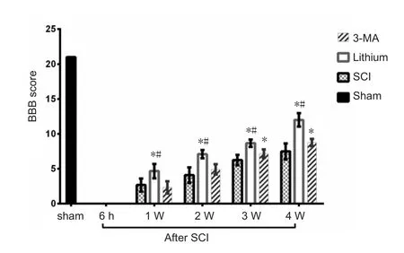
Figure 1 Effects of lithium and 3-MA on motor function in rats with SCI.
ADC values gradually decreased with time in all groups (P< 0.05), and there was no difference among the three groups at 6 hours or 1 week after SCI (P > 0.05). At 2 weeks after SCI, the ADC value was lower in the lithium group than in the SCI and 3-MA groups (P < 0.05), and there was no difference between the SCI and 3-MA groups (P > 0.05). At 3 and 4 weeks after SCI, the ADC value was again lower in the lithium group than in the SCI and 3-MA groups (P < 0.05),and was lower in the 3-MA group than in the SCI group (P< 0.05; Table 2).
Correlation between DTI and neurological function
Pearson correlation analysis showed that FA values were negatively correlated with ADC values in the rat model of spinal cord contusion injury (r = −0.9537, P < 0.05; Figure 2A), consistent with our previous observations (Zhang et al., 2015; He, 2015). FA values were positively and linearly correlated with BBB scores (r = 0.9279, P < 0.05; Figure 2B).ADC values were negatively correlated with BBB scores, and the correlation was linear (r = −0.9173, P < 0.05; Figure 2C).
Immunolabeling for neurons

Figure 2 Correlation between diffusion tensor imaging and neurological function assessment (Pearson correlation analysis).
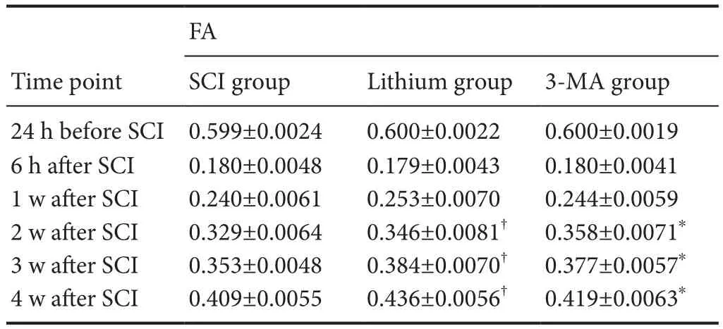
Table 1 FA values at the injury site at different time points after SCI
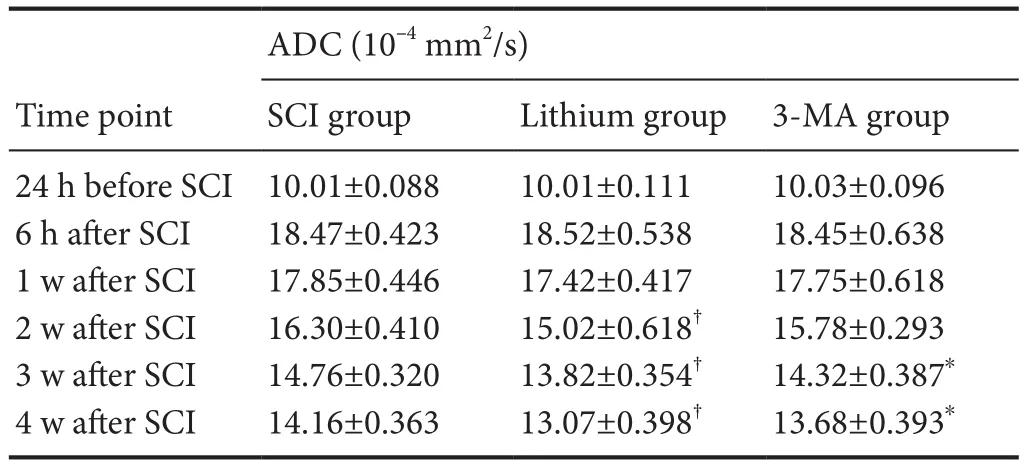
Table 2 ADC values at the injury site at different time points after SCI
Immunohistochemical staining showed that the number of neurons (NeuN+cells) at the site of injury was reduced in the SCI group at 6 hours after SCI, and continued to diminish with time compared with the sham group. In the lithium group, neurons were similarly reduced at 6 hours after SCI but were more numerous than in the SCI and 3-MA groups at 1 and 3 days and 1 week after SCI. In the 3-MA group,neurons were greatly reduced in number compared with the SCI group at 6 hours after SCI and compared with the SCI and lithium groups at 1 and 3 days and 1 week after SCI(Figure 3A). Furthermore, these neurons had an abnormal morphology.
Cell counting showed that the number of neurons in all three experimental groups decreased significantly at 6 hours after SCI compared with the sham group (P < 0.05). The number of neurons in the experimental groups continued to decrease at 1 and 3 days and 1 week after SCI, compared with the previous time point (P < 0.05). More neuronal cells survived in the lithium group than in the SCI group, and more neuronal cells survived in the SCI group than in the 3-MA group (P < 0.05; Figure 3B).
Beclin-1 immunohistochemistry
The Beclin-1+area was larger and more strongly stained,and Beclin-1+cells were more numerous in the SCI group at 6 hours after SCI compared with the sham group. Further expansion of the Beclin-1+area was found at 1 day, but it diminished from 3 days after SCI. In the lithium group, the Beclin-1+area was larger and more intensely stained, and Beclin-1+cells were more numerous at 6 hours after SCI compared with the SCI group. The Beclin-1+area was even larger at 1 day, but it started to diminish from 3 days after SCI, although the staining was still more intense than in the SCI group at 1 week. In the 3-MA group, the staining was slightly more intense than in sham group 1 day after SCI,while it was weaker than in the SCI group at 3 days and 1 week after SCI (Figure 4A).
The relative Beclin-1+area in the SCI group at 6 hours after SCI was significantly greater than 1 (P < 0.05), indicating that it was larger than in the sham group and that the level of autophagy increased after SCI. The relative Beclin-1+area reached a peak at 1 day after SCI and decreased from 3 days after SCI. There was a similar trend in the lithium group,with the relative area increasing from 6 hours after SCI,peaking at 1 day, and decreasing significantly from 3 days (P< 0.05). Compared with the SCI group, the relative Beclin-1+area in the lithium group was greater at 6 hours, 1 and 3 days and 1 week after SCI (P < 0.05). In comparison, the relative Beclin-1+area was significantly smaller in the 3-MA group than in the SCI group at 6 hours and 1 and 3 days after SCI (P < 0.05; Figure 4B).
LC3B immunohistochemistry
The LC3B+area was expanded, the staining intensity was greater, and positive cells were increased in the SCI group at 6 hours after SCI compared with the sham group. Further expansion of the LC3B+area was found at 1 day, but it decreased from 3 days after SCI. The LC3B+area was expanded, the staining intensity was greater, and positive cells were increased in the lithium group at 6 hours after SCI compared with the SCI group. Further expansion of the LC3B+area was found at 1 day, and it shrank from 3 days after SCI, although the staining was still more intense than in the SCI group at 1 week after SCI. In the 3-MA group, 1 day after SCI, the staining was slightly stronger than in the sham group, while it was weaker than in the SCI group at 3 days and 1 week after SCI (Figure 5A).
The quantitative analysis revealed that the relative LC3B+area in the SCI group at 6 hours after SCI was significantly greater than 1 (P < 0.05), indicating that the LC3B+area was larger than in the sham group, and suggesting that the level of autophagy increased after SCI. The relative LC3B+area peaked at 1 day after SCI and decreased from 3 days after SCI. There was a similar trend in the lithium group, with the relative area increasing from 6 hours after SCI, peaking at 1 day, and significantly decreasing from 3 days (P < 0.05).Compared with the SCI group, the relative LC3B+area in the lithium group was greater at 6 hours, 1 and 3 days and 1 week after SCI (P < 0.05). However, the relative LC3B+area was significantly smaller in the 3-MA group than in the SCI group at 6 hours and 1 and 3 days after SCI (P < 0.05; Figure 5B).
Discussion
Advanced evaluation of SCI
Conventional MRI is widely used for patients with SCI.However, it fails to clearly display the degree of injury or the recovery and regeneration of neuronal fibers in the spinal cord after injury. Therefore, in the present study, we used DTI for the three-dimensional reconstruction of white matterfiber bundles.
Based on our previous study, DTI is an objective and accurate method for evaluating recovery following SCI and the effect of therapeutic interventions in complete transection SCI models (Zhang et al., 2015). SCI causes damage to cell membranes and myelin sheaths, leading to the destruction of the molecular diffusion barrier and the unrestricted movement of water (Li et al., 2016). This occurred immediately after SCI. Subsequently, along with glial scar formation,the displacement of water molecules was reduced, and the regeneration of axons forced the water molecules to diffuse primarily in one direction, which was reflected as a gradual increase in the FA value and a decrease in the ADC value.The DTI outcomes we observed in this study were consistent with the pathological changes. The FA and ADC values correlated well with the BBB scores. Lithium promoted recovery following SCI, while 3-MA reduced the therapeutic effectiveness of lithium. Therefore, DTI can accurately reflect axonal necrosis and degeneration, glial cell regeneration and demyelination after SCI, and display changes in the microstructure of the spinal cord in vivo (Zhang et al., 2015; Jirjis et al., 2017).
Autophagy in lithium treatment for SCI
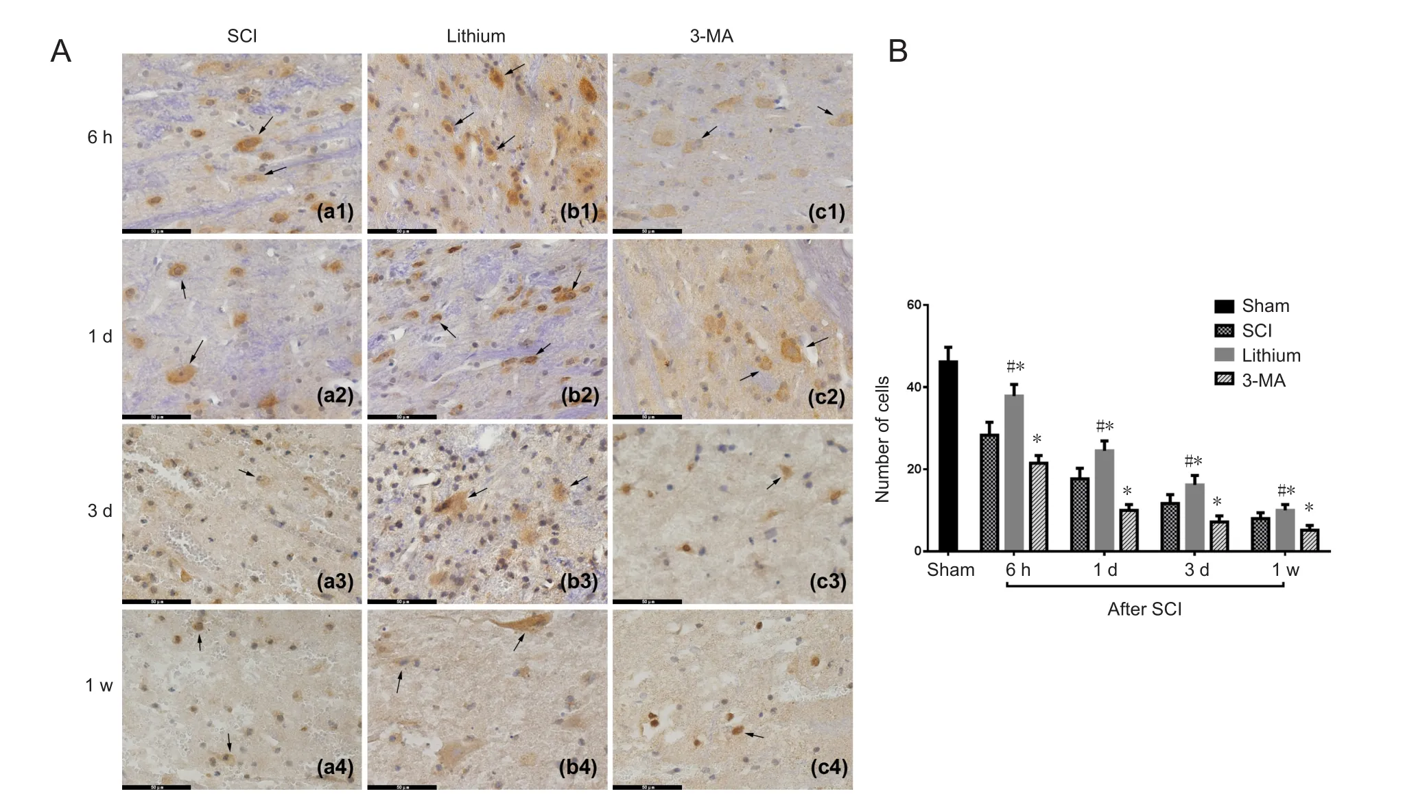
Figure 3 Immunohistochemical staining and counting of neurons (NeuN+ cells) in the injured rat spinal cord at different time points.
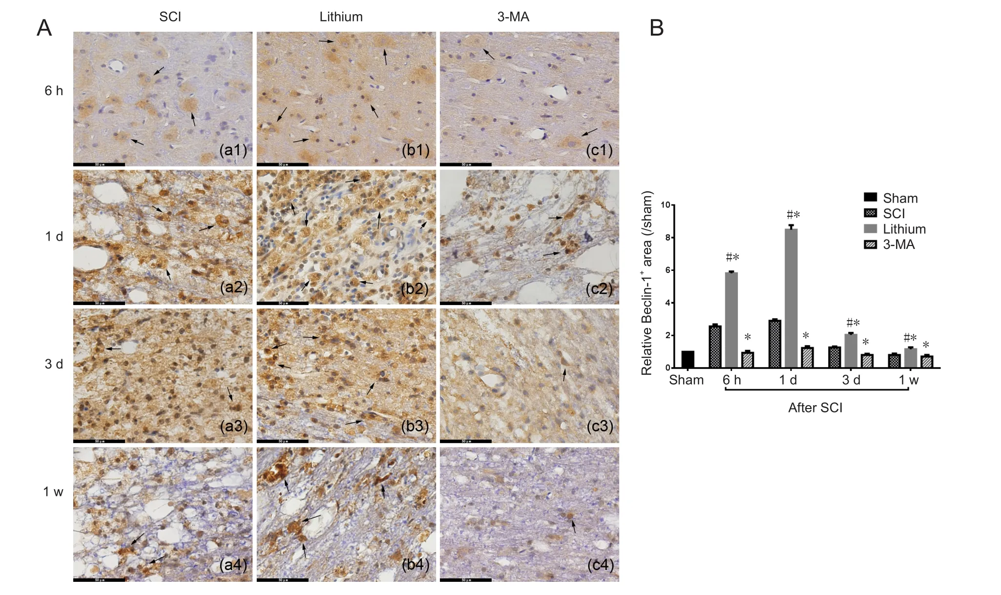
Figure 4 Immunohistochemical staining for Beclin-1 and the relative Beclin-1+ area in the injured spinal cord at different time points.
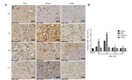
Figure 5 Immunohistochemical staining for LC3B and the relative LC3B+ area in the injured spinal cord at different time points.
Autophagy is an evolutionarily conserved process, and over 30 autophagy-related genes (Atgs) have been identified, of which LC3B (or LC3II) and Beclin-1 are standard markers(Kirisako et al., 1999; Ohsumi, 2001; Mizushima and Yoshimori, 2007; Periyasamy-Thandavan et al., 2009). Accumulating evidence suggests that lithium has neuroprotective properties, suggesting that it may have potential as a new therapy for SCI (Sarkar et al., 2005; Wada et al., 2005; Yan et al., 2007;Pasquali et al., 2009). Although lithium has been widely used for safely and effectively treating neuropsychiatric disorders,it is rarely used for acute SCI (Ohsumi, 2001; Chang et al.,2011; Chen et al., 2013; Kim et al., 2013; Duo and He, 2015;Hou et al., 2015; Seo et al., 2015; Quartini et al., 2016; Zhou et al., 2016; Wu et al., 2018). Therefore, the effectiveness of lithium treatment for acute SCI remained unclear.
The role of autophagy in recovery following injury is still controversial. While some studies have reported that enhanced autophagy improves neuroprotection, others have suggested that the suppression of autophagy is beneficial to recovery (Li et al., 2010; Shimada et al., 2012; O’Donovan et al., 2015; Del Grosso et al., 2016). Previous studies in otherfields have demonstrated that lithium can enhance autophagy, or in contrast, reduce apoptosis and autophagy (Wong et al., 2011; Raja et al., 2015; Guttuso, 2016). Our results show that lithium promotes neurological functional recovery and neural cell survival, which supports the notion that lithium has neuroprotective properties. Furthermore, we observed that these neuroprotective effects were inhibited by 3-MA,which downregulated the autophagy induced by lithium.This implies that lithium reduces neuronal damage and promotes functional recovery by inducing autophagy. Nevertheless, BBB scores were still higher in the 3-MA group than in the SCI group from 3 weeks after SCI, in accordance with the neural cell counting results. This suggests that the mechanisms of autophagy are complex and that other signaling pathways that are not inhibited by 3-MA or activated by lithium play a role in the process (Galluzzi et al., 2017)Furthermore, lithium may also promote neurotrophin secretion, inhibit inflammation or enhance the proliferation of neural progenitor cells (Son et al., 2003; Senatorov et al.,2004; Su et al., 2007; Yasuda et al., 2009; Chi-Tso and Chuang, 2011; Li et al., 2011).
The opposing concept that autophagy aggravates injury may be explained by differences in lithium concentrations and target cells in previous studies. Discrepancies may also be caused by differences in animal models, the therapeutic window and the treatment period. Fang et al. (2016) found that early activated autophagy alleviates spinal cord ischemia/reperfusion injury, while later excessively elevated au-tophagy aggravates the injury. Therefore, autophagy appears to be a dynamic process with differential effects, depending on the time frame and model. Further study is needed to examine the signaling pathways affected by lithium and the dynamic changes in the autophagy pathway.
Summary
Further clinical trials are required to explore the effect of lithium therapy in acute SCI patients. In addition, studies are needed to optimize the time window of treatment, the treatment dose and protocol and to reduce the side effects of lithium.
In conclusion, our findings demonstrate that lithium protects neurons and promotes autophagy in a rat model of acute SCI. DTI is an effective method for evaluating recovery following SCI, and correlates well with neurological functional scores in our rat model of spinal cord contusion injury. The dynamic changes in autophagy after SCI and the effects of lithium on this process need to be investigated further.
Acknowledgments:We are very grateful to Jia-Lin Zhu from Xi’an Jiaotong University of China for technical support.
Author contributions:Data provision and integration: DZ and XHL;study concept and design: DZ and XJH; experimental data analysis: DZ,XZ and XHL; manuscript drafting: DZ; statistical analysis: FW; paper supervisor and instructor: XJH; technical or information support: DZ,XHL, FW and XJH. All authors approved thefinal version of the paper.
Conflicts of interest:None declared.
Financial support:This study was supported by the Beijing Excellent Talent Training Funding in China, No. 2017000021469G215 (to DZ);the Youth Science Foundation of Beijing Tiantan Hospital of China, No.2016-YQN-14 (to DZ); the Natural Science Foundation of Capital Medical University of China, No. PYZ2017082 (to DZ); the Xi’an Science and Technology Project in China, No. 2016048SF/YX04(3) (to XHL). The funding sources had no role in study conception and design, data analysis or interpretation, paper writing or deciding to submit this paper for publication.
Institutional review board statement:All experimental procedures and protocols were approved by the Animal Ethics Committee of Xi’an Jiaotong University of China. All experimental procedures described here were in accordance with the National Institutes of Health (NIH)guidelines for the Care and Use of Laboratory Animals.
Copyright license agreement:The Copyright License Agreement has been signed by all authors before publication.
Data sharing statement:Datasets analyzed during the current study are available from the corresponding author on reasonable request.
Plagiarism check:Checked twice by iThenticate.
Peer review:Externally peer reviewed.
Open access statement:This is an open access journal, and articles are distributed under the terms of the Creative Commons Attribution-Non-Commercial-ShareAlike 4.0 License, which allows others to remix, tweak,and build upon the work non-commercially, as long as appropriate credit is given and the new creations are licensed under the identical terms.
Open peer reviewer:Chang Ho Hwang, University of Ulsan, Republic of Korea.
Additionalfile:Open peer review report 1.
杂志排行
中国神经再生研究(英文版)的其它文章
- Huangqinflavonoid extraction for spinal cord injury in a rat model
- Analysis of transcriptome sequencing of sciatic nerves in Sprague-Dawley rats of different ages
- Exogenous brain-derived neurotrophic factor attenuates cognitive impairment induced by okadaic acid in a rat model of Alzheimer’s disease
- Partial improvement in performance of patients with severe Alzheimer’s disease at an early stage of fornix deep brain stimulation
- Epigenetic marks are modulated by gender and time of the day in the hippocampi of adolescent rats:a preliminary study
- Dysphagia in patients with isolated pontine infarction
