白藜芦醇减轻LPS诱导的牙周膜细胞损伤并抑制TLR4/NF-κB活化
2017-11-29呼海燕刘彩宏
呼海燕 刘彩宏
白藜芦醇减轻LPS诱导的牙周膜细胞损伤并抑制TLR4/NF-κB活化
呼海燕 刘彩宏
目的探究白藜芦醇(RES)对脂多糖(LPS)诱导的人牙周膜细胞(hPDLCs)损伤的保护作用。方法体外培养并鉴定hPDLCs,将培养的hPDLCs随机分为5组:对照组、LPS(10 μg/ml)+RES(0、 30、 60、 90 μmol/L)组,MTT法检测各组细胞增殖能力,ELISA检测各组细胞分泌TNF-α/IL-6水平,PCR和Western blot检测各组细胞TLR4/NF-κB mRNA和蛋白表达。结果分离培养的hPDLCs抗波丝蛋白表达阳性,抗角蛋白表达阴性。与对照组比,LPS处理后细胞增殖能力明显降低,细胞分泌TNF-α/IL-6水平和TLR4/NF-κB mRNA和蛋白表达明显增加; 30~90 μmol/L白藜芦醇预处理后,细胞增殖能力增加(Plt;0.05),细胞分泌TNF-α/IL-6水平、TLR4/NF-κB mRNA以及蛋白表达则下调(Plt;0.05),均呈现一定的浓度依赖性。结论白藜芦醇可抑制TLR4/NF-κB活化并减轻LPS诱导的牙周膜细胞损伤。
白藜芦醇; LPS; 人牙周膜细胞(hPDLCs); TLR4; NF-κB
脂多糖(lipopolysaccharide,LPS)是革兰氏阴性菌的致病因子,它能诱导牙周膜细胞产生炎症因子,进而导致牙周疾病的发生[1]。白藜芦醇(resveratrol,RES)是一种天然抗氧化剂,具有抗肿瘤、抗炎等活性[2],但其在牙周疾病免疫炎症中的报道相对较少,因此,本研究选择人牙周膜细胞(human periodontal ligament cells, hPDLCs)进行体外分离培养,分别通过MTT、ELISA、PCR、Western blot等方法,检测细胞增殖能力,炎性因子分泌和TLR4/NF-κB表达等变化,以探究RES对LPS诱导的hPDLCs损伤的保护作用。
1 材料与方法
1.1 主要试剂与仪器
DMEM培养基(Gibco);胎牛血清(Gemini);兔抗人Toll样受体4(Toll like receptors 4,TLR4)多克隆抗体,兔抗人NF-κB p65多克隆抗体(Sigma);ELISA试剂盒(TNF-α,IL-6)、抗细胞波丝蛋白多克隆抗体、抗细胞角蛋白多克隆抗体、免疫组化染色试剂盒(中杉金桥生物技术公司);酶标仪(Bio-Rad)。
1.2 实验方法
1.2.1 hPDLCs的原代培养 取延安大学附属医院口腔修复科10~15 岁因正畸需要拔除的10 颗健康牙齿,已征得患者本人和家属的同意。牙离体之后,迅速用含有双抗的磷酸缓冲溶液PBS冲洗,置于DMEM培养液(含双抗、10% FBS)中,用双抗液漂洗后,经不含血清的完全培养液的润湿,用手术刀刮取牙根中1/3的牙周膜。将牙周膜组织剪成1 mm×1 mm×1 mm,均匀铺在25 ml的培养瓶中,加入DMEM 培养液,在37 ℃,5% CO2下培养4 h后翻瓶,继续培养,隔天换液,取3~6 代细胞用于后续实验。
1.2.2 免疫细胞化学染色鉴定hPDLCs 取第3代hPDLCs爬片培养,当细胞覆盖至60%左右时,用4%甲醛溶液固定,角蛋白与波形丝蛋白染色,光镜下观察。
1.2.3 绘制hPDLCs生长曲线 取第3 代对数生长期的hPDLCs,接种至96 孔板内,培养24 h后使其贴壁,在培养1~7 d之后分别检测其细胞活性。每孔加入20 μl MTT(5 mg/ml),孵育4 h,吸去上清,加入100 μl DMSO,振荡后,酶标仪检测吸光度值(A490),记录结果,并绘制生长曲线。
1.2.4 MTT检测细胞增殖活性 取第3代对数生长期的hPDLCs,接种至96 孔板内,培养24 h后,加入不同处理的培养基,各浓度设置5 个复孔,分为对照组、LPS组、LPS(10 μg/ml)+RES组(0、 30、 60、 90 μmol/L),培养24 h后,每孔加入20 μl MTT溶液,孵育4 h,弃上清液,加入100 μl DMSO,振荡并检测吸光度值(A490)。
1.2.5 ELISA检测细胞TNF-α/IL-6分泌水平 取第4代对数生长期的hPDLCs,接种于24 孔培养板,按上述分组处理细胞,培养24 h后,收集培养上清液,按照试剂盒说明书检测各组中TNF-α和IL-6的水平。
1.2.6 PCR检测TLR4/NF-κB mRNA表达 取第4代对数生长期的hPDLCs,接种于6孔培养板,按上述分组处理细胞。培养24 h后,Trizol法提取细胞总RNA,并检测其纯度以及完整性。参照逆转录试剂盒说明书合成cDNA,并进行PCR检测,2%~3%琼脂糖凝胶上电泳检测反应产物。引物由上海生工生物工程公司合成。
1.2.7 Western blot检测TLR4/NF-κB蛋白表达 选取第4代对数生长期细胞,参照上述实验处理细胞,24 h后,提取细胞总蛋白,BCA法测定蛋白浓度。经十二烷基磺酸钠-聚丙烯酰胺凝胶电泳(SDS-PAGE),转膜,5%脱脂奶粉封闭,TLR4 (1∶500)、NF-κB p65 (1∶400)、β-actin (1∶1 000)等一抗, 4 ℃孵育过夜,PBST洗膜3 次,二抗室温孵育1 h,PBST洗膜3 次,暗室曝光和压片等步骤,检测TLR4和NF-κB蛋白表达。
1.3 统计学分析
采用SPSS 17.0软件分析,组间比较采用单因素方差分析,两两比较采用LSD-t检验,Plt;0.05为差异为具有统计学意义。
2 结 果
2.1 hPDLCs原代培养及鉴定
组织块法培养5~8 d之后可见细胞游出,hPDLCs原代细胞呈现星形或长梭形,胞突细长,胞体丰满,核为圆形或椭圆形,传代培养后细胞生长迅速(图 1A)。将第3 代hPDLCs细胞在96 孔板中培养1~7 d, MTT法检测其在4~5 d细胞快速增殖,并在第7 天后速度开始减缓,其生长曲线呈“S”形(图 1B)。免疫细胞化学染色鉴定表明,抗波丝蛋白染色呈阳性(图1C),抗角蛋白染色呈阴性(图 1D),证明细胞来源于中胚层,且无上皮来源细胞混杂。
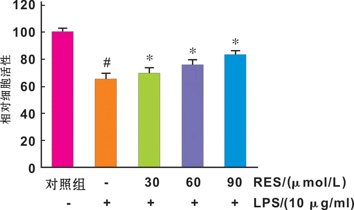
2.2 RES抑制LPS诱导的hPDLCs活性降低
与对照组比较,LPS处理下,hPDLCs细胞活性明显降低。而随着RES浓度增加,细胞活性逐渐增强(Plt;0.05)(图 2)。

A: 第4 代细胞形态(×200); B: 生长曲线; C: 波丝蛋白表达(×100); D: 角蛋白表达(×100)

图 2 LPS和不同浓度RES对细胞增殖活性的影响
Fig 2 Proliferation of hPDLCs treated by LPS and RES(#:vscontrol,Plt;0.05; *vsLPS group,Plt;0.05)
2.3 RES抑制LPS诱导的hPDLCs TNF-α/IL-6分泌增加
ELISA结果显示,LPS明显促进细胞炎性因子TNF-α和IL-6分泌,而RES对hPDLCs细胞TNF-α/IL-6分泌具有明显的抑制作用,并呈现一定的浓度依赖效应(Plt;0.05)(图 3)。

图 3 LPS和RES对细胞TNF-α/IL-6分泌水平的影响
Fig 3 TNF-α and IL-6 levels of hPDLCs treated by LPS and RES(#:vscontrol,Plt;0.05; *:vsLPS group,Plt;0.05)

图 4 LPS和RES对细胞TLR4/NF-κB mRNA表达的影响
Fig 4 TLR4/NF-κB mRNA expression of hPDLCs treated by LPS and RES(#:vscontrol,Plt;0.05; *:vsLPS group,Plt;0.05)
2.4 RES抑制LPS诱导的hPDLCs中TLR4/NF-κB表达上调
PCR结果显示,引物特异性良好。LPS作用下,细胞TLR4与NF-κB mRNA水平均明显增加,而不同浓度RES预处理下,TLR4与NF-κB mRNA的水平均显著降低(Plt;0.05)(图 4~5)。
Western blot结果显示,LPS作用下,细胞TLR4与NF-κB蛋白表达显著升高(Plt;0.01),而不同浓度RES预处理下,TLR4和NF-κB p65蛋白的表达均显著下降,并呈现一定的浓度依赖性(Plt;0.05)。

图 5 LPS和RES对细胞TLR4/NF-κB p65蛋白表达的影响
Fig 5 TLR4/NF-κB expression of hPDLCs treated by LPS and RES(#:vscontrol,Plt;0.05; *:vsLPS group,Plt;0.05)
3 讨 论
LPS作为革兰阴性厌氧菌中的一种致病物质,对牙周组织细胞具有很强的毒性作用[3],被认为是牙周炎症的重要病因之一。研究表明,LPS对hPDLCs具有细胞毒性作用。本研究显示,LPS作用下,hPDLCs活性明显下降。
RES是一种多酚类化合物,作为肿瘤的化学预防剂,它也是预防和治疗动脉粥样硬化、心脑血管疾病的活性物质。近年来,国内外关于白藜芦醇在一些疾病中抗炎活性也有报道[4-5]。有研究显示,RES(50 μmol/L)时能促进人软骨细胞的增殖[4]。本研究表明,在LPS诱导损伤的hPDLCs中加入RES处理后,其细胞活性明显增加,并呈现剂量依赖效应。
研究表明,TLR4是模式识别受体的一种,在机体免疫与炎症反应中发挥关键作用,能激活细胞内炎症反应相关信号通路[6]。NF-κB作为TLR4信号通路的下游因子,调控着多种细胞因子的表达,在炎症损伤和细胞再生方面均有重要作用[7]。TLR4下游因子NF-κB被激活之后,调节TNF-α、IL-6的释放[8]。TNF-α是促进牙周炎发生的重要炎性因子,它能限制牙周组织的修复[9]。研究表明,在牙周膜细胞中,RES有效抑制了损伤细胞中炎性因子IL-1β,IL-6和TNF-α的表达[10]。本研究显示,RES可明显抑制LPS诱导的TNF-α与IL-6水平升高。
Murakami等[11]研究表明,RES有较强的抗自由基活性,并能抑制牙龈卟啉单胞菌诱导的巨噬细胞中NF-κB的活化。临床研究显示,慢性牙周炎患者的牙龈组织中TLR4的表达高于健康人[12],这提示我们TLR4/NF-κB通路可能与牙周炎症密切相关。本研究亦证实,RES可显著降低LPS诱导的hPDLCs TLR4/NF-κB mRNA和蛋白的表达。
综上所述,本文通过LPS和不同浓度RES作用,检测了hPDLCs增殖活性,炎性因子TNF-α/IL-6的分泌和TLR4/NF-κB的表达,发现白藜芦醇可通过抑制TLR4/NF-κB活化减轻LPS诱导的牙周膜细胞损伤。此外,牙周膜细胞炎症因子的表达是受很多机制调控的,多条信号通路都可能参与其中,各信号通路之间还可能存在相互作用,本实验结果提示,TLR4/NF-κB 信号通路参与了白藜芦醇抑制脂多糖诱导的牙周膜细胞炎症因子表达的过程,但其具体机制还需进一步研究。
[1] Gölz L, Memmert S, Rath-Deschner B, et al. Hypoxia andP.gingivalissynergistically induce HIF-1 and NF-κB activation in PDL cells and periodontal diseases[J]. Mediators Inflamm, 2015, 2015: 438085.
[2] 吴明珠, 郭俊明. 白藜芦醇及其衍生物抗炎药理作用研究进展[J]. 畜牧与饲料科学, 2011,32(12):74-75.
[3] Kato H, Taguchi Y, Tominaga K, et al. Porphyromonas gingivalis LPS inhibits osteoblastic differentiation and promotes pro-inflammatory cytokine production in human periodontal ligament stem cells[J]. Arch Oral Biol, 2014, 59(2):167-175.
[4] Andrade JM, Paraíso AF, de Oliveira MV, et al. Resveratrol attenuates hepatic steatosis in high-fat fed mice by decreasing lipogenesis and inflammation[J]. Nutrition, 2014, 30(7-8):915-919.
[5] Csaki C, Keshishzadeh N, Fischer K, et al. Regulation of inflammation signalling by resveratrol in human chondrocytesinvitro[J]. Biochem Pharmacol, 2008, 75(3):677-687.
[6] Tang L,Zhou XD,Wang Q, et al. Expression of TRAF6 and pro-inflammatory cytokines through activation of TLR2, TLR4, NOD1, and NOD2 in human periodontal ligament fibroblasts[J]. Arch Oral Biol, 2011, 56(10):1064-1072.
[7] Lisboa RA,Andrade MV, Cunha-Melo JR. Toll-like receptor activation and mechanical force stimulation promote the secretion of matrix metalloproteinases 1, 3 and 10 of human periodontal fibroblasts via p38, JNK and NF-kB[J]. Arch Oral Biol, 2013, 58(6):731-739.
[8] Chi XP, Ouyang XY, Wang YX. Hydrogen sulfide synergistically upregulates Porphyromonas gingivalis lipopolysaccharide-induced expression of IL-6 and IL-8 via NF-κB signalling in periodontal fibroblasts[J]. Arch Oral Biol, 2014, 59(9):954-961.
[9] Liu W, Konermann A, Guo T, et al. Canonical Wnt signaling differently modulates osteogenic differentiation of mesenchymal stem cells derived from bone marrow and from periodontal ligament under inflammatory conditions[J]. Biochim Biophys Acta, 2014, 1840(3):1125-1134.
[10]Rizzo A, Bevilacqua N, Guida L, et al. Effect of resveratrol and modulation of cytokine production on human periodontal ligament cells[J]. Cytokine, 2012, 60(1):197-204.
[11]Murakami Y, Kawata A, Ito S, et al. The Radical Scavenging Activity and Cytotoxicity of Resveratrol, Orcinol and 4-Allylphenol and their Inhibitory Effects on Cox-2 Gene Expression and Nf-κb Activation in RAW264.7 Cells Stimulated with Porphyromonas gingivalis-fimbriae[J]. In Vivo, 2015, 29(3):341-349.
[12]Sun Y, Guo QM, Liu DL, et al.Invivoexpression of Toll-like receptor 2, Toll-like receptor 4, CSF2 and LY64 in Chinese chronic periodontitis patients[J]. Oral Dis, 2010, 16(4):343-350.
(收稿: 2016-06-04 修回: 2016-10-19)
ResveratrolattenuatesLPS-inducedcellinjuryandTLR4/NF-κBactivationinhumanperiodontalligamentcells
HUHaiyan,LIUCaihong.
716000,RepairDepartmentofStomatology,YananUniversityAffiliatedHosptial,China
Objective: To investigate the protective effect of resveratrol on lipopolysaccharide (LPS)-induced cell injury in human periodontal ligament cells(hPDLCs).MethodshPDLCs were cultured and identified. The cultured hPDLCs were divided into 5 groups: control group and LPS(10 μg/ml)+RES(0/30, 60 and 90 μmol/L respectively) groups. The cell proliferation was detected by MTT assay. The secretion of TNF-α and IL-6 of hPDLCs was detected by ELISA kit. The expression of TLR4/NF-κB mRNA and protein was determined by PCR and Western blot analyses, respectively.ResultsThe cultured cells were negative for cytokeratin and positive for vimentin staining. Compared with the control group, cell proliferation was decreased, the secretion of TNF-α/IL-6 levels and the expression of TLR4/NF-κB mRNA and protein were increased after treatment with LPS. Whereas, with 30-90 μmol/L resveratrol pretreatment, the proliferation ability of hPDLCs was enhanced(Plt;0.05), the secretion levels of TNF-α and IL-6 and the expression of TLR4/ NF-κB mRNA and protein were reduced(Plt;0.01) in a dose-dependent manner.ConclusionResveratrol may attenuate LPS-induced cell injury by inhibiting TLR4/NF-κB pathway in hPDLCs.
Resveratrol;LPS;Humanperiodontalligamentcells(hPDLCs);TLR4;NF-κB
716000, 延安大学附属医院口腔修复科
呼海燕 E-mail: shidasda@163.com
R781.4, R282.71
A
10.3969/j.issn.1001-3733.2017.01.019
