Neuroprotective effects of ganoderma lucidum polysaccharides against oxidative stress-induced neuronal apoptosis
2017-08-07XinzhiSunYingLiaoWeiLiLimeiGuo
Xin-zhi Sun, Ying Liao, Wei Li Li-mei Guo
1 Department of Orthopedics, the First Af filiated Hospital of Zhengzhou University, Zhengzhou, Henan Province, China
2 Department of Public Security Technology, Railway Police College, Zhengzhou, Henan Province, China
3 Department of Pathology, Peking University Health Science Center, Beijing, China
Neuroprotective effects of ganoderma lucidum polysaccharides against oxidative stress-induced neuronal apoptosis
Xin-zhi Sun1,#, Ying Liao2,3,#, Wei Li2, Li-mei Guo3,*
1 Department of Orthopedics, the First Af filiated Hospital of Zhengzhou University, Zhengzhou, Henan Province, China
2 Department of Public Security Technology, Railway Police College, Zhengzhou, Henan Province, China
3 Department of Pathology, Peking University Health Science Center, Beijing, China
Graphical Abstract

Ganoderma lucidum polysaccharides have protective effects against apoptosis in neurons exposed to ischemia/reperfusion injury, but the mechanisms are unclear.e goal of this study was to investigate the underlying mechanisms of the effects of ganoderma lucidum polysaccharides against oxidative stress-induced neuronal apoptosis. Hydrogen peroxide (H2O2) was used to induce apoptosis in cultured cerebellar granule cells. In these cells, ganoderma lucidum polysaccharides remarkably suppressed H2O2-induced apoptosis, decreased expression of caspase-3, Bax and Bim and increased that of Bcl-2.ese findings suggested that ganoderma lucidum polysaccharides regulate expression of apoptosis-associated proteins, inhibit oxidative stress-induced neuronal apoptosis and, therefore, have significant neuroprotective effects.
nerve regeneration; brain injury; H2O2; cerebellar granule cells; Bim; Bax; Bcl-2; cytochrome C; caspase-3; neural regeneration
Introduction
Oxidative stress-induced brain damage has been implicated in many neurodegenerative disorders, such as Parkinson’s disease, Alzheimer’s disease, amyotrophic lateral sclerosis, Huntington’s disease and stroke (Dawson and Dawson, 2003; Veurink et al., 2003; Malkus et al., 2009). Accumulating evidence has suggested that oxidative stress associated with excessive production of reactive oxygen species profound affects neurodegenerative pathogenesis (de Vries et al., 2008; Zolezzi et al., 2013; Newland et al., 2016). Reactive oxygen species can cause lipid peroxidation, protein denaturation and DNA/RNA damage (Ye et al., 2009; Collado et al., 2012; Zou et al., 2015).Reactive oxygen species induce several signal transduction pathways, including intrinsic and extrinsic caspase activation, which may lead to excessive cell apoptosis and expression of inflammatory genes (Chan, 2001; Allen and Bayraktutan, 2009). Oxidative stress triggers apoptosis through activation of many signaling molecules, including kinases and proteases (Tan et al., 1998; Andersen, 2004; Kaul et al., 2005). Hydrogen peroxide (H2O2) is used as a stressor to induce oxidative stress in experimental models (Brown et al., 2013; Sies, 2014) and to stimulate apoptotic and necrotic pathways (Clement et al., 1998).
Pharmaceutical compounds extracted from mushrooms showed benefits for a variety of conditions such as cancers, immunologic disorders and neurodegenerative diseases (Wang et al., 1997; Wasser and Weis, 1999; Cheung et al., 2000).Ganoderma lucidum(G.lucidum) belongs to the polyporaceae family of Basidiomycota, a type of mushroom widely used as a traditional medicine for thousands of years, especially in Asia (Ji et al., 2007). A variety of bioactive chemicals, such as polysaccharides, triterpenoids and proteins, can be extracted from the fruiting bodies, cultured mycelia and spores of G.lucidum(Mizushina et al., 1999). Clinical trials and other experimental studies indicated that the active compounds isolated from its fruiting body, known as “Lingzhi,” participate in a variety of biological processes, showing anti-inflammatory, antioxidant, anti-tumor and immunomodulatory activities (Lakshmi et al., 2003; Lin and Zhang, 2004; Zhao et al., 2012; Pan et al., 2013; Ferreira et al., 2015). G.lucidumpolysaccharides (GLPS) were shown to be neuroprotective, increasing viability in cerebral cortical neurons exposed to ischemia/ reperfusion and in models for traumatic spinal cord injury (Zhao et al., 2004; Gokce et al., 2015).is evidence indicated that GLPS is a potentially promising drug candidate. However, the roles of GLPS in modulating oxidative stress-induced neuronal apoptosis have been poorly understood.e aim of our study was to investigate whether GLPS would protect cultured cerebellar granule neurons from apoptosis induced by H2O2.
Materials and Methods
Cell culture
Rat cerebellar granule cells (CGCs) were prepared from 7 or 8-day-old Sprague-Dawley rat pups, as previously described (D’Mello et al., 1993). All experimental procedures were performed in accordance with the Guideline for the Care and Use of Laboratory Animals of the Animal Research Ethics Committee of Peking University Health Science Center (China) andunder the principles and guidelines of the National Institutes of Health Guide for the Care and Use of Laboratory Animals. All efforts were made to minimize the number and suffering of the animals used in the experiments.
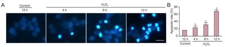
Figure 1 H2O2induced apoptosis of cultured CGCs.

Figure 2 H2O2treatment altered protein expression in cerebellar granule cells.
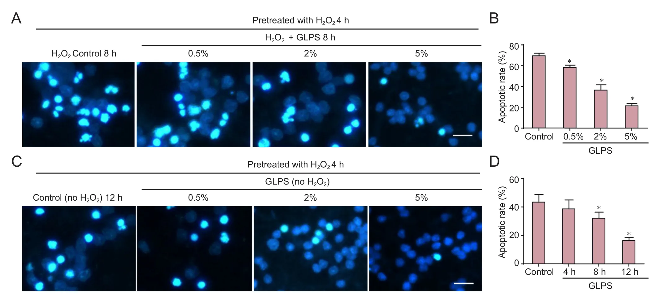
Figure 3 GLPS administration suppressed H2O2-induced apoptosis of cerebellar granule cells (light microscopy).

Figure 4 GLPS administration regulated H2O2-induced protein alterations in cerebellar granule cells.
Briefly, neurons were dissociated from freshly dissected cerebella by mechanical disruption in the presence of trypsin (Life Technologies, Carlsbad, CA, USA) and DNase (Life Technologies) and then seeded at a density of 1.5 × 10⁶cells/mL in basal modified Eagle’s media (Life Technologies) containing 10% fetal bovine serum (Life Technologies) and potassium (Sigma-Aldrich, St. Louis, MO, USA) at concentrations causing membrane depolarization (25 mM KCl).e GLPS that was administered (0.5%, 2% or 5% (w/v); at 4, 8 or 12 hours) contained 56.9% carbohydrate and 32.45% protein and was from Lvgu Biotech (Fuzhou, China). Aer 7 days of culturingin vitro, CGCs were incubated in the presence or absence of 50 μM H2O2(at 4, 8, 12 hours; Sigma-Aldrich), with or without GLPS, for the indicated time points. More details of specific treatments are in the figure legends.e control for both H2O2and GLPS was water.
Western blot assay
Western blotting was performed as previously described (Yan et al., 2015). Briefly, neuronal lysates were separated usingsodium dodecyl sulfate-polyacrylamide gel electrophoresis and protein bands electrophoretically transferred to a polyvinylidene difluoride membrane. Membranes were blocked in Tris-buffered saline with 5% milk and 0.05% Tween-20 and then probed with primary antibodies at 4°C overnight.e following primary antibodies were used: rabbit anti-cytochrome c, rabbit anti-cleaved caspase-3, mouse anti-Bax, rabbit anti-Bim, mouse anti-Bcl-2 and mouse anti-β-tubulin, each diluted 1:1,000 (all primary antibodies from Cell Signaling Technology, Danvers, MA, USA). Appropriate horseradish peroxidase-conjugated secondary antibodies (Jackson ImmunoResearch, West Grove, PA, USA) were used to detect reactive bands with the enhanced chemiluminescence (ECL) and ECL-plus systems (GE Healthcare, Chalfont St. Giles, UK). To quantify bands on western blots, the average intensity of the pixels in a background-selected region was calculated and subtracted from each pixel in the sample. To correct for deviations, densitometry values obtained within the linear range of detection were normalized to those for β-tubulin.
Quantification of neuronal apoptosis
CGC apoptosis was quantified as previously described (Linseman et al., 2004). In brief, CGCs were cultured in 24-well plates and incubated with various treatments. After incubation for the indicated times, CGCs were stained with the DNA dye Hoechst 33258 (Sigma, 5 μg/mL) to visualize nuclear morphology. Apoptosis was quantified by scoring the percentage of neuronal cells with condensed or fragmented nuclei. Neurons were counted from three randomly chosen fields per well, under a light microscope (BX51WI light microscope; Olympus, Tokyo, Japan). To obtain unbiased results, experiments were performed in a blinded manner and cells were scored by investigators without knowledge of their prior treatments. All experiments were repeated at least three times and over 500 neurons were counted for each treatment group.
Statistical analysis
Statistical analyses were performed using SPSS 16.0 soware (SPSS, Chicago, IL, USA). All statistical data are expressed as the mean ± SEM of at least three independent experiments (n≥ 3).e statistical significance of differences was analyzed using Student’st-test between two groups and one-way analysis of variance with Student-Newman-Keulspost hoctest for comparisons among more than two groups. A value ofP< 0.05 was considered statistically significant.
Results
Oxidative stress significantly induced neuronal apoptosis in cultured CGCs
H2O2is commonly used to induce cell apoptosis, secondary to free radicals (Medina et al., 2002). It is therefore considered a useful agent for generating models of oxidative stress (Chang et al., 2003). Accordingly, we used 50 μM H2O2for an experimental apoptosis model. First, cultured neurons were treated with H2O2for 4, 8 or 12 hours, then stained with Hoechst 33258 to view apoptotic cells. H2O2administration markedly induced neuronal apoptosis, in a time-dependent manner. Apoptotic neurons were those with nuclear shrinkage, chromatin condensation or fragmentation (Figure 1A). Apoptosis began as early as 4 hours aer H2O2treatment and, 12 hours later, the apoptotic rate had reached nearly 70% (Figure 1B). Next, we investigated which proteins were activated during apoptosis. We found that cytosolic cytochrome c levels were increased, and caspase-3 was cleaved (Figure 2A).e statistical data are shown inFigure 2B.ese data indicated that H2O2activated the mitochondrial apoptotic pathway, involving BH3-only family proteins. BH3-only proteins are critical for neuronal apoptosis (Happo et al., 2012; Doerflinger et al., 2015), and can be divided into two classes: (1) pro-apoptotic proteins, such as Bax, Bad and Bim; and (2) anti-apoptotic proteins, such as Bcl-2 and Bcl-xl. As shown inFigure 2C, Bim and Bax levels were increased and those of Bcl-2 were decreased, all changing in a time-dependent manner.e statistical data are shown inFigure 2D. All findings suggested that H2O2significantly induced neuronal apoptosis in CGCs.
GLPS markedly suppressed oxidative stress-induced apoptosis
Compounds extracted from G.lucidumshowed anti-cancer, antioxidant and liver protective effects. GLPS was neuroprotective against traumatic spinal cord injury in rats (Gokce et al., 2015). However, the role of GLPS in modulating oxidative stress-induced apoptosis in cultured cerebellar granule cells remains unknown. In this study, we found that apoptosis induced by H2O2was suppressed in a dose-dependent manner by GLPS administration (Figure 3A).e statistical data are shown inFigure 3B. Neurons were pre-treated with H2O2for 4 hours. Cells were then washed and incubated with GLPS for an additional 4, 8 or 12 hours. The results showed that cells underwent apoptosis, even at 12 hours aer H2O2administration. However, GLPS addition significantly protected neurons from apoptosis, in a time-dependent manner (Figure 3C).e statistical data are shown inFigure 3D. We then examined whether GLPS suppressed apoptosis through inhibition of the mitochondrial pathway. As shown inFigure 4A, levels of activated caspase-3, induced by H2O2, were suppressed by GLPS (Figure 4B). Furthermore, the increased levels of Bax and Bim were also attenuated and the decreased level of Bcl-2 was increased in H2O2treated cells also receiving GLPS (Figure 4CandD).ese results indicated that GLPS suppressed H2O2-induced apoptosis.
Discussion
In this study, we demonstrated oxidative stress-induced apoptosis of cultured cerebellar granule cells. H2O2increased cleavage and, therefore, activation of caspase-3, cytochrome c release, upregulation of the pro-apoptotic proteins Bax and Bim and downregulation of the anti-apoptotic protein Bcl-2, ultimately causing apoptosis in CGCs. GLPS administration significantly suppressed these processes, thus inhibiting H2O2-induced neuronal apoptosis. This elucidation of the neuroprotective mechanisms of GLPS may contribute to clinical use of active compounds isolated from G.lucidum. G.lucidumhas been used as a preventive medicine in Asiafor thousands of years (Shiao, 2003). Polysaccharides, isolated from G.lucidumfruiting bodies, have antioxidant (Liu et al., 2010), immunomodulatory (Bao et al., 2001) and antitumor properties (Cao and Lin, 2006). Moreover, polysaccharides were protective against cerebral ischemic injury (Zhou et al., 2010) and traumatic spinal cord injury in rats (Gokce et al., 2015). GLPS induced neuronal differentiation of pheochromocytoma cell cultures and protected PC12 neurons from apoptosis, by the Erk1/2 and the CREB signaling pathways (Cheung et al., 2000). G.lucidumextracts decreased inflammatory mediator production by activated microglia and protected dopaminergic neurons against inflammatory and oxidative damage (Zhang et al., 2011). Furthermore, G.lucidumspores preserved injured spinal motor neurons by modulating expression of proteins important for axonal regeneration (Zhang et al., 2006).ese findings suggested that polysaccharides isolated from G.lucidumhad both neuroprotective and antioxidant properties. To the best of our knowledge, ours is the first report on the neuroprotective effects of GLPS against oxidative stress-induced apoptosis in cultured cerebellar granule cells.
H2O2induced apoptosis in neuronal and non-neuronal cells, an effect associated with its cytotoxicity (Mailly et al., 1999; Kumar et al., 2001; Chang et al., 2003). During the late stages of apoptosis, DNA fragmentation occurs, following reactive oxygen species generation, caspase-3 activation and mitochondrial dysfunction (Yang et al., 2004). Moreover, oxidative stress-induced neurotoxicity involves a mitochondria dependent apoptotic pathway, including cytochrome c release, caspase-3 activation and changes in the Bax/Bcl-2 ratio (Brune et al., 2003; Cunha-Oliveira et al., 2007; Lai et al., 2011; Radi et al., 2014), all effects demonstrated in our study. Bcl-2 can counteract the pro-apoptotic effect of Bax/Bim by forming a heterodimer (Kobayashi et al., 1998). During apoptosis, increased Bax translocates to the mitochondria, leading to decreased mitochondrial membrane potential (Linseman et al., 2004; Cunha-Oliveira et al., 2006). Bim forms heterodimers with Bcl-2, releasing Bax from Bcl-2, thus enabling its mitochondrial translocation (Letai et al., 2002) and Bim also directly activates Bax for apoptosis (Gavathiotis et al., 2008; Du et al., 2011). Our results showed that GLPS addition markedly suppressed all signal transduction processes induced by oxidative stress. That is, GLPS inhibited caspase-3 activation, suppressed Bax/Bim upregulation and prevented Bcl-2 downregulation, thus ultimately preventing H2O2-induced apoptosis in CGCs. Among the BH-3-only pro-apoptotic proteins, Bid, Noxa and Puma were reported to mediate apoptosis (Ren et al., 2010). The regulation of these proteins by GLPS, also protecting against oxidative stress-induced neuronal apoptosis, should be investigated in the future.
In summary, our study characterized protection by GLPS against oxidative stress-induced neurotoxicity, contributing new insights into the neuroprotective mechanisms of G.lucidum.ese findings provided potential new evidence supporting clinical use of G.lucidumto treat neurodegenerative diseases involving oxidative stress.
Author contributions:YL performed research, collected data and analyzed and interpreted data. XZS and WL performed research and wrote the paper. LMG designed research and revised the manuscript. All authors approved the final version of the paper.
Conflicts of interest:None declared.
Research ethics:
Open access statement:
Contributor agreement:A statement of “Publishing Agreement” has been signed by an authorized author on behalf of all authors prior to publication.Plagiarism check:This paper has been checked twice with duplication-checking soware ienticate.
Peer review:A double-blind and stringent peer review process has been performed to ensure the integrity, quality and significance of this paper.
Allen CL, Bayraktutan U (2009) Oxidative stress and its role in the pathogenesis of ischaemic stroke. Int J stroke 4:461-470.
Andersen JK (2004) Oxidative stress in neurodegeneration: cause or consequence? Nat Med 10 Suppl:S18-25.
Bao X, Liu C, Fang J, Li X (2001) Structural and immunological studies of a major polysaccharide from spores of Ganoderma lucidum (Fr.) Karst. Carbohydr Res 332:67-74.
Brown JD, Day AM, Taylor SR, Tomalin LE, Morgan BA, Veal EA (2013) A peroxiredoxin promotes H2O2 signaling and oxidative stress resistance by oxidizing a thioredoxin family protein. Cell Rep 5:1425-1435.
Brune B, Zhou J, von Knethen A (2003) Nitric oxide, oxidative stress, and apoptosis. Kidney Int S22-24.
Cao QZ, Lin ZB (2006) Ganoderma lucidum polysaccharides peptide inhibits the growth of vascular endothelial cell and the induction of VEGF in human lung cancer cell. Life Sci 78:1457-1463.
Chan PH (2001) Reactive oxygen radicals in signaling and damage in the ischemic brain. J Cereb Blood Flow Metab 21:2-14.
Cheung WM, Hui WS, Chu PW, Chiu SW, Ip NY (2000) Ganoderma extract activates MAP kinases and induces the neuronal differentiation of rat pheochromocytoma PC12 cells. FEBS Lett 486:291-296.
Clement MV, Ponton A, Pervaiz S (1998) Apoptosis induced by hydrogen peroxide is mediated by decreased superoxide anion concentration and reduction of intracellular milieu. FEBS Lett 440:13-18.
Collado R, Oliver I, Tormos C, Egea M, Miguel A, Cerda C, Ivars D, Borrego S, Carbonell F, Saez GT (2012) Early ROS-mediated DNA damage and oxidative stress biomarkers in Monoclonal B Lymphocytosis. Cancer Lett 317:144-149.
Cunha-Oliveira T, Rego AC, Garrido J, Borges F, Macedo T, Oliveira CR (2007) Street heroin induces mitochondrial dysfunction and apoptosis in rat cortical neurons. J Neurochem 101:543-554.
Cunha-Oliveira T, Rego AC, Cardoso SM, Borges F, Swerdlow RH, Macedo T, de Oliveira CR (2006) Mitochondrial dysfunction and caspase activation in rat cortical neurons treated with cocaine or amphetamine. Brain Res 1089:44-54.
D’Mello SR, Galli C, Ciotti T, Calissano P (1993) Induction of apoptosis in cerebellar granule neurons by low potassium: inhibition of death by insulin-like growth factoriand cAMP. Proc Natl Acad Sci U S A 90:10989-10993.
Dawson TM, Dawson VL (2003) Molecular pathways of neurodegeneration in Parkinson’s disease. Science 302:819-822.
de Vries HE, Witte M, Hondius D, Rozemuller AJ, Drukarch B, Hoozemans J, van Horssen J (2008) Nrf2-induced antioxidant protection: a promising target to counteract ROS-mediated damage in neurodegenerative disease? Free Radic Biol Med 45:1375-1383.
Doerflinger M, Glab JA, Puthalakath H (2015) BH3-only proteins: a 20-year stock-take. FEBS J 282:1006-1016.
Du H, Wolf J, Schafer B, Moldoveanu T, Chipuk JE, Kuwana T (2011) BH3 domains other than Bim and Bid can directly activate Bax/Bak. J Biol Chem 286:491-501.
Ferreira IC, Heleno SA, Reis FS, Stojkovic D, Queiroz MJ, Vasconcelos MH, Sokovic M (2015) Chemical features of Ganoderma polysaccharides with antioxidant, antitumor and antimicrobial activities. Phytochemistry 114:38-55.
Gavathiotis E, Suzuki M, Davis ML, Pitter K, Bird GH, Katz SG, Tu HC, Kim H, Cheng EH, Tjandra N, Walensky LD (2008) BAX activation is initiated at a novel interaction site. Nature 455:1076-1081.
Gokce EC, Kahveci R, Atanur OM, Gurer B, Aksoy N, Gokce A, Sargon MF, Cemil B, Erdogan B, Kahveci O (2015) Neuroprotective effects of Ganoderma lucidum polysaccharides against traumatic spinal cord injury in rats. Injury 46:2146-2155.
Happo L, Strasser A, Cory S (2012) BH3-only proteins in apoptosis at a glance. Journal of cell science 125:1081-1087.
Ji Z, Tang Q, Zhang J, Yang Y, Jia W, Pan Y (2007) Immunomodulation of RAW264.7 macrophages by GLIS, a proteopolysaccharide from Ganoderma lucidum. J Ethnopharmacol 112:445-450.
Kaul S, Anantharam V, Yang Y, Choi CJ, Kanthasamy A, Kanthasamy AG (2005) Tyrosine phosphorylation regulates the proteolytic activation of protein kinase Cdelta in dopaminergic neuronal cells. J Biol Chem 280:28721-28730.
Kobayashi T, Ruan S, Clodi K, Kliche KO, Shiku H, AndreeffM, Zhang W (1998) Overexpression of Bax gene sensitizes K562 erythroleukemia cells to apoptosis induced by selective chemotherapeutic agents. Oncogene 16:1587-1591.
Kumar S, Bharti A, Mishra NC, Raina D, Kharbanda S, Saxena S, Kufe D (2001) Targeting of the c-Abl tyrosine kinase to mitochondria in the necrotic cell death response to oxidative stress. J Biol Chem 276:17281-17285.
Lai B, Pu H, Cao Q, Jing H, Liu X (2011) Activation of caspase-3 and c-Jun NH2-terminal kinase signaling pathways involving heroin-induced neuronal apoptosis. Neurosci Lett 502:209-213.
Lakshmi B, Ajith TA, Sheena N, Gunapalan N, Janardhanan KK (2003) Antiperoxidative, anti-inflammatory, and antimutagenic activities of ethanol extract of the mycelium of Ganoderma lucidum occurring in South India. Teratog Carcinog Mutagen 1:85-97.
Letai A, Bassik MC, Walensky LD, Sorcinelli MD, Weiler S, Korsmeyer SJ (2002) Distinct BH3 domains either sensitize or activate mitochondrial apoptosis, serving as prototype cancer therapeutics. Cancer Cell 2:183-192.
Lin ZB, Zhang HN (2004) Anti-tumor and immunoregulatory activities of Ganoderma lucidum and its possible mechanisms. Acta Pharmacol Sinica 25:1387-1395.
Linseman DA, Butts BD, Precht TA, Phelps RA, Le SS, Laessig TA, Bouchard RJ, Florez-McClure ML, Heidenreich KA (2004) Glycogen synthase kinase-3beta phosphorylates Bax and promotes its mitochondrial localization during neuronal apoptosis. J Neurosci 24:9993-10002.
Liu W, Wang H, Pang X, Yao W, Gao X (2010) Characterization and antioxidant activity of two low-molecular-weight polysaccharides purified from the fruiting bodies of Ganoderma lucidum. Int J Biol Macromol 46:451-457.
Mailly F, Marin P, Israel M, Glowinski J, Premont J (1999) Increase in external glutamate and NMDA receptor activation contribute to H2O2-induced neuronal apoptosis. J Neurochem 73:1181-1188.
Malkus KA, Tsika E, Ischiropoulos H (2009) Oxidative modifications, mitochondrial dysfunction, and impaired protein degradation in Parkinson’s disease: how neurons are lost in the Bermuda triangle. Mol Neurodegener 4:24.
Medina S, Martinez M, Hernanz A (2002) Antioxidants inhibit the human cortical neuron apoptosis induced by hydrogen peroxide, tumor necrosis factor alpha, dopamine and beta-amyloid peptide 1-42. Free Radic Res 36:1179-1184.
Mizushina Y, Takahashi N, Hanashima L, Koshino H, Esumi Y, Uzawa J, Sugawara F, Sakaguchi K (1999) Lucidenic acid O and lactone, new terpene inhibitors of eukaryotic DNA polymerases from a basidiomycete, Ganoderma lucidum. Bioorg Med Chem 7:2047-2052.
Newland B, WolffP, Zhou D, Wang W, Zhang H, Rosser A, Wang W, Werner C (2016) Synthesis of ROS scavenging microspheres from a dopamine containing poly(beta-amino ester) for applications for neurodegenerative disorders. Biomater Sci 4:400-404.
Pan K, Jiang Q, Liu G, Miao X, Zhong D (2013) Optimization extraction of Ganoderma lucidum polysaccharides and its immunity and antioxidant activities. Int J Biol Macromol 55:301-306.
Radi E, Formichi P, Battisti C, Federico A (2014) Apoptosis and oxidative stress in neurodegenerative diseases. J Alzheimers Dis 42 Suppl 3:S125-152.
Ren D, Tu HC, Kim H, Wang GX, Bean GR, Takeuchi O, Jeffers JR, Zambetti GP, Hsieh JJ, Cheng EH (2010) BID, BIM, and PUMA are essential for activation of the BAX- and BAK-dependent cell death program. Science 330:1390-1393.
Shiao MS (2003) Natural products of the medicinal fungus Ganoderma lucidum: occurrence, biological activities, and pharmacological functions. Chem Rec 3:172-180.
Sies H (2014) Role of metabolic H2O2generation: redox signaling and oxidative stress. J Biol Chem 289:8735-8741.
Tan S, Wood M, Maher P (1998) Oxidative stress induces a form of programmed cell death with characteristics of both apoptosis and necrosis in neuronal cells. J Neurochem 71:95-105.
Veurink G, Fuller SJ, Atwood CS, Martins RN (2003) Genetics, lifestyle and the roles of amyloid beta and oxidative stress in Alzheimer’s disease. Ann human Biol 30:639-667.
Wang SY, Hsu ML, Hsu HC, Tzeng CH, Lee SS, Shiao MS, Ho CK (1997)e anti-tumor effect of Ganoderma lucidum is mediated by cytokines released from activated macrophages and T lymphocytes. Int J Cancer 70:699-705.
Yan YY, Wang XM, Jiang Y, Chen H, He JT, Mang J, Shao YK, Xu ZX (2015)e role of Rho/Rho-kinase pathway and the neuroprotective effects of fasudil in chronic cerebral ischemia. Neural Regen Res 10:1441-1449.
Yang Y, Kaul S, Zhang D, Anantharam V, Kanthasamy AG (2004) Suppression of caspase-3-dependent proteolytic activation of protein kinase C delta by small interfering RNA prevents MPP+-induced dopaminergic degeneration. Mol Cell Neurosci 25:406-421.
Ye J, Han Y, Wang C, Yu W (2009) Cytoprotective effect of polypeptide from Chlamys farreri on neuroblastoma (SH-SY5Y) cells following HO exposure involves scavenging ROS and inhibition JNK phosphorylation. J Neurochem 111:441-451.
Zhang R, Xu S, Cai Y, Zhou M, Zuo X, Chan P (2011) Ganoderma lucidum Protects Dopaminergic Neuron Degeneration through Inhibition of Microglial Activation. Evid Based Complement Alternat Med 2011:156810.
Zhang W, Zeng YS, Wang Y, Liu W, Cheng JJ, Chen SJ (2006) [Primary study on proteomics about Ganoderma lucidium spores promoting survival and axon regeneration of injured spinal motor neurons in rats. Zhongxiyi Jiehe Xuebao 4:298-302.
Zhao HB, Lin SQ, Liu JH, Lin ZB (2004) Polysaccharide extract isolated from ganoderma lucidum protects rat cerebral cortical neurons from hypoxia/reoxygenation injury. J Pharmacol Sci 95:294-298.
Zhao W, Jiang X, Deng W, Lai Y, Wu M, Zhang Z (2012) Antioxidant activities of Ganoderma lucidum polysaccharides and their role on DNA damage in mice induced by cobalt-60 gamma-irradiation. Food Chem Toxicol 50:303-309.
Zhou ZY, Tang YP, Xiang J, Wua P, Jin HM, Wang Z, Mori M, Cai DF (2010) Neuroprotective effects of water-soluble Ganoderma lucidum polysaccharides on cerebral ischemic injury in rats. J Ethnopharmacol 131:154-164.
Zolezzi JM, Silva-Alvarez C, Ordenes D, Godoy JA, Carvajal FJ, Santos MJ, Inestrosa NC (2013) Peroxisome proliferator-activated receptor (PPAR) gamma and PPARalpha agonists modulate mitochondrial fusion-fission dynamics: relevance to reactive oxygen species (ROS)-related neurodegenerative disorders? PLoS One 8:e64019.
Zou Y, Li YJ, Yang S, Zhang QW (2015) Knowledge base, research front and hot spot analysis of Cu-Zn superoxide dismutase. Zhongguo Zuzhi Gongcheng Yanjiu 19:6861-6867.
Copyedited by Doctrow SR, Pack M, Wang J, Li CH, Qiu Y, Song LP, Zhao M
How to cite this article: Sun XZ, Liao Y, Li W, Guo LM (2017) Neuroprotective eects of ganoderma lucidum polysaccharides against oxidative stress-induced neuronal apoptosis. Neural Regen Res 12(6):953-958.
*Correspondence to:
Li-mei Guo, M.D., guolimei16@sohu.com.
orcid:
0000-0002-4472-2705
(Li-mei Guo)
10.4103/1673-5374.208590
Accepted: 2017-04-30
杂志排行
中国神经再生研究(英文版)的其它文章
- Synaptosomal-associated protein 25 may be an intervention target for improving sensory and locomotor functions after spinal cord contusion
- On the role of endogenous neurotoxins and neuroprotection in Parkinson’s disease
- Interfacing peripheral nerve with macro-sieve electrodes following spinal cord injury
- Mechanisms underlying the promotion of functional recovery by deferoxamine after spinal cord injury in rats
- Galantamine protects against beta amyloid peptide-induced DNA damage in a model for Alzheimer’s disease
- Dual and multi-drug delivery nanoparticles towards neuronal survival and synaptic repair