Genetic diversity in Ebola virus: Phylogenetic and in silico structural studies of Ebola viral proteins
2016-11-14AlbaGrifoniAlessandraLoPrestiMartaGiovanettiCarlaMontesanoMassimoAmicosanteVittorioColizziAlessiaLaiGianguglielmoZehenderEleonoraCellaSilviaAngelettiMassimoCiccozzi
Alba Grifoni, Alessandra Lo Presti, Marta Giovanetti,, Carla Montesano, Massimo Amicosante,4, Vittorio Colizzi, Alessia Lai, Gianguglielmo Zehender, Eleonora Cella,6, Silvia Angeletti, Massimo Ciccozzi,8*
1ProxAgen Ltd, Sofia, Bulgaria
2Department of Infectious Parasitic and Immunomediated Diseases, National Institute of Health, Rome, Italy
3Department of Biology, University of Rome 'Tor Vergata', Rome, Italy
4Department of Biomedicine and Prevention, University of Rome 'Tor Vergata', Rome, Italy
5Laboratory of Infectious Diseases and Tropical Medicine, University of Milan
6Department of Public Health and Infectious Diseases, Sapienza University of Rome, Rome, Italy
7Clinical Pathology and Microbiology Laboratory, University Hospital Campus Bio-Medico of Rome, Rome, Italy
8University Campus Bio-Medico, Rome, Italy
Genetic diversity in Ebola virus: Phylogenetic and in silico structural studies of Ebola viral proteins
Alba Grifoni1, Alessandra Lo Presti2, Marta Giovanetti2,3, Carla Montesano3, Massimo Amicosante1,4, Vittorio Colizzi3, Alessia Lai5, Gianguglielmo Zehender5, Eleonora Cella2,6, Silvia Angeletti7, Massimo Ciccozzi2,8*
1ProxAgen Ltd, Sofia, Bulgaria
2Department of Infectious Parasitic and Immunomediated Diseases, National Institute of Health, Rome, Italy
3Department of Biology, University of Rome 'Tor Vergata', Rome, Italy
4Department of Biomedicine and Prevention, University of Rome 'Tor Vergata', Rome, Italy
5Laboratory of Infectious Diseases and Tropical Medicine, University of Milan
6Department of Public Health and Infectious Diseases, Sapienza University of Rome, Rome, Italy
7Clinical Pathology and Microbiology Laboratory, University Hospital Campus Bio-Medico of Rome, Rome, Italy
8University Campus Bio-Medico, Rome, Italy
Accepted 15 March 2016
Available online 20 April 2016
Ebola virus
Objective: To explore the genetic diversity and the modifi cation of antibody response in the recent outbreak of Ebola Virus. Methods: Sequences retrieved from public databases, the selective pressure analysis and the homology modelling based on the all protein (nucleoprotein, VP35,VP40, soluble glycoprotein, small soluble glycoprotein, VP30, VP24 and polymerase) were used.
Results: Structural proteins VP24, VP30, VP35 and VP40 showed relative conserved sequences making them suitable target candidates for antiviral treatment. On the contrary, nucleoprotein,polymerase and soluble glycoprotein have high mutation frequency. Conclusions: Data from this study point out important aspects of Ebola virus sequence variability that for epitope and vaccine design should be considered for appropriate targeting of conserved protein regions.
1. Introduction
Zaire Ebola virus (EBOV), a member of the Filoviridae family, is a virulent Category A pathogen that causes considerable morbidity and mortality. The EBOV genome is a linear, non-segmented, singlestranded RNA approximately of 19 kb. The virus is filamentous and pleomorphic with a mean unit length of 1 200 nm[1]. The viral genome encodes for a nucleoprotein (NP), a glycoprotein (GP), a RNA dependent RNA polymerase (L), and four structural proteins termed VP24, VP30, VP35 and VP40[2-4]. The structural proteins, VP40 and VP24, represent viral matrix proteins connecting the nucleocapsid to the viral envelope. NP, VP30 and L proteins are of fundamental importance in the replication and transcription of the Ebola genome[2,5]. The envelope GP is an integral membrane protein, which forms spike-like protrusions on the surface of the virion. Recently, surface GP level regulated by RNA editing mechanism has shown a fundamental role in EBOV pathogenicity and viral immune escape[6].
EBOV causes Ebola viral disease, characterized by fever, malaise,and other nonspecific symptoms such as myalgia, headache,vomiting, and diarrhea. About 30%-50% patients manifest hemorrhagic symptoms. Moreover, in some severe cases multiorgan dysfunction, including hepatic damage, renal failure, and central nervous system involvement occur, leading to shock and death[7]. ‘Cytokine storm’ with immune suppression of CD4 and CD8 lymphocytes is a candidate mechanism for production of the terminal hemorrhagic fever[8].
EBOV was first identified in 1976 during the epidemic of hemorrhagic fever in Zaire, now Democratic Republic of Congo,with the epicenter of the outbreak in Yambuku. Zaire EBOV appeared again in Democratic Republic of Congo in 1977 near Yambuku and subsequent outbreaks among humans have occurred in west-central Africa in distinct waves during 1994-1997 and 2001-2005[9]. The recent and ongoing outbreak of EBOV Disease began in December 2013 in forested areas of Southeastern Guinea aff ecting additionally the West African countries of Liberia, Nigeria, and Sierra Leone. In Sierra Leone, a total of 8 698 confirmed cases with 3 587 confi rmed deaths were reported in the Ebola Situation Report of 2 September 2015, of which 302 (221 deaths) among health care workers[10,11]. A signifi cant decline in both Ebola cases and deaths was observed until April 2015, although, sporadic outbreaks and deaths continue to occur, including infection among health care workers[10]. In Africa, EBOV disease infection have been documented through the handling of infected chimpanzees, gorillas,fruit bats, monkeys, forest antelope and porcupines found ill or dead or in the rainforest. Ebola spreads to the community through personto-person transmission, with infection resulting from direct contact with the blood, secretions, organs or other bodily fl uids, and indirect contact with environments contaminated[12].
Although the knowledge of clinical and pathogenic aspects of Ebola viral disease has recently improved the role of antibody response in viral clearance and protection against EBOV in humans is not fully understood. Fatal EBOV infection is characterized by a defective innate immune response, leading to uncontrolled release of infl ammatory mediators and chemokines in the late stage of the disease, and correlates with the collapse of adaptive immunity with massive T and B lymphocyte apoptosis. Immune protection seems to be associated with the development of both cellular and humoral immunity[13-17].
Several amino acid diff erences have been characterized in the recent Ebola outbreak. A better knowledge of the viral protein structure modifi cations represent the key point for drug design and vaccination[18].
In this study, the selective pressure analysis was carried out to detect the presence of sites under positive selective pressure that could represent candidate ‘hot spot’ with a crucial rule in the viral escape and evolution. Homology modelling analysis has been performed to evaluate the virus evolution consequences in the protein recognition by host immune response. We previously performed these analyses considering only the contribution of GP protein[18].
In this paper, the study is extended on all EBOV genome transcripts to evaluate new targets for therapeutic and vaccine strategies. Bioinformatics and immune-informatic approaches can provide new insights into the pathogen’s evolution, genetic diversity and heterogeneity and the related protective immune response against the virus to evaluate new targets for therapeutic and vaccine strategies.
2. Materials and methods
2.1. Sequence data set and phylogenetic analysis
Seven diff erent dataset were built, one for each protein [NP, VP35,VP40, soluble glycoprotein (sGP), VP30, VP24, L] downloading a number of sequences that ranged from 91 for NP and VP40 proteins to 101 to sGP protein. The small soluble glycoprotein (ssGP) was not investigated due to the fact that it is a portion with the same reading frame of the GP, already described[19]. All the sequences with known sampling date and geographical location were obtained from the National Center for Biotechnology Information (http:// www.ncbi.nlm.nih.gov/).
The sampling dates for the sequences in the dataset ranged from 1976 to 2014. All datasets were used to perform the selective pressure and the homology modeling analysis. All the sequences were aligned using ClustalⅩ software and edited by using Bio-Edit software v. 7.0[19]. The best-fitting nucleotide substitution models were chosen with the hierarchical LRT strategy described by Swoff ord & Sullivan[20], as implemented in the MODELTEST v. 3.7 program[21].
2.2. Selective pressure analysis
Comparison of relative fi xation rates of synonymous (silent) and non-synonymous (amino acid-altering) mutations provide a means for understanding the mechanisms of molecular sequence evolution. The non-synonymous/synonymous rate ratio (ω=dN/dS) is an important indicator of selective pressure at the protein level, with ω =1 meaning neutral mutations, ω<1 purifying selection, and ω>1 diversifying positive selection.
The CODEML program implemented in the PAML 3.14 software package (http://abacus.gene.ucl.ac.uk/software/paml.html)[22] was used to investigate the adaptive evolution of the diff erent data set of EBOV.
Six models of codon substitution: M0(one-ratio), M1a(nearly neutral), M2a(positive selection), M3(discrete), M7(beta), and M8(beta and omega) were used in this analysis[23]. Since these models are nested, we used codon-substitution models to fi t the model to the data using the likelihood ratio test (LRT)[24]. The M3model, with three dN/dS(ω) classes, allows ω to vary among sites by defi ning a set number of discrete site categories, each with its own ω value. Through maximum-likelihood optimization, it is possible to estimate the ω and P values and the fraction of sites in the aligned data set that falls into a given category. Finally, the algorithm calculates the a posteriori probability of each codon belonging to a particular site category. Using the M3model, sites with a posterior probabilityexceeding 90% and a ω value >1.0 were designated as being‘positive selection sites’[23]. The site rate variation was evaluated comparing M0with M3, while positive selection was evaluated comparing M1with M2. The Bayes empirical Bayes approach implemented in M2aand M8was used instead to determine the positively selected sites by calculating the posterior probabilities of ω classes for each site[25]. It is worth noting that PAML LRTs have been reported to be conservative for short sequences (eg. positive selection could be underestimated), although the Bayesian prediction of sites under positive selection is largely unaff ected by sequence length[25,26]. The dN/dSrate (ω) was also estimated by the ML approach implemented in the program HyPhy to enforce the previous analysis[27]. Two diff erent algorithms estimated site-specifi c positive and negative selection: the fi xed eff ect likelihood and random eff ect likelihood. The fixed effect likelihood fits a ω rate to every site and uses the likelihood ratio to test if dN=dS. The random effect likelihood is a variant of the Nielsen-Yang approach which assumes that a discrete distribution of rates exists across sites and allows both dSand dNto vary independently site by site. The three methods have been described in more detail elsewhere[28]. In order to select sites under selective pressure and keep our test conservative, a P value of≤ 0.1 or a posterior probability of ≥ 0.9 as relaxed critical values was assumed.
onary analysis, the
equence Accession Number: NC_002549 was used to trace the exact position of the amino acids found under selection.
2.3. Amino acid mutation frequency analysis
Alignment between the Ebola protein reference sequences derived from NC_002549 (NP: P18272, GP: Q05320, VP24: Q05322, VP30: Q05323, VP35: Q05127, VP40: Q05128, SGP: P60170, SSGP: Q9YMG2) and protein sequences of the new Ebola epidemic have been performed evaluating all the amino acid mutation frequencies and positions.
2.4. Homology modelling
The L protein has been excluded by the homology modelling study because it consists of more than 2 000 amino acid residue and it cannot be modelled. Amino acid mutation with a frequency lower than 0.1 has not been considered. A model of all the Ebola protein, considering the sequences of the new epidemic, has been generated using diff erent homology modelling servers. The servers have been tested using VP24 because its structure has been already resolved. The obtained models have been compared with VP24 crystallography structure (PDB ID:4M0Q) and only the servers able to retrieve an RMSD ≤ 0.5 have been chosen for further analyses. Phyre2 server[29] showed the best performance combining homology modelling and de novo modelling strategies in comparison with other homology modelling servers such as RaptorⅩ, I-Tasser and SWISS-MODEL[30-32] GP protein could not be properly modelled using the diff erent servers. Thus, loop modelling of the GP crystal structure in complex with a neutralizing antibody has been performed (PDB ID: 3CSY). Loops modelling of GP to further perform sGP and ssGP homology modelling experiments have been performed on YASARA (Yet Another Scientifi c Artifi cial Reality Application, http://www.yasara.com). All the resulting models obtained have been ‘repaired’ to obtain best protein quality and Alanine scanning has been performed using FoldⅩ tools implemented in YASARA as previously reported[33].
3. Results
3.1. Evolutionary analysis
Genetic variability was determined by nucleotide sequencing of fragments ranging from 753 nt for the VP24 protein to 6 636 nt for the L protein. The Alfa parameter of the gamma distribution for all the protein analyzed was <1, showing as this distribution has a characteristic L-shape and suggesting a nucleotide substitution rate heterogeneity across sites.
Likelihood values and parameter estimates obtained from diff erent data sets with site under selective pressure are listed in Table 1. Estimates of the transition/transversion rate ratio (ts/tv) are quite homogeneous among models in each data set and thus are not shown in Table 1.
The average non-synonymous/synonymous substitution rate ratio in VP40 protein ranges from 0.150 to 0.218 whereas in L protein from 0.089 to 0.097. These values suggests that in VP40 protein nonsynonymous mutation has about 15 to 22 percent much chances as a synonymous mutation of being fi xed in the population, whereas in L protein about from 9 to 10 percent for non-synonymous mutation to be fi xed over time (Table 1).
Nevertheless having these proteins a ω ratio < 1, the purifying selection dominates their evolution.
However, models that allow for positively selected sites ( M2, M3and M8) signifi cantly fi t the data better that their counterpart models for estimating neutral and negatively selected sites, as suggested by the LRT (M1, M0and M7respectively). The comparison between these models suggest the presence of positively selected sites with a proportion of 3% for VP40 protein and a small percentage proportion(<1%) for L protein.
Two statistically supported sites, under positive selection, each one with a probability >98%, respectively at amino acidic position 247(F, L); 324 (I, L, V) were found for VP40 protein. Only one positive site, statistically supported, with a probability > 99% at amino acidic position 1610 (F, L), for the L, have been found. Negative selected sites statistically supported, for all the protein of EBOV ranged from 1% to 5.1% for VP35 and L, were respectively found.
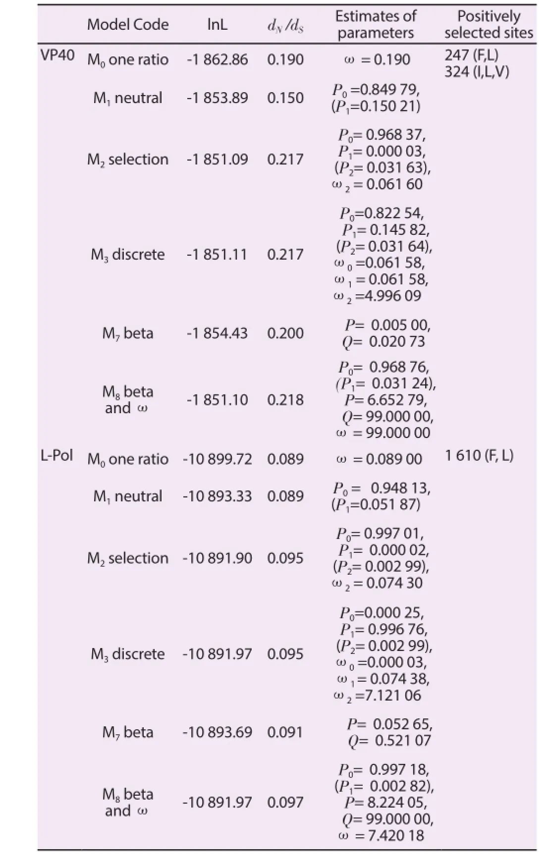
Table 1Likelihood values and parameters estimates for the selection analysis of the VP40 and L-polymerase gene proteins.
3.2. Homology modelling strategy
L amino acid sequence has been analyzed; however, no structure modelling has been performed because the protein has more than 2 000 amino acid residues. Of the overall 2 212 amino acid positions,53 sites are mutated (2.4%) and, among them, 24 have a frequency higher than 0.1. Structural proteins NP, VP35, VP24, VP30 and glycoproteins sGP and ssGP have been therefore analyzed for mutation frequency higher than 0.1 and for the consequences that each amino acid mutated positions could induce on their structures(Figure 1-7 and Table 2).
NP mutation analysis shows that 32 out of 739 amino acid residues(4.33%) are mutated. Among them 13 out of 32 mutation have a frequency mutation higher than 0.1 (Figure 1). NP mutation in amino acid position 8 is a destabilizing mutation, while mutations in amino acid positions 11, 478, 517, 525, 527, 534, 708 are stabilizing mutations. Overall eight out thirteen frequent mutations induce a conformational change in NP structure (Table 2).

Figure 1. Ebola protein modelled structure of NP and amino acid positions with a mutation frequency higher than 0.1.Six out of 340 amino acid positions are mutated in VP35 (1.8%). Among them amino acid positions 12, 41, 68, and 204 have a mutation frequency higher than 0.1 and alanine scanning results show that are all stabilizing mutations (Figure 2 and Table 2).
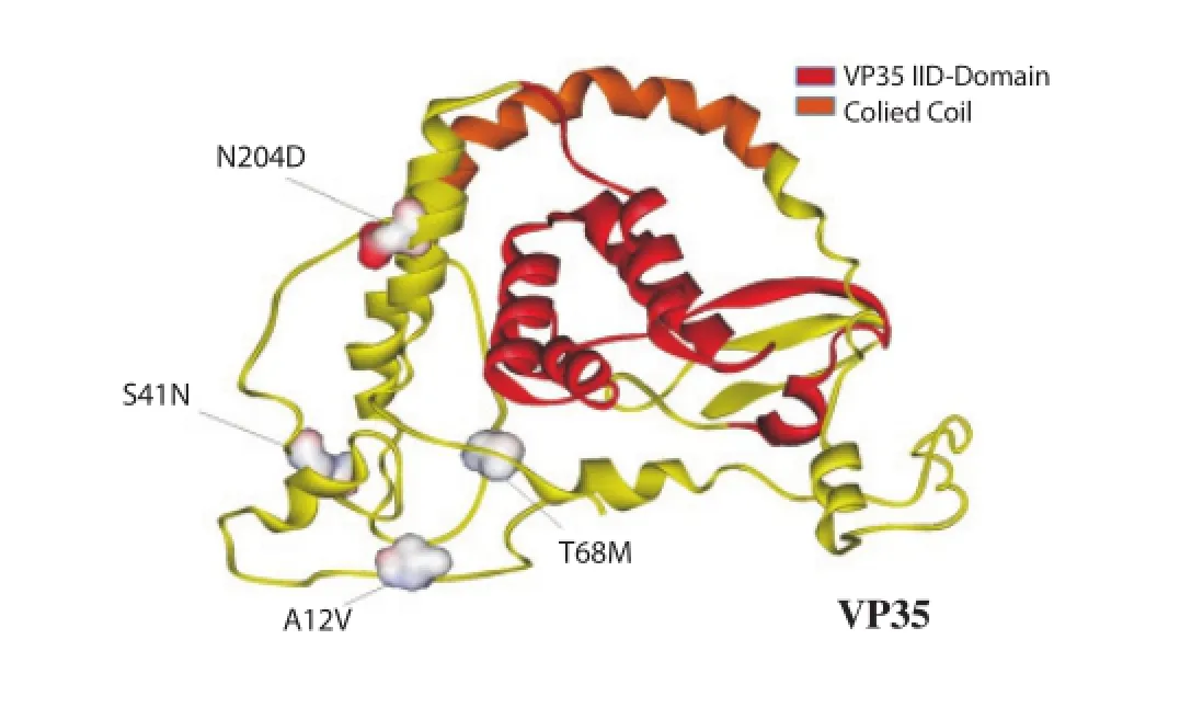
Figure 2. Ebola protein modelled structure of VP35 and amino acid positions with a mutation frequency higher than 0.1.
Structural proteins VP24 and VP30 have structures highly conserved (Figure 3 and 4) with a low number of amino acid mutated positions (VP24=0.8% and VP30= 1.5%). None of the amino acid mutations for each position have a frequency higher than 0.1.
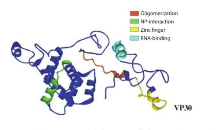
Figure 3. Ebola protein modelled structure of of VP30 and amino acid positions with a mutation frequency higher than 0.1.
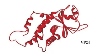
Figure 4. Ebola protein modelled structure of VP24 and amino acid positions with a mutation frequency higher than 0.1.
Seven out of 326 amino acid positions are mutated in VP40 structural protein (2.1%). Among them, amino acid positions 20 and 324 have a mutation frequency higher than 0.1. Alanine scanning analysis shows a stabilizing mutation only in amino acid position 324, which is located in the VP40 membrane-binding region (Figure 5 and Table 2).
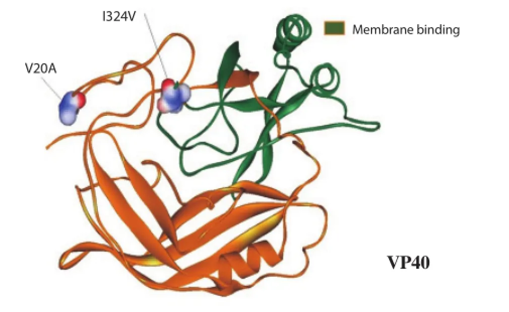
Figure 5. Ebola protein modelled structure of VP40 and amino acid positions with a mutation frequency higher than 0.1.
5.8% of mutated amino acid positions (21 out of 364), fi ve of them with a frequency higher than 0.1 as shown in Figure 6. Specifi cally,amino acid positions 82, 262 and 314 are located in the smallsecreted GP portion, while amino acid positions 331 and 336 are located in the Δ-peptide. In addition, in amino acid position 314 the mutation is stabilizing while only amino acid positions 331 and 336 have destabilizing mutations (Table 2). Due to sequence overlapping with GP1,2, ssGP mutation analysis has been extracted by our previous results on GP[8]. Among them, 6 out of 14 mutations have a frequency mutation higher than 0.1 (Figure 7).
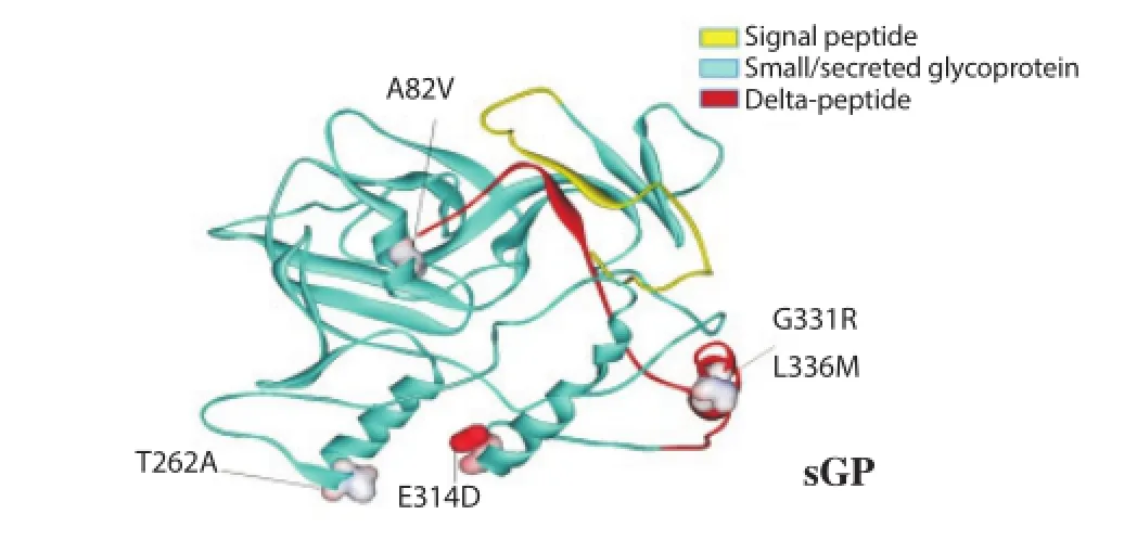
Figure 6. Ebola protein modelled structure of sGP and amino acid positions with a mutation frequency higher than 0.1.
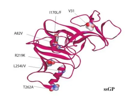
Figure 7. Ebola protein modelled structure of ssGP and amino acid positions with a mutation frequency higher than 0.1.
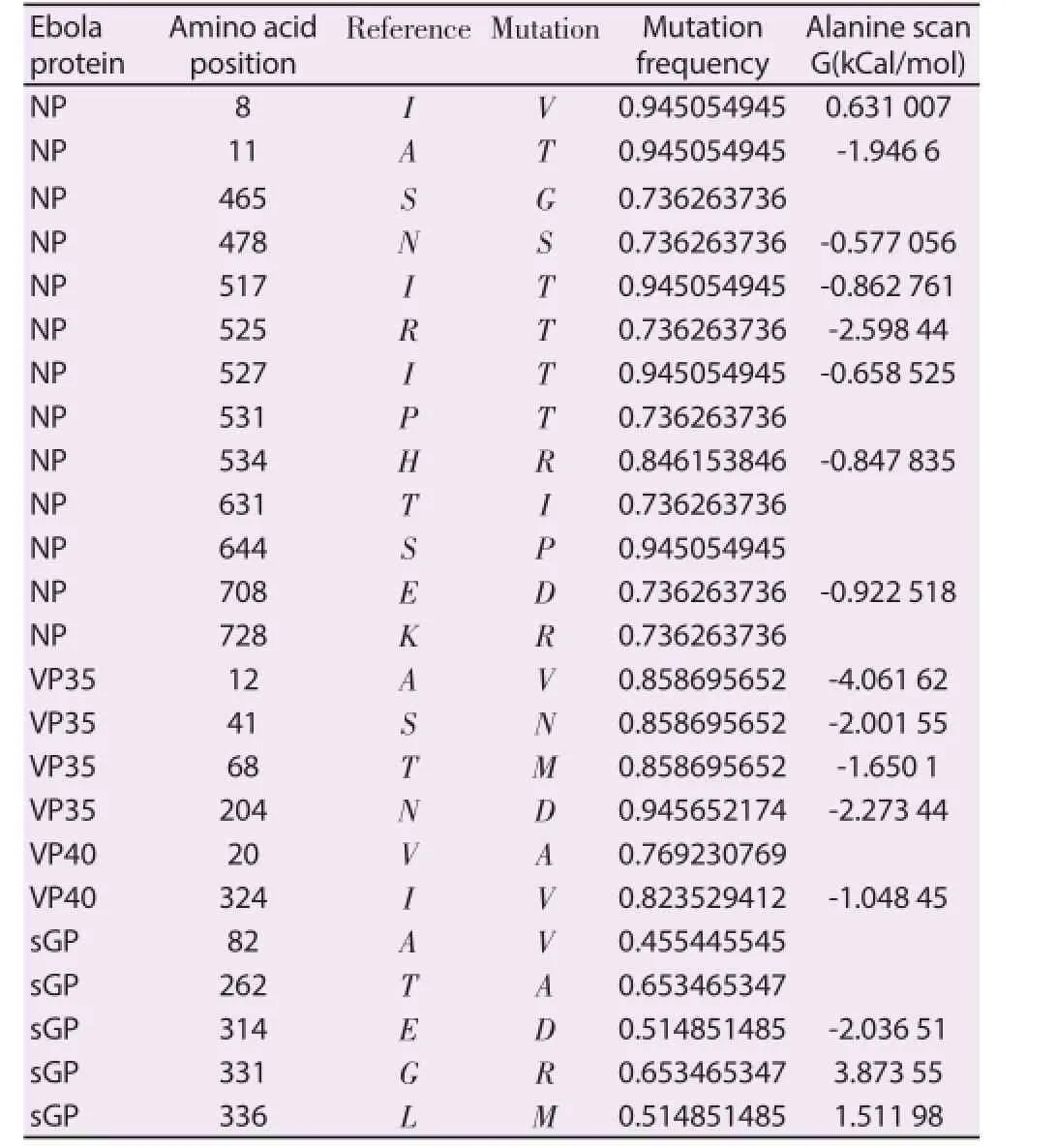
Table 2Ebola proteins mutated amino acid positions.
4. Disscussion
Many factors may influence the circulation and the genetic evolution of EBOV strains, including Virus infectivity, pre-existing immunity in the population, and antigenic variability of the virus. Antigenic variation, in particular, may play an important role in the ability of these viruses to escape the human immune response.
In this context, the analysis of the EBOV proteins gene evolutionis important not only because some of the protein are an important target for the immune response during EBOV infection, but also to better understand the evolutionary dynamic of this virus.
This paper studied the evolution of EBOV proteins using ML techniques. In this analysis only two of the analyzed proteins,showed a high average ratio of non-synonymous to synonymous nucleotide substitution (VP40 and L proteins) ranging from 0.08 to 0.22. As this value is not higher than the threshold of ω >1,is therefore not indicative of positive selection[19]. Moreover, the alpha parameter value was below 1, meaning that, most of the sites along the gene may be invariable because they are under strong purifying selection. A high proportion of amino acids can be largely invariable, probably because amino acid substitutions are not tolerated or selected for (i.e. strong purifying selection). Because the average ω seems usually not sensitive enough to detect Darwinian selection at the molecular level, we used in this case the codon substitution models to detect sites under positive selection. By using these methods, we have identifi ed three sites under positive selection in two diff erent gene proteins (VP40 and L). These results can indicates that although in these proteins a high proportion of amino acids can be largely invariable probably due at structural and functional constraints, adaptive evolution may occur at certain sites of the genome. On the contrary, several amino acid diff erences have been characterized in the recent Ebola outbreak in EBOV G glycoprotein[18]. A better knowledge of the viral protein structure modifications is the key point for drug design and vaccination. Beside the fact that diff erent studies have been focusing on Ebola GP, also VP24 VP30 VP35 and VP40 structural proteins have been recently investigated as potential drug targets[34-37]. Therefore, the evaluation of the genetic heterogeneity in these proteins assumes an increased importance not only in the viral immune escape but also in the drug design. In this study, structural proteins VP24, VP30,VP35 and VP40 show relatively conserved sequences making them suitable target candidates for antiviral treatment as previously suggested[36,37]. On the contrary, NP, L and soluble GP have high mutation frequency. In this context, NP and GP have been chosen as vaccine candidates and for antibody epitopes design as they are the most abundant protein in the infected cells[38-41], but with the frequency of mutation that characterized them attention should be used in the target region.
In conclusion, this study pointed out important aspects of EBOV sequence variability that for epitope and vaccine design should be taken in consideration for appropriate targeting of conserved protein regions.
Acknowledgements
This work was supported by National Institute of Health and Proxagent Ltd in collaboration with University of Rome ‘Tor Vergata’.
Conflict of interest
We declare that we have no confl ict of interest.
References
[1] Kuhn JH, Andersen KG, Baize S, Bao Y, Bavari S, Berthet N, et al. Nomenclature- and database-compatible names for the two Ebola virus variants that emerged in Guinea and the Democratic Republic of the Congo in 2014. Viruses 2014; 6(11): 4760-4799.
[2] Feldmann H, Kiley MP. Classification, structure, and replication of fi loviruses. Curr Top Microbiol Immunol 1999; 235: 1-21.
[3] Feldmann H, Volchkov VE, Volchkova VA, Stroher U, Klenk HD. Biosynthesis and role of fi loviral glycoproteins. J Gen Virol 2001; 82(Pt 12): 2839-2848.
[4] Stahelin RV. Membrane binding and bending in Ebola VP40 assembly and egress. Front Microbiol 2014; 5: 300.
[5] Rougeron V, Feldmann H, Grard G, Becker S, Leroy EM. Ebola and marburg haemorrhagic fever. J Clin Virol 2015; 64: 111-119.
[6] Volchkova VA, Dolnik O, Martinez MJ, Reynard O, Volchkov VE. RNA editing of the GP eene of Ebola virus is an important pathogenicity factor. J Infect Dis 2015; 212(Suppl 2): S226-S233.
6月22—23日,宁都县两天降雨量超过200 mm的有12个乡镇。暴雨洪水造成4.554万人受灾,紧急转移人口 629人,倒塌房屋 126间,县防办发布预警信息1 650条。东韶乡、安福乡、东山坝镇、钓峰乡、长胜镇、石上镇、青塘镇、洛口镇等8个乡镇出现了房屋倒塌,群众在房屋倒塌前得到转移,避免直接伤亡236人。
[7] Azarian T, Lo Presti A, Giovanetti M, Cella E, Rife B, Lai A, et al. Impact of spatial dispersion, evolution, and selection on Ebola Zaire virus epidemic waves. Sci Rep 2015; 5: 10170.
[8] Wauquier N, Becquart P, Padilla C, Baize S, Leroy EM. Human fatal Zaire Ebola virus infection is associated with an aberrant innate immunity and with massive lymphocyte apoptosis. PLoS Negl Trop Dis 2010; 4(10): e837.
[9] Pourrut X, Kumulungui B, Wittmann T, Moussavou G, Delicat A, Yaba P, et al. The natural history of Ebola virus in Africa. Microbes Infect 2005; 7(7-8): 1005-1014.
[10] World Health Organization, Weekly Epidemiological Record. Seasonal infl uenza vaccine composition for tropical and subtropical countries: WHO Expert group meeting, 23-24 April 2015. [Online] Available at: http://www.who.int/wer/2015/wer9036.pdf?ua=1.
[11] World Health Organization. Ebola Situation Report - 2 September 2015.[Online] Available at: http://apps.who.int/ebola/current-situation/ebolasituation-report-2-september-2015.
[12] Gire SK, Goba A, Andersen KG, Sealfon RS, Park DJ, Kanneh L, et al. Genomic surveillance elucidates Ebola virus origin and transmission during the 2014 outbreak. Science 2014; 345(6202): 1369-1372.
[13] Audet J, Kobinger GP. Immune evasion in ebolavirus infections. Viral Immunol 2015; 28(1): 10-18.
[14] Gupta M, Mahanty S, Ahmed R, Rollin PE. Monocyte-derived human macrophages and peripheral blood mononuclear cells infected with Ebola virus secrete MIP-1alpha and TNF-alpha and inhibit poly-IC-induced IFN-alpha in vitro. Virology 2001; 284(1): 20-25.
[15] Takada A, Ebihara H, Jones S, Feldmann H, Kawaoka Y. Protective effi cacy of neutralizing antibodies against Ebola virus infection. Vaccine 2007; 25(6): 993-999.
[16] Wilson JA, Hevey M, Bakken R, Guest S, Bray M, Schmaljohn AL, et al. Epitopes involved in antibody-mediated protection from Ebola virus. Science 2000; 287(5458): 1664-1666.
[17] Wong G, Kobinger GP, Qiu X. Characterization of host immune responses in Ebola virus infections. Expert Rev Clin Immunol 2014;10(6): 781-790.
[18] Giovanetti M, Grifoni A, Lo Presti A, Cella E, Montesano C, Zehender G, et al. Amino acid mutations in Ebola virus glycoprotein of the 2014 epidemic. J Med Virol 2015; 87(6): 893-898.
[19] Ciccozzi M, Babakir-Mina M, Lo Presti A, Farchi F, Zehender G,Ebranati E, et al. Genetic variability of the small t antigen of the novel KI, WU and MC polyomaviruses. Arch Virol 2010; 155(9): 1433-1438.
[20] Wilgenbusch JC, Swoff ord D. Inferring evolutionary trees with PAUP*. Curr Protoc Bioinformatics 2003; DOI:10.1002/0471250953.bi0604s00.
[21] Posada D, Crandall KA. MODELTEST: testing the model of DNA substitution. Bioinformatics 1998; 14(9): 817-818.
[22] Yang Z. PAML: a program package for phylogenetic analysis by maximum likelihood. Comput Appl Biosci 1997; 13(5): 555-556.
[23] Yang Z, Nielsen R, Goldman N, Pedersen AM. Codon-substitution models for heterogeneous selection pressure at amino acid sites. Genetics 2000; 155(1): 431-449.
[24] Nielsen R, Yang Z. Likelihood models for detecting positively selected amino acid sites and applications to the HIV-1 envelope gene. Genetics 1998; 148(3): 929-936.
[25] Yang Z, Wong WS, Nielsen R. Bayes empirical bayes inference of amino acid sites under positive selection. Mol Biol Evol 2005; 22(4): 1107-1118.
[26] Anisimova M, Bielawski JP, Yang Z. Accuracy and power of bayes prediction of amino acid sites under positive selection. Mol Biol Evol 2002; 19(6): 950-958.
[27] Pond SL, Frost SD, Muse SV. HyPhy: hypothesis testing using phylogenies. Bioinformatics 2005; 21(5): 676-679.
[28] Kosakovsky Pond, Frost SD. Not so diff erent after all: a comparison of methods for detecting amino acid sites under selection. Mol Biol Evol 2005; 22(5): 1208-1222.
[29] Kelley LA, Sternberg MJ. Protein structure prediction on the Web: a case study using the Phyre server. Nat Protoc 2009; 4(3): 363-371.
[30] Biasini M, Bienert S, Waterhouse A, Arnold K, Studer G, Schmidt T, et al. SWISS-MODEL: modelling protein tertiary and quaternary structure using evolutionary information. Nucleic Acids Res 2014; 42(W1): W252-W258.
[31] Kallberg M, Wang H, Wang S, Peng J, Wang Z, Lu H, et al. Templatebased protein structure modeling using the RaptorX web server. Nat Protoc 2012; 7(8): 1511-1522.
[32] Zhang Y. I-TASSER server for protein 3D structure prediction. BMC Bioinformatics 2008; 9(1): 40.
[33] Van Durme J, Delgado J, Stricher F, Serrano L, Schymkowitz J, Rousseau F. A graphical interface for the FoldⅩ forcefield. Bioinformatics 2011; 27(12): 1711-1712.
[34] Audet J, Wong G, Wang H, Lu G, Gao GF, Kobinger G, et al. Molecular characterization of the monoclonal antibodies composing ZMAb: a protective cocktail against Ebola virus. Sci Rep 2014; 4: 6881.
[35] Clinton TR, Weinstock MT, Jacobsen MT, Szabo-Fresnais N, Pandya MJ, Whitby FG, et al. Design and characterization of ebolavirus GP prehairpin intermediate mimics as drug targets. Protein Sci 2015; 24(4): 446-463.
[36] Hernandez H, Marceau C, Halliday H, Callison J, Borisevich V, Escaff re O, et al. Development and characterization of broadly cross-reactive monoclonal antibodies against all known Ebola virus species. J Infect Dis 2015; 212(Suppl 2): S410-S413.
[37] Raj U, Varadwaj PK. Flavonoids as multi-target inhibitors for proteins associated with Ebola virus: in-silico discovery using virtual screening and molecular docking studies. Interdiscip Sci Com Life Sci 2015: DOI:10.1007/s12539-014-0246-5.
[38] Dziubanska PJ, Derewenda U, Ellena JF, Engel DA, Derewenda ZS. The structure of the C-terminal domain of the Zaire Ebola virus nucleoprotein. Acta Crystallogr D Biol Crystallogr 2014; 70(9): 2420-2429.
[39] Matassov D, Marzi A, Latham T, Xu R, Ota-Setlik A, Feldmann F, et al. Vaccination with a highly attenuated recombinant vesicular stomatitis virus vector protects against challenge with a lethal dose of Ebola virus. J Infect Dis 2015; 212(Suppl 2): S443-S451.
[40] Tsuda Y, Caposio P, Parkins CJ, Botto S, Messaoudi I, Cicin-Sain L,et al. A replicating cytomegalovirus-based vaccine encoding a single Ebola virus nucleoprotein CTL epitope confers protection against Ebola virus. PLoS Negl Trop Dis 2011; 5(8): e1275.
[41] Wong G, Qiu X, Ebihara H, Feldmann H, Kobinger GP. Characterization of a bivalent vaccine capable of inducing protection against both Ebola and cross-clade H5N1 infl uenza in mice. J Infect Dis 2015; 212(Suppl 2): S435-S442.
ent heading
10.1016/j.apjtm.2016.03.016
15 January 2016
Prof. Massimo Ciccozzi, Department of Infectious Parasitic and Immunomediated Diseases, National Institute of Health, Rome, Italy.
Phone: +390649903187
e-mail: ciccozzi@iss.it
in revised form 20 February 2016
ARTICLE INFO
Article history:
Proteins
Evolutionary analysis
猜你喜欢
杂志排行
Asian Pacific Journal of Tropical Medicine的其它文章
- Determination of ligand cluster and binding site within VP40 of Ebola virus: clue for drug development
- Clinacanthus nutans: a review of the medicinal uses, pharmacology and phytochemistry
- Current perspectives on dengue episode in Malaysia
- Etiological agents causing leptospirosis in Sri Lanka: A review
- Phylogeny of Murray Valley encephalitis virus in Australia and Papua New Guinea
- Dengue outbreak in Swat and Mansehra, Pakistan 2013; an epidemiological and diagnostic perspective
