Mechanism of Japanese encephalitis virus genotypes replacement based on human, porcine and mosquito-originated cell lines model
2016-11-14LoanPhuongDoTrangMinhBuiNgaThiPhan
Loan Phuong Do, Trang Minh Bui, Nga Thi Phan
National Institute of Hygiene and Epidemiology, Hanoi 10000, Vietnam
Mechanism of Japanese encephalitis virus genotypes replacement based on human, porcine and mosquito-originated cell lines model
Loan Phuong Do, Trang Minh Bui, Nga Thi Phan*
National Institute of Hygiene and Epidemiology, Hanoi 10000, Vietnam
Accepted 15 March 2016
Available online 20 April 2016
Japanese encephalitis virus Genotype Ⅰ
Genotype Ⅲ
Multiplication
Shift genotype
Objective: To examine the multiplication efficiency Japanese encephalitis virus (JEV)genotype Ⅰ (GⅠ) and genotype Ⅲ (GⅢ) of diff erent cell lines which originated from human,porcine, mosquitoes in order to prove mechanism of JEV GⅠ replacement JEV GⅢ since it emerging in nature recent decades. Methods: The mixture of GI and GIII JEV isolates was inoculated on human rhabdomyosarcoma (RD), pig kidney epithelial (PS) and Aedes albopictus C6/36 clone (C6/36) which originated from human, porcine and mosquitoes, respectively. Plaque assays were performed to calculate virus titer and real-time RT-PCR with GI and GIII specifi c primer sets to quantify the number of GI and GIII RNA copies. Results: The highest virus titer reached at the 3rd day of post infection when GⅠ and GⅢ mixture was inoculated on RD and PS and that of C6/36 was at the 4thday. JEVs were amplifi ed and maintained by C6/36 cells after 10 passages whereas that by RD and PS only limited within 8 and 6 passages,respectively. GⅠ strain amplifi ed and maintained more effi ciently on C6/36 and PS but not RD, whereas GⅢ strain amplified and maintained more efficiently on RD. Conclusions: There is a correlation between the multiplication effi ciency of GⅠ and GⅢ JEV strains when these two genotype strains co-infected on diff erent cell lines with the predominance of GⅠstrains in C6/36 and PS and the limited detection of GⅠ strains in RD cells proving a possible mechanism of shift JEV genotypes in nature recent decades since GⅠ emerging.
1. Introduction
Japanese encephalitis virus (JEV) is a mosquito-borne virus of the genus Flavivirus in the family Flaviviridae which has circulated widely in Asian and western Pacific countries, and in northern Australia[1,2]. Approximately 67 900 cases of JE typically occur annually and 10 000-15 000 JEV-related human deaths are reported annually[2,3]. JEV is transmitted to susceptible reservoirs by arthropods with the mainly involvement of mosquitoes, especially Culex tritaeniorhynchus whereas pigs, horses, birds and bats are the primary natural hosts of JEV. Humans are the dead-end host in which JEV could not be transmitted from humans to humans.
The JEV genome consists of a single-stranded positive-sense RNA in which the open reading frame encodes a large polyprotein. The N-terminal region of the polyprotein encodes the structural proteins: Capsid (C)-pre-membrane (prM)-Envelope (E) and the nonstructural proteins (NS) (NS1-NS2A-NS2B-NS3-NS4-NS5)[4]. JEV is classifi ed into fi ve genotypes based on the nucleotide sequences of the C/prM and E protein genes[5]. However, all fi ve genotypes have been detected in mosquitoes and reservoirs while the number of genotypes detected in humans is limited. While JEV genotypesⅠ and Ⅲ are predominant in humans, only two of JEV genotypeⅡ strains were detected so far; one in Australia and the other in Korea[5,6].
In humans, genotype Ⅰ (GⅠ) strains have displaced genotypeⅢ (GⅢ) strains to become the predominant genotype in many countries[7]. The emergence of JEV GⅠ strains was fi rst identifi ed in mosquitoes and pigs in northern Asian countries, Korea in 1993 and Japan in 1994[8-10] and in mosquitoes in China in 1979[11]. However,GⅠ strains have mainly been isolated from mosquitoes and swine while very few have been isolated from humans[11,12]. Therefore, the JEV GⅠ strains are supposed more adapting to mosquitoes and pigs than to humans[14]. Among twelve haplotypes that are defi ned based on the four amino acid residues in the E protein (sites 123, 209, 227,and 408), the haplotypes and the host ranges of the GⅠ isolates are narrower than those of the GⅢ isolates[15].
To identify the adaptation of JEV GⅠ and GⅢ strains in diff erent host species experimentally, we conducted the study to examine the multiplication of JEV GⅠ and GⅢ strains when these two genotypes co-infected in different cell lines which originate from human,porcine and mosquitoes. This study aims to prove an evidence of the mechanism shift JEV genotypes in nature recent decades since JEV GⅠ emerging in most of Asian countries where JEV GⅢ circulating before.
2. Material and methods
2.1. Cell and viruses
Pig kidney epithelial (PS); human rhabdomyosarcoma (RD);mosquito Aedes albopictus C6/36 clone (C6/36), baby hamster kidney-21 cells were maintained in minimum essential medium(MEM) supplemented with 10% fetal bovine serum. The cells were incubated at 37 ℃ with 5% CO2except C6/36 cell was incubated at 28 ℃. The Japanese encephalitis virus (JEV) strain 93VN118 and 94VN141 were originally detected from mosquitoes in 1993 and 1994 respectively. Their genotypes were identifi ed as genotype Ⅰ-b(GⅠ-b) (94VN141) and genotype Ⅲ (GⅢ) (93VN118) (GenBank accession number AB93331 and AB93332)[12]. Those virus strains were chosen because they are the first JEV GⅠ and the last JEV GⅢ to be detected from mosquitoes in the Northern Vietnam. Virus stocks were stored at -80 ℃ until examined.
2.2. Viral infection
The virus titers of two isolates were determined by plaque assay,and then diluted to be at the same titer (100 PFU/mL) in MEM with 2% fetal calf serum (FCS).
The mixture of two strains whose titer was 100 PFU/mL was inoculated on the monolayer of RD, PS and C3/36 cell lines for 60 min at 37 ℃ and 28 ℃ respectively. The un-absorbed viruses were removed from cells. The infected RD, PS cells were incubated at 37 ℃ in a humidifi ed 5% CO2atmosphere and C6/36 in 28 ℃. The supernatants were harvested continuously for 5 d after inoculation in order to determine time-point when virus titers reached peak by using plaque assay. The virus mixture was then inoculated continuously 10 passages on RD, PS and C3/36 cell lines. The supernatants were harvested at the determined peak day in every passage and stored at -80 ℃. Plaque assays were performed to determine which cell line could maintain and amplify JEV effi ciently and TaqMan RT-PCR were performed to determine which genotype amplifi ed effi ciently by each cell line.
2.3. Plaque assay
Serial ten-fold dilutions of JEV GⅠ and GⅢ mixture at each determined time-point were prepared in chilled MEM with 2% FCS from 10-1to 10-6. Diluted virus samples were inoculated on three cell lines and then MEM with 2% FCS plus with 1.25 methylcellulose was overlaid after viral absorption for 90 min. Each dilution was performed in triplicate. The baby hamster kidney-21 cells were incubated at 37 ℃ with 5% CO2for 7 d. Cells then were stained with 0.1% crystal violet after saline formal fi xation for demonstration of plaques. The excess stain was washed off with tap water; air-dried. The number of plaques was measured in each well and calculated the virus titer. Plaque assays were repeated three times at the same conditions.
2.4. RNase treatment and nucleic acid extraction from supernatant
In order to measure only RNAs of JEV on the cell, before RNA extraction, the infected supernatants were treated with RNase. One unit of Rnase A (Qiagen Science, Germantown, MD, USA) diluted 1:10 or 1:100 with nuclease free water was added to 200 μL of each sample and incubated at 37 ℃ for 30 min. Rnase A acitivity,thereafter was inhibited by 18U of Rnase inhibitor (Qiagen) and incubated at room temperature for 30 min. The RNAs were then extracted using QIAamp Viral RNA Mini Kit (Qiagen) according to the manufacturer’s instructions.
2.5. TaqMan real-time RT-PCR assay
JEV GⅠ and GⅢ strains quantifi cation was performed separately after each passage with a specifi c primer set with SuperScript® ⅢPlatinum® One-Step qRT-PCR Kit w/o ROX (Invitrogen, Carlsbad,CA, USA) as described previously.
F o r GⅠ d e t e c t i o n, f o r w a r d p r i m e r (5’-GGGGACAAGCAGAT TAACCA-3’), reverse primer(5’-G A AG G C ACC ACC A A AC AC T T-3’) w e r e u s e d at a final concentration of 0.2 μM and probe (FAMTCAACAACT T TGAAAGGGGC-36TAMSp) at a final concentration of 0.1 μM. The same primer concentration was used for GⅢ, primer set with forward primer (5’-CCTTGCAAAATTCCGATTGT-3’), reverse primer (5’-TGAGC TCCC T TCAAAGTCGT-3’) and probe (FAMCTGGTGACAGTGAACCCCTT-36TAMSp). A Eppendorf Mastercycler ep Realplex (Eppendorf AG, Hamburg, Germany), was used for analysis. The thermal profi le included incubation at 50 ℃for 15 min, at 95 ℃ for 2 min for reverse transcription. DNA was amplifi ed with 40 cycles at 95 ℃ for 15 s, 60 ℃ for 30 s.
3. Results
3.1. Determination of time-point of virus titer peak on different cell-lines
To determine the time point when virus titers in the supernatant were the highest, we inoculated the virus mixture on three celllines: PS, RD and C6/36. With the same virus titer inoculation (100 PFU/mL) and the same virus mixture volume (100 μL), the highest mixture virus titers reached after 3 d post infection (p.i.) on PS(1.3×10³PFU/mL) and RD (1.6×10⁵PFU/mL), and 4 d p.i. on C6/36 (7.2×10⁵) when measured by plaques assay (Figure 1).

Figure 1. Multiplication kinetics by time post infection of JEV mixture of strains representative of GⅠ and GⅢ genotype in C6/36, RD and PS cell lines.
3.2. Most efficient cell-line for JEV multiplication
When continuously inoculated 10 passages on cell-lines, the virus was maintained by C6/36 during 10 passages and the highest virus titer reached after the fi fth passage (1.2×10⁷PFU/mL) but not by PS or RD. The virus titer could not be determined after the 6thpassage when virus mixture was inoculated on PS and after the 8thpassage when the virus mixture was inoculated on RD (Figure 2).
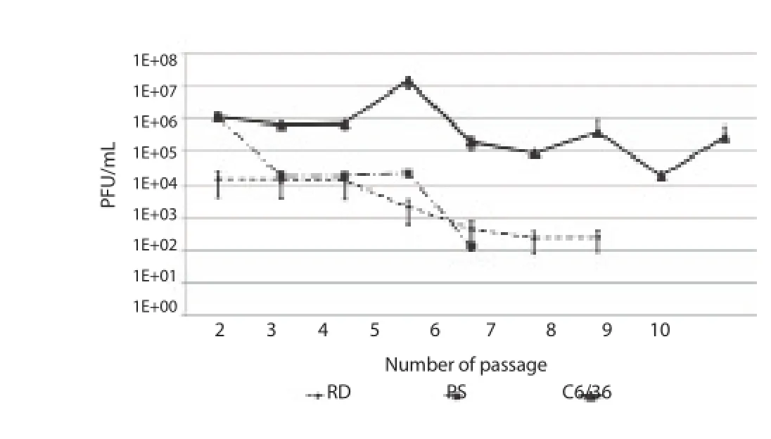
Figure 2. Multiplication kinetics within ten passages of JEV mixture of strains representative of GⅠ and GⅢ genotype in C6/36, RD and PS cells.
3.3. Multiplication of GⅠ and GⅢ strains in human, porcine and mosquito cells
After 93VN118 strain (GⅢ) and 94VN141 strain (GⅠ) was mixed and continuously passage using PS, RD and C6/36, the numbers of RNA copy of each strain after each passage were quantified indirectly through Ct value (the higher RNA copies, the lower Ct value) by real-time RT-PCR using GⅠ/GⅢ specific genotype primer sets.
In C6/36, Ct value of 94VN141 strain (GⅠ) was lower than 93VN118 strain (GⅢ) meaning that the virus titer of GⅠ strain was higher than that of GⅢ strain at all 10 passages (Figure 3a). Although GⅠ strain was amplifi ed more effi ciently in C6/36 than GⅢ strain but both genotype strains were maintained during 10 passages because its RNA copies still could be detected by real-time RT-PCR at 10thpassage.
Figure 3. Multiplication kinetics of strains representative of GⅠ and GⅢgenotype when co-infected in C6/36, PS and RD cells.a: C6/36 cell line; b: PS cell line; c: RD cell line.
On PS cell-line, both GⅠ and GⅢ decreased gradually after each passage but the dead-end circle point of GⅢ strain was after 6thpassage and that of GⅠ strain was after 7thand 8thpassage (Figure 3b) meaning that GⅠ strain was maintained longer than GⅢ by RDcell-line.
On RD cell-line, the GⅠ strain decreased gradually and disappeared after 5thpassage gradually but GⅢ strain increased and got peak at 3rdpassage and decreased gradually after that. However, GⅢ strain was maintained longer than GⅠ strain when the dead-end point was after 8thpassage (Figure 3c).
4. Discussion
The study was examined the amplification and maintenance capacity of three diff erent cell-lines which originated from humans,porcine and mosquitoes, the three main components of JEV cycle in nature to the most two predominant JEV genotypes. The virus titer reached peak on C6/36 1 d later than that on RD and PS. It is worthy to note that multiplication of GⅠ-b strains increased when the temperature increased[7] and the incubation temperature of C6/36 cells is 28 ℃ and that of RD and PS cell is 37 ℃. GⅠ-b strains were also proven to be temperate genotype whereas GⅢ is signifi cantly associated with temperate climates[16].
C6/36 was the most effective tool to isolate JEV since the JEV strains could multiply and maintain high titer after 10 passages whereas the multiplication of JEV was limited within 6 to 8 passages when viruses were inoculated on PS and RD. When two genotype strains (GⅠ and GⅢ) were co-infected in vitro, GⅠ-b strain amplifi ed more effi ciently than GⅢ strains on C6/36 and PS cells whereas GⅢ strain amplifi ed more effi ciently on RD cells than GⅠ-b strain showed that GⅠ-b strains increased viral multiplication on C6/36 cells compared to GⅠ-a strains as well as other genotype strains[7]. GⅠ-b strain could not maintain itself on RD cells, and disappeared after 5 passages. Although there were differences between in vitro and natural condition such as C6/36 cells lack a functional RNA interference response[7], the results provided some pieces of experimental explanation to the predominant and replacement of GⅠ strains to GⅢ strains in mosquitoes and pigs[7,17]and very few GⅠ strains have been isolated from humans[12,13].
There is a correlation between the high multiplication effi ciency of GⅠ JEV strains when two genotype strains, GⅠ and GⅢ, were coinfected on porcine and mosquito cells with the predominant of GⅠstrains in mosquitoes and porcine and the limited detection of GⅠstrains in humans. The high multiplication effi ciency of GⅢ strains on humans also corrected well with the predominant of GⅢ strain in humans.
Conflict of interest statement
We declare that we have no confl ict of interest.
Acknowledgments
This research is funded by Vietnam National Foundation for Science and Technology Development (NAFOSTED) under grant number: 106.16-2011.68.
[1] Erlanger TE, Weiss S, Keiser J, Utzinger J, Wiedenmayer K. Past, present,and future of Japanese encephalitis. Emerg Infect Dis 2009; 15(1): 1-7.
[2] Solomon T. Control of Japanese encephalitis-within our grasp? N Engl J Med 2006; 355(9): 869-871.
[3] Campbell GL, Hills SL, Fischer M, Jacobson JA, Hoke CH, Hombach JM, et al. Estimated global incidence of Japanese encephalitis: a systematic review. Bull World Health Organ 2011; 89(10): 766-774.
[4] Lindenbach BD, Murray CL, Thiel HJ, Rice CM. Flaviviridae: The viruses and their replication. In: Knipe DM, Howley PM. (eds.), Fields virology, 6th Edition. New York: Lippincott Williams & Wilkins; 2013.
[5] Williams DT, Wang LF, Daniels PW, Mackenzie JS. Molecular characterization of the fi rst Australian isolate of Japanese encephalitis virus, the FU strain. J Gen Virol 2000; 81(10): 2471-2480.
[6] Schuh AJ, Li L, Tesh RB, Innis BL, Barrett ADT. Genetic characterization of early isolates of Japanese encephalitis virus: genotype II has been circulating since at least 1951. J Gen Virol 2010; 91(Pt 1): 95-102.
[7] Schuh AJ, Ward MJ, Brown AJL, Barrett ADT. Dynamics of the emergence and establishment of a newly dominant genotype of Japanese encephalitis virus throughout Asia. J Virol 2014; 88(8): 4522-4532.
[8] Yun SM, Cho JE, Ju YR, Kim SY, Ryou J, Han MG, et al. Molecular epidemiology of Japanese encephalitis virus circulating in South Korea,1983-2005. Virol J 2010; 7: 127.
[9] Tang WF, Ogawa M, Eshitac Y, Aonoa H, Makino Y . Molecular evolution of Japanese encephalitis virus isolates from swine in Oita, Japan during 1980-2009. Infect Genet Evol 2010; 10(2): 329-336.
[10] Ma SP, Yoshida Y, Makinoet Y, Tadano M, Ono T, Ogawa M. Short report: a major genotype of Japanese encephalitis virus currently circulating in Japan. Am J Trop Med Hyg 2003; 69(2): 151-154.
[11] Wang HY, Takasaki T, Fu SH, Sun XH, Zhang HL, Wang ZX, et al. Molecular epidemiological analysis of Japanese encephalitis virus in China. J Gen Virol 2007; 88(Pt 3): 885-894.
[12] Do LP, Bui TM, Hasebe F, Morita K, Phan NT. Molecular epidemiology of Japanese encephalitis in northern Vietnam, 1964-2011: genotype replacement. Virol J 2015; 12: 51.
[13] Wang L, Fu S, Fu SH, Zhang HL, Ye XF, Yu DS, Deng Z. Identification and isolation of genotype-Ⅰ Japanese encephalitis virus from encephalitis patients. Virol J 2010. 7: 345.
[14] Nga PT, del Carmen Parquet M, Cuong VD, Ma SP, Hasebe F, Inoue S,et al. Shift in Japanese encephalitis virus (JEV) genotype circulating in northern Vietnam: implications for frequent introductions of JEV from Southeast Asia to East Asia. J Gen Virol 2004; 85(Pt 6): 1625-1631.
[15] Han N, Adams J, Chen P, Guo ZY, Zhong XF, Fang W, et al.Comparison of genotypes Ⅰ and Ⅲ in Japanese encephalitis virus reveals distinct differences in their genetic and host diversity. J Virol 2014; 88(19): 11469-11479.
[16] Schuh AJ, Ward MJ, Brown AJ, Barrett AD. Phylogeography of Japanese encephalitis virus: genotype is associated with climate. PLoS Negl Trop Dis 2013; 7(8): e2411.
[17] Pan XL, Liu H, Wang HY, Fu SH, Liu HZ, Zhang HL, et al. Emergence of genotype Ⅰ of Japanese encephalitis virus as the dominant genotype in Asia. J Virol 2011. 85(19): 9847-9853.
ent heading
10.1016/j.apjtm.2016.03.007
15 January 2016
Nga Thi Phan, National Institute of Hygiene and Epidemiology, Hanoi 10000, Vietnam.
E-mail: phannganihe@gmail.com
Fountadion project: This research is funded by Vietnam National Foundation for Science and Technology Development (NAFOSTED) under grant number: 106.16-2011.68.
in revised form 20 February 2016
ARTICLE INFO
Article history:
杂志排行

Asian Pacific Journal of Tropical Medicine的其它文章
- Determination of ligand cluster and binding site within VP40 of Ebola virus: clue for drug development
- Clinacanthus nutans: a review of the medicinal uses, pharmacology and phytochemistry
- Current perspectives on dengue episode in Malaysia
- Etiological agents causing leptospirosis in Sri Lanka: A review
- Phylogeny of Murray Valley encephalitis virus in Australia and Papua New Guinea
- Dengue outbreak in Swat and Mansehra, Pakistan 2013; an epidemiological and diagnostic perspective