Perfusion of gastrodin in abdom inal aorta for alleviating spinal cord ischem ia reperfusion injury
2016-06-29HuaFangJingChaoZhangMiaoYangHuaFengLiJianPingZhangFangXiangZhangQuanYunWangRuRongWangJinLiu
Hua Fang, Jing-Chao Zhang, Miao Yang, Hua-Feng Li, Jian-Ping Zhang*, Fang-Xiang Zhang, Quan-Yun Wang, Ru-Rong Wang, Jin Liu
1Department of Anesthesiology, Guizhou Provincial People's Hospital, Guiyang 550002, Guizhou Province, China2Department of Anesthesiology, Affiliated People's Hospital of Guizhou Medical University, Guiyang 550002, Guizhou Province, China3Department of Anesthesiology, West China Second University Hospital, Chengdu 610041, Sichuan Province, China4Department of Anesthesiology, West C na Hospital, Sichuan University, Chengdu 610041, Sichuan Province, China
Perfusion of gastrodin in abdom inal aorta for alleviating spinal cord ischem ia reperfusion injury
Hua Fang1,2, Jing-Chao Zhang1,2, Miao Yang1,2, Hua-Feng Li3, Jian-Ping Zhang1,2*, Fang-Xiang Zhang1,2, Quan-Yun Wang4, Ru-Rong Wang4, Jin Liu4
1Department of Anesthesiology, Guizhou Provincial People's Hospital, Guiyang 550002, Guizhou Province, China
2Department of Anesthesiology, Affiliated People's Hospital of Guizhou Medical University, Guiyang 550002, Guizhou Province, China
3Department of Anesthesiology, West China Second University Hospital, Chengdu 610041, Sichuan Province, China
4Department of Anesthesiology, West C na Hospital, Sichuan University, Chengdu 610041, Sichuan Province, China
AR T ICLE IN FO
Article history:
Received 15 April 2016
Received in revised form 16 May 2016 Accepted 15 June 2016
Available online 20 July 2016
Keywords:
Gastrodin
Spinal cord ischem ia reperfusion injury
Mitochondria
Motor evoked potential
ABSTRACT Objective: To observe the effects of perfusion of the gastrodin in abdom inal aorta for alleviating the spinal cord ischemia reperfusion injury (SCIRI). Methods: A total of 36 New Zealand white rabbits were divided random ly into sham-operated group (group S), control group (group C) and gastrodin group (group G), 12 rabbits for each group. Aorta abdom inalis infrarenalis blocking method was applied to establish the SCIRI model. The changes of motor evoked potentials (MEPs) before the ischemia and on 30 min, 60 min, 6 h, 12 h and 24 h of reperfusion of the gastrodin were respectively recorded, and the neurologic function score before the ischemia, on the 6 h, 12 h and 24 h of the reperfusion of the gastrodin were assessed. And the changes of the concentration of serum neuron specific enolase (NSE), interleukin (IL)-l β and IL-8 were measured before the ischem ia, after 45 m in of ischem ia, and on 30 m in,60 m in, 6 h, 12 h and 24 h of reperfusion of gastrodin. Then the levels of spinal cord nerve cells mitochondrial superoxide dismutase (SOD), reactive oxygen species (ROS), glutathione peroxidase (GSH-PX), malondialdehyde (MDA), total antioxidant capacity (T-AOC) and mitochondrial swelling degree (MSD) were tested and the histopathologic changes in spinal cord tissues were observed. Results: The levels of the NSE, IL-lβ, IL-8, ROS, MDA and MSD of group C were all significantly elevated after the ischem ia (P<0.01); the levels of the spinal nerve cell mitochondria SOD, GSH-PX and T-AOC were all significantly reduced (P<0.01), MEPs and spinal cord tissue pathology were damaged significantly (P<0.01). The rate of motor neuron abnormalities and the damages of spinal cord tissue pathology of group G were significantly milder than those of group C (P<0.01); the levels of NSE, IL-lβ, IL-8,ROS, MDA and MSD were significantly lower than those of group C (P< 0.01), but the levels of SOD, GSH-PX and T-AOC were all significantly higher than those of group C (P<0.01), and the recovery of neurologic function score during the reperfusion of gastrodin was significantly faster than group C (P<0.01). Conclusions: Perfusion of the gastrodin in abdominal aorta can alleviate the spinal cord ischem iare perfusion injury by promoting the m itochondrial antioxidant capacity and inhibiting the inflammatory reaction.
E-mail: albatrossa@126.com
Tel: 13765169137
Foundation project: It was supported by National Natural Science Foundation of China (Grant number: 30672025); Science and Technology Department of Guizhou Province Foundation Project (Grant number: QinkeheSY[2013]3063,QinhekeJ[2013]2179, QinkeheLH[2014]7021).
1. Introduction
During the period of spinal cord ischem ia reperfusion injury(SCIRI), the reducing of the mitochondrial antioxidant capacity and the changes of the m itochondrial membrane structure are closely related to the incidence of functional disorder of the hind-limb nerve[1,2]. The gastrodin has certain protective effects on cerebral ischem ia-reperfusion injury through its anti-inflammatory effect and anti-oxidation effect[3,4]. And our previous study has confirmed that the perfusion of 100 mg/kg of gastrodin in partial abdominal aorta can facilitate the recovery of the spinal hind-lime function[5]through improving the spinal cord m icrocirculation function. Inorder to further study the protection mechanism of gastrodin to the spinal cord, the BL-420S experimental system was applied to realtime monitor the change rule of the motor evoked potentials (MEPs)after the perfusion of gastrodin in partial abdominal aorta, and we continue to observe the effects of gastrodin on the m itochondrial antioxidant capacity and the inflammatory reaction of the SCIRI and discuss the its mechanism of action.
2. Materials and methods
2.1. Primary reagents
Gastrodin injection (Batch number: H20066464, produced by Shanghai Modern Hasen Pharmaceutical Co., Ltd. 200 mg/bottle);neuron-specific enolase (NSE), interleukin (IL)-lβ, IL-8 and ELISA kits were all purchased from USCN Business Co., Ltd.; the kits of glutathione peroxidase (GSH-PX), malondialdehyde (MDA), total anti-oxidation capacity (T-AOC), superoxide dismutase (SOD) and reactive oxygen species (ROS) were all purchased from Shanghai Yanji Biological Technology Co., Ltd.
2.2. Experimental animals
The selected 36 SPF grades of New Zealand white rabbits of either gender with 4-6 months and 2-2.5 kg of body mass were provided by Laboratory Animal Center of Sichuan University. The animals were fed in separate cages. The rabbits were fed freely with the standard m ixed feeding stuffs and water in the room temperature of 20 ℃-25 ℃. In the process of the experiment, the handling of animals was strictly abided by the Regulation of Experimental Animals,and approved by Ethics Comm ittee of Sichuan University. This experiment was operated and finished at the Experimental Center of Sichuan University. The experiment animals were fasted 12 h before the operation.
2.3. Grouping and model establishment
The rabbits were divided into 3 groups through the random number table, which were control group (group C), gastrodin group (group G) and sham-operated group (group S), 12 for each group. The SCIRI model was established through referring to the reference[5],which was the injection of the concentration of 30 mg/kg of 1.5% pentobarbital sodium and 0.25 mg/kg of vecuronium bromide on the ear marginal vein to anaesthetize the trachea cannula, and then the breathing machine was connected for mechanical ventilation. The disinfection towers were put on the left iliac region, and then the extradural catheter was inserted into the left arteria femoralis extended to the 2 cm under the initial point of the left renal artery. The operation was carried step by step into the enterocoelia after the abdomen was disinfected and the disinfection towers were put on the surface of the abdomen, and the gauzed pad soaked with the normal saline was used to cover the visceral organs of enterocoelia. And then the revealed abdom inal aorta was separated to 0.5 cm under the initial point of left renal artery, and the medium artery clamp was used temporarily to clip abdominal aorta after the injection of 1 mg/kg of heparin on the ear marginal vein to make sure the pulse of the abdom inal aorta was stopped under the clipped point. The clamp was removed after 45 m in of the blocking of blood flow of the abdominal aorta, and the lumbar SCIRI was confirmed; the average arterial pressure, pulse oxygen saturation, electrocardiogram and arterial blood gas were monitored and the anus temperature of 36-37 ℃ was maintained in the process of the operation. After blocking abdominal aorta, the patients in group G were injected with 100 mg/ kg of gastrodin injection immediately in the abdominal aorta through the catheter for the 5 m in pretreatment of the ischemia of spinal cord tissues. Group C was perfused with the equal capacity of normal saline. And group S was only for surgery operation w ithout blocking the abdominal aorta.
2.4. Observational index
2.4.1. Neurologic function scores
Two observers who didn’t know the grouping conditions evaluated and recorded the neurologic function scores before ischemia, after 6 h, 12 h and 24 h of perfusion of the gastrodin, and tested 3 times for each rabbits, and calculated the average value, after the general observation of the animals. The neurologic function scores included: (1) The evaluation of hind limb reflex function was measured through 24 h reperfusion and referenced to the standard of Reuter[6],the more serious the functional disorder of movement reflection, the higher score the Reuter; (2) The hindlimb motor function scores was referenced to the grading standard of Jacobs[7] to measure the hind limb movement function.
2.4.2. Measurement of MEPs
The BL-420S experimental system (produced by Chengdu Techman Software Co., LTD.) was applied to record the hind limb MEPs respectively before the ischem ia, on 30 m in, 60 m in, 6 h, 12 h and 24 h of reperfusion referred to the reference[8]. Recording method was as follows. Firstly, for recording, a 3.0 mm circular hole on the right hind limb movement projection area of the rabbits was drilled by using the dental bur (equal to the left 2.5 mm of the sagittal line of the center of the skull and 2.5 mm upside of the lambdoidal suture) and the endocranium was revealed, then diameter of 2.0 mm of the aseptic unipolar recording electrode silver ball was used torecord electrode wave of the endocranium by connecting with it. The recorded onset latency (OL) included the beginning time point of the stimulation to the time point of the appearance of the peak of N wave (unit: ms); the recorded interpeak amplitude (IPA) included from the peak of the N wave to valley of the P wave (unit: μv). Secondly, in terms of stimulation, two 0.05 mm of aseptic yinqiu unipolar recording electrode were inserted into the left calf muscle (the distance was 2.5 cm), and the electric pulse stimulation with the stimulus frequency of 4Hz and the intensity of 5 mA was conducted. The brain bone flap was restored and the scalp was sutured after the experiment.
2.4.3. Detection of blood index
A total of 2 m L of the femoral venous blood at the time points before ischemia, at 45 min after ischemia, 30 min, 60 min, 6 h, 12 h and 24 h of reperfusion of gastrodin was extracted for centrifugation,and then the supernate was collected to detect the concentrations of the NSE, IL-lβ and IL-8 through liquid double antibody sandw ich ELLSA method by Elx800 ELISA reader (produced by Bio-TEK Co., Ltd.).
2.4.4. Detection of spinal tissue index
L3 - L4 segmental spinal cord tissues were extracted respectively before the ischemia, at 45 min after the ischemia, 30 min and 60 min of reperfusion of the gastrodin, and a part of the spinal cord tissues were fastened to 10% of the formalin for the pathological detection of spinal cord tissue; according to reference[9,10]; another part of the spinal cord tissues was used for the preparation of the mitochondria in nerve cells of spinal cord tissues: 1:9(w/v) of the separating medium (0.225 mol/L D-mannitol pH7.4, 0.075 mol/L sucrose, 10 mol/L Tris-HCI and 0.05 mol/L EDTA) was added into the spinal cord tissues under the ice condition of 0-4 ℃, after the ultrasonic refining and the 600× g 5 m in centrifugation, the suspended sediment of the separating medium was again ultrasonic refined. The supernate was centrifuged for 5 min at 600× g in the temperature of 4 ℃, then it was again centrifuged for 10 min at 10 000× g in the temperature of 4 ℃, and the concentration of m itochondria suspension protein was detected through the coomassie brilliant blue method. According to the operation instruction of the kits, the 6405 type ultraviolet spectrophotometer (produced by Jenway Co.,Ltd) was applied to detect the m itochondria ROS, MDA, GSH-PX, SOD and T-AOC in nerve cells of spinal cord tissues and the detect the lum inance value of m itochondria suspension on 520 nm as the index of mitochondrial swelling degree (MSD). The abnormal motor neuron and the abnormal motor neuron of spinal Ⅷ-Ⅺ area were calculated according to the formula to calculate the rate of the motor neuron abnormality[5,8]. And the formula was the rate of motor neuron abnormalities (%) = abnormal number of neurons in selected field of vision of every slice section/ total number of neurons in selected field of vision 100%.
2.5. Statistical analysis
The software SPSS16.0 was used for statistical analysis, the measurement data were expressed as mean ± SD, and the one-way analysis of variance was used for the comparison among groups. P<0.05 was statistically different.
3. Results
3.1. General conditions
In the process of the operation, there was no accidental death of experiment animals, and they were fully awake after 2 h of the operation w ithout infections.
3.2. Changes of hind limb NFS on different time points of reperfusion
Jacobs scores of 3 groups were all five and Reuter scores of 3 groups were all zero before the ischemia. Jacobs score of the changes of hind lamb NFS on different time points of reperfusion of group C was significantly reduced compared to that of group C before the ischem ia and group S (P<0.01). Group G was significantly higher than group C in terms of Jacobs scores on different time points of reperfusion (P<0.01). Reuter scores of the changes of hind lamb NFS on different time points of reperfusion of group C was significantly increased compared to that of group C before the ischem ia and group S (P<0.01). Group G was significantly lower than group C in terms of Reuter scores on different time points of reperfusion (Table 1).
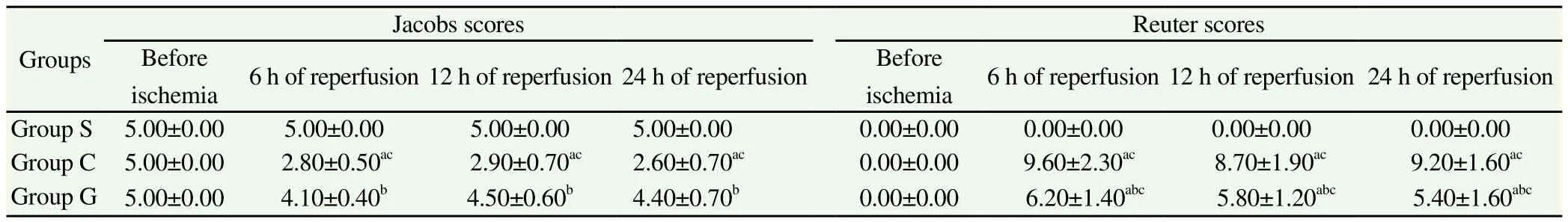
Table 1 Changes of hind limb NFS on different time points of reperfusion (n=12).
3.3. Changes of concentration of serum NSE, IL-lβ and IL-8
The concentration of NSE, IL-lβ and IL-8 of group S had no significant changes. The concentration of NSE, IL-lβ and IL-8 of group C on 45 m in of ischem ia and on different time points of reperfusion were significantly elevated compared to that before ischemia and group S (P<0.01). The concentration of NSE, IL-lβ and IL-8 of group G on the reperfusion of 60 m in recovered to the level before the ischemia, the concentration of NSE, IL-lβ and IL-8 of group G at 45 min of the ischemia and at the different time points of reperfusion were all significantly lower than that of group C (P<0.01) (Table 2).
3.4. Changes of MEPs
The MEPs, OL and IPA of group S had no significant changes. The MEPs wave forms of group C and group G were all disappeared on 15 min of the ischemia. The MEPs OL of group C was significantly increased and its IPA was significantly decreased compared to that of group C before the ischem ia and group S on different time points of reperfusion, (P<0.01). On 30 m in of reperfusion, MEPs OL and IPA levels of group G nearly returned to the levels before ischemia;its MEPs OL levels were significantly lower and its IPA levels were significantly higher than that of group C on different time points of reperfusion (P<0.01) (Table 3).
3.5. Changes of indexes of spinal cord tissues on different time points
The SOD, GSH-PX, T-AOC, MDA, MSD, ROS and the rate of motor neuron abnormalities of group S had no significantly changes. The m itochondrial swelling was presented as the decrease of the value of light absorption on 520 nm. The SOD, GSH-PX, MSD and T-AOC of group C after the ischem ia of spinal cord were all significantly decreased (P<0.01), and the rate of motor neuron abnormalities, ROS and MDA were significantly increased (P<0.01),compared to that before the ischemia and that of group S; the SOD,GSH-PX, MSD and T-AOC were significantly decreased in a further step (P<0.01) during the reperfusion, and the rate of motor neuron abnormalities, ROS and MDA were continually significantly increased (P<0.01); the SOD, GSH-PX, MSD and T-AOC of group G were all significantly decreased (P<0.05) and the rate of motor neuron abnormalities, ROS and MDA were all significantly increased (P<0.05) on 45 min of the ischemia; the levels of SOD,GSH-PX, MSD, T-AOC, ROS and MDA of group G on 30 m in of reperfusion recovered to those before the ischemia. The SOD, GSHPX, MSD and T-AOC of group G on 45 m in of ischem ia, 30 m in and 60 min of reperfusion were significantly higher than those of group C, but the ROS, the rate of motor neuron abnormalities and MDA were significantly lower than those of in group C (P<0.01) (Table 4).

Table 2 Changes of concentration of serum NSE, IL-lβ and IL-8 on different time points (n=12).

Table 3 Changes of MEPs, OL and IPA on different time points.
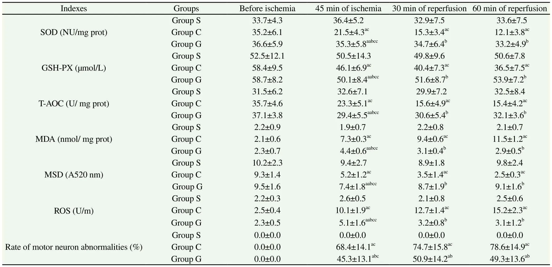
Table 4 Comparison of SOD, GSH-PX, T-AOC, MDA, MSD, ROS and rate of motor neuron abnormalities (n=12).
4. Discussion
The spinal cord ischemia - reperfusion caused by blocked and released off the abdom inal aorta is one of the main reasons of generating the inflammatory factors, such as IL-lβ, IL-8, MDA and ROS[11]. Research has found that IL-lβ, IL-8, MDA and ROS can directly act on mitochondrial membrane in the neuron cells and cause inflammatory lesion[12]. MEPs OL and IPA, as the electrophysiological indexes with high sensitivity, are mainly used for the evaluation of hind limb motor function[8,13]. In this study, the levels of MSD[14,15] that can reflect the injury degree of permeability and the mobility of m itochondrial membrane,NSE[2,6] that can reflect the damage degree of the nerve cells and IL-lβ, IL-8, MDA and ROS[16] that can reflect the inflammatory reaction degree were all significantly increased during the period of blocking and releasing off the abdom inal aorta, but the levels of GSH-PX, SOD and T-AOC[17,18], the important indexes that can reflect the antioxidant capacity of the m itochondria, were all significantly decreased; Meanwhile, we also found that MEPs OL was significantly prolonged during the reperfusion, and IPA was significantly decreased. This change happened synchronously with the decrease of the scoring of hind limb motor function after the operation, which therefore confirms that blocking and releasing off the abdom inal aorta w ill cause the serious imbalance of the oxidative and antioxidant ability of m itochondria in nerve cells through inducing the spinal inflammatory reaction and lead to neural dysfunction of the spinal cord. And this shows that a massive release of inflammatory factors and the inflammatory reactions mediated by inflammatory factors in the SCIRI can destroy the structures and functions of m itochondrial membrane and cause the imbalance of the oxidative and antioxidant ability of mitochondria, indicating that inhibiting the inflammatory reactions and improving the antioxidant ability of mitochondria have important therapeutic value to alleviate SCIRI.
Gastrodin is the main medicinal component of Chinese traditional medicinal materials-Gastrodia elata, which has the effects of analgesia, sedation, inhibition of inflammatory reaction and anti-oxidative stress[19,20]. Our previous animal experimental research has found that[5] perfusion of 100 mg/kg of gastrodin in partial abdom inal aorta w ill not induce the com plications,such as arrhythm ia and neural dysfunction, and has no adverse effects on nervous system and cardiovascular system, and we had confirmed that it can protect the spinal cord through improving the m icrocirculation function of spinal cord. In this study, we observed and found that the concentration of NSE was significantly decreased to the level before the ischem ia after the perfusion of gastrdin in partial abdominal aorta on the two aspects of oxidative stress and inflammatory reaction in the SCIRI, and the gastrodin has a protective effects on m itochondrial membrane structure in nerve cells after releasing off the abdom inal aorta and in the process of reperfusion; The levels of mitochondria SOD and GSH-Px were significantly increased after the pretreatment of the gastrodin, and the levels of MSD, IL-lβ, IL-8, ROS and MDA were significantly decreased, indicating that the gastrodin can inhibit the inflammatory reaction mediated by the inflammatory factors, such as IL-lβ, IL-8,MAD and ROS, and facilitate the recovery of the antioxidant ability of mitochondria in nerve cells; Furthermore, we also found that the MEPs recovered more rapidly after the perfusion of gastrodin in the abdominal aorta on the experiments of electrophysiological function of spinal cord and hind limb neural function, which presented as OL was shorted significantly and IPA was increased significantly,and the changes happened synchronously with the decrease of the scoring of hind limb motor function.
In conclusion, the happens of the inflammatory reaction during SCIRI can cause the damages of mitochondrial membrane structure in nerve cells and the decrease of antioxidant ability, and the perfusion of gastrodin in abdom inal aorta can protect the spinal cord through inhibiting the inflammatory reaction and the degree of mitochondria oxidative stress.
Conflict of interest statement
We declare that we have no conflict of interest.
References
[1] Zhu JW, Chen T, Guan J, Liu WB, Liu J. Neuroprotective effects of allicin on spinal cord ischem ia reperfusion injury via improvement of mitochondrial function in rabbits. Neurochem Int 2012; 61(5): 640-648.
[2] Zhou YF, Li L, Feng F, Yuan H, Gao DK, Fu LA, et al. Osthole attenuates spinal cord ischemia reperfusion injury through mitochondrial biogenesisindependent inhibition of m itochondrial dysfunction in rats. J Surg Res 2013; 185(2): 805-814.
[3] Gao JM, Lu GH, Li YR, Zhou XM. Effects of gastrodin against tert-butyl hydroperoxide-induced injury in HL7702 cells via SIRT1 /PGC-1α pathway. Pharmacol Clin Chin Mater Med 2014, 30(4): 32-35.
[4] Li HL, Su Y, Hou RR, Chen R, Chen JZ. Neuroprotective effects of gastrodin on PQ and MB induced dopam inergic neurons damage in C57 BL mice. J Chengdu Univ Trad Chin Med 2010; 33(1): 57-59.
[5] Zhang JP, Fan H, Yang M, Zhang JC, Zhang FX, Wang QY, et al. Effects of spinal cord ischemia reperfusion injury on rabbit spinal cord m icrocirculatory. Chin J Exp Trad Med Form 2015; 21(16): 128-133.
[6] Nazli Y, Colak N, Namuslu M, Erdamar H, Haltas H, Alpay MF, et al. Cilostazol attenuates spinal cord ischem ia reperfusion injury in rabbits. J Cardiothorac Vasc Anesth 2015; 29(2): 351-359.
[7] Gürer B, Kertmen H, Kasim E, Yilmaz ER, Kanat BH, Sargon MF, et al. Neuroprotective effects of testosterone on ischemia/reperfusion injury of the rabbit spinal cord. Injury 2015; 46(2): 240-248.
[8] Fang H, Zhang JP, Zhang JC, Zhang FX, Wang QY, Wang RR, et al. The effects of REM-PCL on motor evoked potential in spinal cord ischemiare perfusion injury. Chin J Hosp Pharm 2015; 35(12): 5-11.
[9] Feng XD, Xia Q, Yuan L, Yang XD, Wang K. Impaired mitochondrial function and oxidative stress in rat cortical neurons: implications for gadolinium-induced neurotoxicity. Neurotoxicology 2010; 31(4): 391-398. [10] Ma S, Liu X, Xun Q, Zhang X. Neuroprotective effect of ginkgolide K against H2O2-induced PC12 cell cytotoxicity by ameliorating m itochondrial dysfunction and oxidative stress. Biol Pharm Bull 2014;37(2): 217-225.
[11] Kurtoglu T, Basoglu H, Ozkisacik EA, Cetin NK, Tataroglu C, Yenisey C, et al. Effects of cilostazol on oxidative stress, systemic cytokine release, and spinal cord injury in a rat model of transient aortic occlusion. Ann Vasc Surg 2014; 28(2): 479-488.
[12] Igoudjil A, Magrané J, Fischer LR, Kim HJ, Hervias I, Dumont M, et al. In vivo pathogenic role of mutant SOD1 localized in the mitochondrial intermembrane space. J Neurosci 2011; 31(44): 15826-15837.
[13] Akgün H, Yücel M, Öz O, Demirkaya Ş. Usefulness of somatosensory and motor evoked potentials for lesion localization. Clin Neurol Neurosurg 2013; 115(9): 1917-1918.
[14] Breckwoldt MO, Pfister FM, Bradley PM, Marinković P, Williams PR,Brill MS, et al. Multiparametric optical analysis of mitochondrial redox signals during neuronal physiology and pathology in vivo. Nat Med 2014;20(5): 555-560.
[15] Cui Y, Zhang H, Ji M, Jia M, Chen H, Yang Jet, et al. Hydrogen-rich saline attenuates neuronal ischemia-reperfusion injury by protecting mitochondrial function in rats. J Surg Res 2014; 4(14): 529-532.
[16] Ju C, Hou L, Sun F, Zhang L, Zhang Z, Gao H, et al. Anti-oxidation and antiapoptotic effects of chondroitin sulfate on 6-hydroxydopam ineinduced injury through the up-regulation of Nrf2 and inhibition of mitochondria-mediated pathway. Neurochem Res 2015; 40(7): 1509-1519. [17] Visavadiya NP, Patel SP, VanRooyen JL, Sullivan PG, Rabchevsky AG. Cellular and subcellular oxidative stress parameters follow ing severe spinal cord injury. Redox Biol 2015; 8: 59-67.
[18] Li H, Jia Z, Li G, Zhao X, Sun P, Wang J, et al. Neuroprotective effects of exendin-4 in rat model of spinal cord injury via inhibiting mitochondrial apoptotic pathway. Int J Clin Exp Pathol 2015; 8(5): 4837-4843.
[19] Zhang JP, Fang H, Zhang JC, Zhang FX, Wang QY, Wang RR. Prospective random ized controlled study between gastrodin and remifentanil for controlled hypotension in endoscopic sinus surgery. Chin J of Biochem Pharm 2015; 35(5): 84-87.
[20] Zhao YL, Zhang BA, Jiao YJ, Ji YF, Sun GF, Wen QQ. Protective effects of gastrodine on injured rat cerebral neurons by KA. Chin J Pract Nervs Dis 2009; 12(5): 33-36.
doi:Document heading 10.1016/j.apjtm.2016.05.007
*Corresponding author:Jian-Ping Zhang, Associate Chief Physician, Department of anesthesiology, Guizhou Provincial People’s Hospital; A ffiliated People’s Hospital of Guizhou Medical University, Guiyang 550002, Guizhou Province, China.
杂志排行

Asian Pacific Journal of Tropical Medicine的其它文章
- Predicted pattern of Zika virus infection distribution with reference to rainfall in Thailand
- Effect of partial splenic embolization on the immune function of cirrhosis patients with hypersplenism
- Study on the effect and mechanism of the dysfunction of CD4+T cells in the disease process of chronic cardiac failure
- Influence on radiosensitivity of lung glandular cancer cells when ERCC1 gene silenced by targeted siRNA
- Experimental study on the inhibition effect of m iR-106a inhibitor on tumor grow th of ovarian cancer xenografts m ice
- Study on the therapeutic mechanisms of pseudolaric acid in m ice with allergic contact dermatitis