红细胞分布宽度在急性肺血栓栓塞症患者长期随访中的作用
2016-04-06奚群英王勇柳志红赵智慧罗勤顾晴熊长明倪新海
奚群英,王勇,柳志红,赵智慧,罗勤,顾晴,熊长明,倪新海
红细胞分布宽度在急性肺血栓栓塞症患者长期随访中的作用
奚群英,王勇*,柳志红,赵智慧,罗勤,顾晴,熊长明,倪新海
摘要
关键词肺血栓;高血压,肺性;红细胞分布宽度
作者单位:100037 北京市,中国医学科学院 北京协和医学院 国家心血管病中心 阜外医院 心内科
Effect of Red Cell Distribution Width on Long-term Follow-up Study in Patients With Acute Pulmonary Thromboembolism
XI Qun-ying, WANG Yong, LIU Zhi-hong, ZHAO Zhi-hui, LUO Qin, GU Qing, XIONG Chang-ming, NI Xin-hai.
Department of Cardiology, Cardiovascular Institute and Fu Wai Hospital, CAMS and PUMC, Beijing (100037), China
Corresponding Author: LIU Zhi-hong, Email:liuzhihong@fuwai.com
Abstract
Objective:To explore the effect of red blood cell distribution width (RDW) on long-term follow-up study in patients with acute pulmonary thromboembolism (APE).
Methods: A total of 214 consecutive patients with the first episode of APE admitted in our hospital from 2009-01 to 2012-12 were enrolled. The patients were divided into 2 groups: RDW≤15% group, n=202 and RDW>15% group, n=12. Baseline RDW was measured at admission, the follow-up study was conducted at 3, 6, 12 months thereafter, and then at once per year. The major primary end point was chronic thromboembolic pulmonary hypertension (CTEPH). The independent predictor for CTEPH occurrence was studied by uni- and multivariate logistic regression analysis and the predictive capability of RDWwas evaluated by ROC curve.
Results: All patients finished the follow-up study at the mean of (31±17) months. The overall occurrence rate of CTEPH was 7.5% (16/214), which was higher in RDW>15% group than that in RDW≤15% group (33.3% vs 5.9%, P=0.002). Multivariate logistic regression analysis indicated that with adjusted clinical data and other predictors, RDW>15% was still the strong predictor for CTEPH occurrence (OR=7.916, 95% CI 1.474-42.500, P=0.016). Adding RDW to the evaluating model, the predictive capability could be significantly improved by ROC curve (AUC increased from 0.856 to 0.901, P< 0.01).
Conclusion: Elevated RDW is the independent predictor for CTEPH occurrence in APE patients, which is helpful to estimate the prognosis and treatment strategy in APE patients.
Key words Pulmonary embolism; Hypertension, pulmonary; Red blood cell distribution width
(Chinese Circulation Journal, 2016,31:65.)
慢性血栓栓塞性肺动脉高压(CTEPH)定义为:在有效抗凝至少3个月后,(1)右心导管检查平均肺动脉压≥25 mmHg(1 mmHg=0.133 kPa), 同时肺毛细血管楔压≤15 mmHg,肺血管阻力>2 wood单位; (2) 弹性肺动脉(包括主肺动脉及叶、段、亚段肺动脉)中存在多发慢性/机化的血栓/栓子[1]。根据不同研究者的报道,急性肺血栓栓塞症(APE)事件后发生CTEPH的几率为0.4%~5.1%[2, 3],而在反复发作的肺血栓栓塞症(PE)患者此比例上升至10%[4]。CTEPH是PE事件的不良结局,可引起进行性的右心功能障碍,进而导致死亡[5]。既往的研究提示,对APE患者进行强化的抗凝治疗可能减少后续CTEPH的发生[6]。可靠的CTEPH预测因子可帮助临床医生监测和识别可能发生CTEPH的高危患者,这部分患者有可能在强化的治疗中获益。
红细胞分布宽度(RDW)是反映红细胞异质性的一个参数,在血液常规检查中即可获得,通常用于贫血的鉴别诊断。近期的一系列研究表明,RDW是多种心肺血管疾病如冠心病[7]、心力衰竭[8]、肺动脉高压[9, 10]、PE[11,12]的预后预测因子。既往的研究表明,入院时RDW增高是APE短期死亡率的独立预测因子[11,12]。RDW在APE后的长期随访事件中的预测能力尚不明确。
1 资料与方法
筛选了2009-01至2012-12在阜外医院心内科肺血管病房住院的首次APE患者214例,平均年龄(61±14)岁, 男性97例(45%)。根据RDW是否增高分为RDW≤15%组(n=202)和RDW>15%组(n=12)。PE的诊断依据为病史、体征、胸部X线片、心电图、动脉血气分析、下肢超声、计算机断层摄影术(CT)肺血管成像结果。诊断标准为CT肺血管成像见至少1段以上的肺动脉内充盈缺损。诊断流程按照既往报道的方法进行[13]。有危险因素的APE定义为患者有以下因素:恶性肿瘤、长期制动史、慢性心力衰竭、慢性呼吸衰竭、近期内大创伤、近期内外科手术、妊娠、口服避孕药或激素替代治疗,无上述因素的患者定义为不明原因的APE。排除标准: (1)既往有PE、深静脉血栓病史; (2)预期寿命<6个月; (3)3个月内的输血史; (4)正在接受血液透析治疗。资料收集包括基线临床特征、病史、实验室检测数据、接受的治疗方案和终点事件。研究经阜外医院伦理委员会批准,患者均签署知情同意书。
APE诊治:明确为APE或有中、高度PE可能性的患者即给予低分子量肝素或普通肝素抗凝。明确诊断APE且同时有心原性休克或持续存在的低血压时,如无溶栓禁忌证,予以重组组织型纤溶酶原激活剂(rt-PA,50 mg或100 mg 静脉内输注2 h)溶栓治疗。溶栓治疗后继续口服维生素K拮抗剂,使国际标准化比值在2.0~3.0。有危险因素的APE患者至少抗凝3个月;不明原因的APE患者至少6个月。随访时间为APE后第3、6、12个月,此后每年1次。如患者有任何症状或体征提示CTEPH,则接受进一步的检查(包括超声心动图、肺通气/灌注扫描、右心导管检查及肺动脉造影、CT肺血管成像),以明确是否存在CTEPH。
实验室检测:患者在入院时或次日早晨抽血。经肘前静脉抽取血液样本注入乙二胺四乙酸抗凝管或普通生化管中。常规血液学指标包括RDW由自动血液分析仪XE-2100 (Sysmex公司, 日本)取得。大内皮素-1、N末端B型利钠肽原(NT-proBNP)均以酶联免疫试剂盒(Biomedica Medizinprodukte GmbH& Co KG公司, 奥地利)检测。其他生化指标由自动生化分析仪(Beckman Coulter公司,美国)检测。我院实验室的RDW正常参考值为10%~15%。
统计学分析:数据分析均采用SPSS 16.0统计学软件。连续性资料以均数±标准差(±s)表示,分类资料以数量和所占比例表示。两组间分类资料的比较采用卡方检验,连续性资料的比较采用student’s t检验。单变量和多变量Logistic回归分析确定CTEPH的独立预测因子。Kaplan- Meier 生存分析比较两组的存活率。受试者工作特征 (ROC) 曲线评价RDW的预测能力。P<0.05为差异有统计学意义。
2 结果
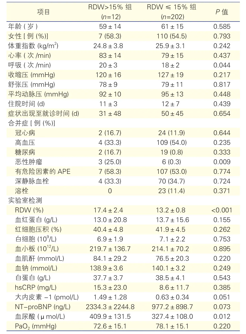
两组患者的基线临床资料情况(表1):RDW>15%组患者的呼吸频率(P=0.044)、血尿酸水平(P=0.012)、恶性肿瘤病史比例(P=0.009)均高于RDW ≤ 15%组。
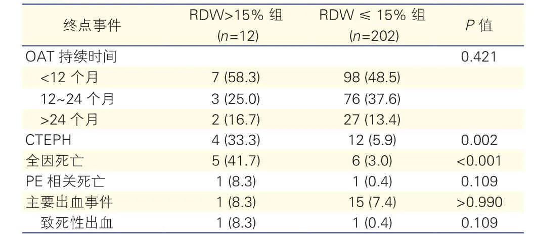
随访终点事件:全部患者完成随访,平均随访时间(31±17)个月,CTEPH 的发生率是7.5% (16/214)。RDW>15%组CTEPH发生率(P=0.002)、全因死亡率(P<0.001)均明显高于RDW≤15%组(表2)。RDW值异常增高与全因死亡增加相关(风险比=7.370,95%可信区间:1.291~42.066,P=0.025)。Kaplan-Meier分析显示,RDW≤15%组患者的生存率明显高于RDW>15%组(P<0.001)。

表1 两组患者基线临床资料情况(?±s)

表2 两组患者随访终点事件情况[例(%)]
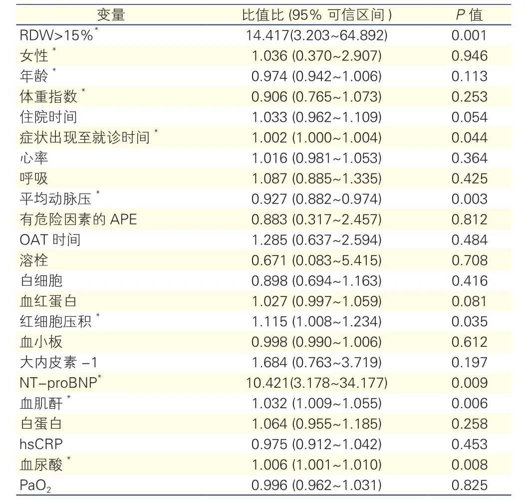
单变量Logistic回归分析(表3):RDW>15% 与CTEPH发生显著相关(P=0.001)。与CTEPH发生相关的其他因素包括症状出现至就诊的时间较长(P=0.044),就诊时平均动脉压较低(P=0.003) ,NT-proBNP水平 (P=0.009) 、红细胞压积(P=0.035)、血肌酐(P=0.006) 、血尿酸(P=0.008) 异常等。

表3 发生CTEPH的单变量Logistic回归分析结果
多变量Logistic回归分析(表4):在校正临床资料和其他预测因素后,RDW>15%仍是强有力的发生CTEPH独立预测因子(P=0.016)。发生CTEPH的其他独立预测因子包括异常的NT-proBNP水平(P=0.001)、症状出现至就诊的时间较长(P=0.034)、就诊时平均动脉压较低(P=0.028)等。

表4 发生CTEPH的多变量Logistic回归分析结果
ROC曲线下面积:以RDW单独预测发生CTEPH的ROC曲线下面积为0.730(95%可信区间:0.622~0.839,P=0.002);在预测模型中去掉RDW,ROC曲线下面积为0.856(95%可信区间:0.740~0.971,P<0.001);在预测模型中加入RDW,ROC曲线下面积为0.901(95%可信区间:0.837~0.965,P<0.001)。在预测模型中增加RDW能显著增加模型的预测能力(ROC曲线下面积从0.856增加至0.901,P<0.01)。
3 讨论
RDW是血液常规检查中的报告项目,其值为红细胞体积的标准差/平均红细胞体积。既往的研究显示RDW可预测多种心肺血管疾病的预后。近期的两项研究表明,RDW可预测APE的早期死亡风险[11, 12]。我们的研究结果表明RDW可预测APE的长期随访事件。PE是临床常见病,虽多数患者经治疗后血液动力学恢复正常,但少部分患者发展为CTEPH。本研究中,CTEPH 的发生率是7.4%,高于其他研究的报道[2, 3],可能与研究人群(本研究患者症状出现至就诊的时间较长)、口服抗凝药时间和随访时间不同有关。CTEPH是PE后的不良事件,导致患者右心功能障碍、生活质量低下和死亡。虽然肺动脉血栓内膜切除术是治疗CTEPH的有效方法,但20%~40%的患者因血栓在远端肺血管或合并手术禁忌证等而不能接受手术治疗;且有10%~20%的患者术后残余肺动脉高压[14]。既往的研究提示,CTEPH的发生率可能随着对APE患者的强化治疗而减少[6]。因此,筛选出可能会发展为CTEPH的APE患者至关重要。本研究发现基线RDW值增高是预测发生CTEPH的强有力指标。由于RDW是血常规检查报告中所包含的常规项目且经济、方便,因此对于临床判断APE转归及决定治疗策略,特别是对广大的基层医院具有重要的临床意义。
为何RDW能预测心肺血管疾病预后的机制尚不明确。目前认为心肺血管疾病患者存在的营养不良(包括铁缺乏)[15-18]、炎症[19-21]、氧化应激[22]可能是RDW值的影响因素。本研究显示RDW值增高患者有更高的血尿酸水平,且恶性肿瘤史也更多。此二者都会引起机体的炎症状态,可能也影响了RDW水平。
综上所述,RDW水平增高是APE后发生CTEPH的独立预测因子,有助于判断APE患者的转归及决定治疗策略。
参考文献
[1] Galie N, Hoeper MM, Humbert M, et al. Guidelines for the diagnosis and treatment of pulmonary hypertension: The Task Force for the Diagnosis and Treatment of Pulmonary Hypertension of the European Society of Cardiology (ESC) and the European Respiratory Society (ERS), Endorsed by the International Society of Heart and Lung Transplantation (ISHLT). Eur Heart J, 2009, 30: 2493-2537.
[2] Becattini C, Agnelli G, Pesavento R, et al. Incidence of chronic thromboembolic pulmonary hypertension after a first episode of pulmonary embolism. Chest, 2006, 130: 172-175.
[3] Pengo V, Lensing AW, Prins MH, et al. Incidence of chronic thromboembolic pulmonary hypertension after pulmonary embolism. N Engl J Med, 2004, 350: 2257-2264.
[4] Haythe J. Chronic thromboembolic pulmonary hypertension: a review of current practice. Prog Cardiovasc Dis, 2012, 55: 134-143.
[5] 柳志红. 慢性血栓栓塞性肺动脉高压的处理. 中国循环杂志, 2011, 26: 243-244.
[6] Kline JA, Steuerwald MT, Marchick MR, et al. Prospective evaluation of right ventricular function and functional status 6 months after acute submassive pulmonary embolism: frequency of persistent or subsequent elevation in estimated pulmonary artery pressure. Chest, 2009, 136: 1202-1210.
[7] Dabbah S, Hammerman H, Markiewicz W, et al. Relation between red cell distribution width and clinical outcomes after acute myocardial infarction. Am J Cardiol, 2010, 105: 312-317.
[8] Borne Y, Smith JG, Melander O, et al. Red cell distribution width and risk for first hospitalization due to heart failure: apopulation-based cohort study. Eur J Heart Fail, 2011, 13: 1355-1361.
[9] Hampole CV, Mehrotra AK, Thenappan T, et al. Usefulness of red cell distribution width as a prognostic marker in pulmonary hypertension. Am J Cardiol, 2009, 104: 868-872.
[10] 李震南, 柳志红, 顾晴, 等.红细胞分布宽度与特发性肺动脉高压病情和预后的相关性分析.中国循环杂志, 2013, 28: 111-114.
[11] Ozsu S, Abul Y, Gunaydin S, et al. Prognostic value of red cell distribution width in patients with pulmonary embolism. Clin Appl Thromb Hemost, 2014, 20: 365-370.
[12] Zorlu A, Bektasoglu G, Guven FM, et al. Usefulness of admission red cell distribution width as a predictor of early mortality in patients with acute pulmonary embolism. Am J Cardiol, 2012, 109: 128-134.
[13] Piazza G, Goldhaber SZ. Chronic thromboembolic pulmonary hypertension. N Engl J Med, 2011, 364: 351-360.
[14] Wang Y, Liu ZH, Zhang HL, et al. Association of elevated NT-proBNP with recurrent thromboembolic events after acute pulmonary embolism. Thromb Res, 2012, 129: 688-692.
[15] Mayer E. Surgical and post-operative treatment of chronic thromboembolic pulmonary hypertension. Eur Respir Rev, 2010, 19: 64-67.
[16] Allen LA, Felker GM, Mehra MR, et al. Validation and potential mechanisms of red cell distribution width as a prognostic marker in heart failure. J Card Fail, 2010, 16: 230-238.
[17] Forhecz Z, Gombos T, Borgulya G, et al. Red cell distribution width in heart failure: prediction of clinical events and relationship with markers of ineffective erythropoiesis, inflammation, renal function, and mutritional state. Am Heart J, 2009, 158: 659-666.
[18] Decker I, Ghosh S, Comhair SA, et al. High levels of zincprotoporphyrin identify iron metabolic abnormalities in pulmonary arterial hypertension. Clin Transl Sci, 2011, 4: 253-258.
[19] Rhodes CJ, Howard LS, Busbridge M, et al. Iron deficiency and raised hepcidin in idiopathic pulmonary arterial hypertension: clinical prevalence, outcomes, and mechanistic insights. J Am Coll Cardiol, 2011, 58: 300-309.
[20] de Gonzalo-Calvo D, de Luxan-Delgado B, Rodriguez-Gonzalez S, et al. Interleukin 6, soluble tumor necrosis factor receptor I and red blood cell distribution width as biological markers of functional dependence in an elderly population: atranslational approach. Cytokine, 2012, 58: 193-198.
[21] Lippi G, Targher G, Montagnana M, et al. Relation between red blood cell distribution width and inflammatory biomarkers in a large cohort of unselected outpatients. Arch Pathol Lab Med, 2009, 133: 628-632.
[22] Semba RD, Patel KV, Ferrucci L, et al. Serum antioxidants and inflammation predict red cell distribution width in older women: the Women's Health and Aging Study I. Clin Nutr, 2010, 29: 600-604.
(编辑:王宝茹)
临床研究
收稿日期:(2015-08-27)
中图分类号:R543.2
文献标识码:A
文章编号:1000-3614(2016)01-0065-04
doi:10.3969/j.issn.1000-3614.2016.01.014
作者简介:奚群英 主治医师 博士 研究方向:肺血管病 Email:xiqunyinghz@163.com 通讯作者:柳志红 Email: liuzhihong@fuwai.com*为共同第一作者
基金项目:国家“十一五”科技支撑计划项目(2006BAIOIA06)
目的:探讨红细胞分布宽度(RDW)在急性肺血栓栓塞症(APE)患者长期随访中的作用。
方法:筛选2009-01至2012-12在阜外医院心内科肺血管病房住院的首次APE患者214例,根据RDW是否增高分为RDW≤15%组(n=202)和RDW>15%组(n=12)。测定基础的RDW值,并在APE后第3、6、12个月随访,此后每年随访1次,主要终点事件为慢性血栓栓塞性肺动脉高压(CTEPH)。用单变量和多变量Logistic回归分析确定CTEPH的独立预测因子,受试者工作特征 (ROC) 曲线评价RDW的预测能力。
结果:全部患者完成随访,平均随访时间(31±17)个月,CTEPH 的发生率是7.5%(16/214)。RDW>15%组CTEPH发生率明显高于RDW≤15%组(33.3% vs 5.9%,P=0.002)。多变量Logistic回归分析:在校正临床资料和其他预测因素后,RDW>15%仍是强有力的发生CTEPH独立预测因子(比值比=7.916, 95%可信区间:1.474~42.500, P=0.016)。在预测模型中增加RDW能显著增加模型的预测能力(ROC曲线下面积从0.856增加至0.901,P<0.01)。
结论:RDW增高是APE后发生CTEPH的独立预测因子,有助于判断APE患者的转归及决定治疗策略。
