蜂胶黄酮对炎症反应单核细胞中NLRP3炎性小体活性的影响
2016-01-12张晓晖,曾伟,陈鹏等
蜂胶黄酮对炎症反应单核细胞中NLRP3炎性小体活性的影响
张晓晖1,曾伟1,陈鹏2,姚树桐2
(1泰山医学院附属医院,山东泰安271000;2泰山医学院基础医学院)
摘要:目的观察蜂胶黄酮对氧化型低密度脂蛋白(ox-LDL)诱导的炎症反应单核细胞中核苷酸结合寡聚化结构域样受体3(NLRP3)炎性小体活性的影响。方法培养单核细胞THP1并分为5个组。实验1、2、3组及模型组加入ox-LDL 100 μg/mL制作单核细胞炎症反应模型;实验1、2、3组在加入ox-LDL前分别给予5、10、15 μg/mL蜂胶黄酮处理1 h;对照组不添加药物。各组培养24 h后,采用Western blotting法检测细胞中的NLRP3蛋白,采用ELISA法检测细胞培养液上清中的IL-1β、IL-18。结果对照组、模型组、实验1组、实验2组、实验3组细胞中NLRP3蛋白相对表达量分别为0.26±0.04、1.12±0.15、0.98±0.11、0.65±0.07、0.52±0.06,模型组与对照组相比,P均<0.05;实验2组、实验3组与模型组相比,P均<0.05。模型组培养液上清中IL-1β、IL-18水平高于对照组,实验2组、实验3组培养液上清中IL-1β、IL-18水平低于模型组(P均<0.05)。结论 蜂胶黄酮可下调ox-LDL诱导的单核细胞炎症反应模型中NLRP3蛋白的表达,降低IL-1β、IL-18分泌水平,抑制NLRP3炎性小体的活性。
关键词:蜂胶黄酮;核苷酸结合寡聚化结构域样受体3;单核细胞;炎症反应;白细胞介素1β;白细胞介素18
doi:10.3969/j.issn.1002-266X.2015.39.004
中图分类号:R541.4文献标志码:A
基金项目:国家自然科学基金资助项目(81202979)。
作者简介:第一张晓晖(1977-),男,本科,主治医师,主要研究方向为动脉粥样硬化的发生发展机制。E-mail: tyfyzxh@163.com
收稿日期:(2015-08-29)
Effects of propolis flavonoids on activation of NLRP3 inflammasome in monocytes
ZHANGXiao-hui1,ZENGWei,CHENPeng,YAOShu-tong
(1AffiliatedHospitalofTaishanMedicalCollege,Taian271000,China)
Abstract:ObjectiveTo explore the effects of propolis flavonoids on the activation of oxidized low-density lipoprotein (ox-LDL) induced nucleotide-binding oligomerization domain-like receptor containing protein 3 (NLRP3) inflammasome in mononuclear cells. MethodsThe monocytes THP1 were cultured and divided into five groups. The experimental groups 1, 2, 3 and the model group were treated with 100 μg/mL ox-LDL to produce mononuclear inflammatory reaction models. The monocytes of the experimental groups 1, 2 and 3 were treated with 5, 10 and 15 μg/mL propolis flavonoids before ox-LDL, and the monocytes of the control group was treated by ox-LDL in the absence of propo1is flavonoids. After 24 h, the expression of NLRP3 was measured by Western blotting. The levels of IL-1β and IL-18 were measured by ELISA. ResultsThe levels of NLRP3 protein in the control group, model group and the experimental groups 1, 2 and 3 were 0.26±0.04, 1.12±0.15, 0.98±0.11, 0.65±0.07 and 0.52±0.06, respectively. The expression of NLRP3 protein was significantly increased in the model group as compared with that in the control group (all P<0.05). The expression of NLRP3 protein in the experimental groups 2 and 3 was significantly decreased as compared with that of the model group (all P<0.05). The levels of IL-1β and IL-18 was significantly higher in the model group as compared with those of the control group, and the levels of IL-1β and IL-18 in the experimental groups 2 and 3 was significantly decreased as compared with those of the model group (all P<0.05). ConclusionPropolis flavonoids may down-regulate the expression of NLRP3 protein in the ox-LDL induced inflammatory response of the mononuclear cells, decrease the levels of IL-1β and IL-18 and inhibit the activation of NLRP3 inflammasome.
Key words: propolis flavonoids; nucleotide-binding oligomerization domain-like receptor containing protein 3; monocytes; inflammatory response; interleukin-1β; interleukin-18
近年来,动脉粥样硬化的发病率逐年上升并呈年轻化趋势,寻找安全有效的抗动脉粥样硬化药物具有重要意义[1]。蜂胶黄酮是从蜂胶中提取出的活性物质,有抗病毒、抗菌、抗氧化、抗肿瘤、调节免疫力及改善微循环等多种生物学活性[2~4]。此外,蜂胶黄酮还能抑制血小板黏附和聚集、保护血管内皮细胞免受炎症损伤,具体机制尚不明确。炎性体是细胞内一类大分子蛋白复合体。核苷酸结合寡聚化结构域样受体3(NLRP3)炎性小体是较为经典的炎性体,它可被相关病原分子及缺血缺氧条件激活,促进IL-1β、IL-18分泌,参与动脉粥样硬化的发生发展[5,6]。2015年2~8月,我们将氧化型低密度脂蛋白(ox-LDL)作用于单核细胞THP1,建立炎症反应模型,观察蜂胶黄酮对THP1细胞中NLRP3炎性小体活性的影响。
1材料与方法
1.1实验材料THP1细胞购自Scien Cell研究实验室;蜂胶黄酮由本实验室制备;ox-LDL购自北京鼎国昌盛生物技术有限责任公司;NLRP3抗体购自Abcam公司;IL-1β、IL-18 ELISA检测试剂盒购自R&D Systems公司。
1.2细胞培养与分组THP1细胞培养于添加10%胎牛血清的1640培养基中,在37 ℃、5%CO2的培养箱中进行无菌培养。培养细胞传3代以后,将细胞分为5个组,分别为对照组、模型组、实验1组、实验2组、实验3组。
1.3动脉粥样硬化单核细胞模型制作及蜂胶黄酮干预方法实验1、2、3组及模型组中加入ox-LDL 100 μg/mL制作单核细胞炎症反应模型;实验1、2、3组在加入ox-LDL前分别给予5、10、15 μg/mL蜂胶黄酮处理1 h;对照组不添加药物。各组培养24 h后收集细胞,进行后续实验。
1.4各组细胞中NLRP3蛋白检测采用Western blotting法。向各组细胞中加入RIPA裂解液200 μL,冰上放置30 min后,4 ℃、12 000 r/min离心15 min。取上清液,用BCA法测定蛋白浓度。制备分离胶及浓缩胶,将蛋白样品与Loading Buffer混合,煮沸5 min。在每个上样孔加入蛋白样品80 μg,进行电泳。取出凝胶,在恒流180 mA下湿转90 min,转于NC膜上。5%脱脂奶粉室温封闭1 h,加入兔抗人NLRP3抗体(1∶1 000),4 ℃过夜,TBST洗膜3次,每次10 min;加入羊抗兔二抗,室温孵育60 min,TBST洗膜3次,每次洗10min。用ECL化学发光液在暗室进行显色、拍片,以NLRP3/GAPDH灰度值表示NLRP3蛋白相对表达量。
1.5各组培养液上清中IL-1β、IL-18检测收集各组细胞培养液上清,采用ELISA法检测IL-1β、IL-18,按试剂盒说明书操作。
1.6统计学方法采用SPSS16.0软件进行统计分析。计量资料以±s表示,组间比较采用方差分析。P<0.05为差异有统计学意义。
2结果
2.1各组细胞中NLRP3蛋白表达比较对照组、模型组、实验1组、实验2组、实验3组细胞中NLRP3蛋白相对表达量分别为0.26±0.04、1.12±0.15、0.98±0.11、0.65±0.07、0.52±0.06,模型组NLRP3蛋白表达量高于对照组(P<0.05);实验2组、实验3组NLRP3蛋白表达量低于模型组(P均<0.05)。
2.2各组培养液上清中IL-1β、IL-18水平比较模型组培养液上清中IL-1β、IL-18水平高于对照组,实验2组、实验3组培养液上清中IL-1β、IL-18水平低于模型组(P均<0.05)。见表1。
表1 各组培养液上清中IL-1β、IL-18
水平比较(pg/mL, ± s)
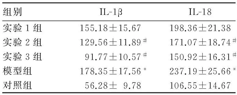
表1 各组培养液上清中IL-1β、IL-18
组别IL-1βIL-18实验1组155.18±15.67198.36±21.38实验2组129.56±11.89#171.07±18.74#实验3组91.77±10.57#150.92±16.31#模型组178.35±17.56*237.19±25.66*对照组56.28±9.78106.55±14.67
注:与对照组相比,*P<0.05;与模型组相比,#P<0.05。
3讨论
蜂胶素是蜜蜂从植物新生芽条或愈伤组织中采集分泌物后,加入蜜蜂的舌腺、蜡腺等腺体分泌物及花粉等混合而成的具有特殊芳香气味的黏胶状物质[7]。蜂胶的化学成分复杂,主要成分为黄酮化合物。蜂胶黄酮具有抗病毒、软化血管、降血脂、调节免疫、改善微循环、抗氧化等广泛的生物学活性[8,9],还有抗动脉粥样硬化的作用。动脉粥样硬化发病机制复杂,目前存在几种学说,包括脂质渗入学说、内皮细胞损伤学说、炎症学说等[10,11],其中脂代谢异常在动脉粥样硬化的形成中发挥重要作用。血液中高水平的低密度脂蛋白(LDL)被活性氧簇(ROS)等自由基氧化后成为ox-LDL,后者参与动脉粥样硬化的形成过程。ox-LDL可被血液中的单核细胞吞噬,被吞噬的ox-LDL在细胞中不易被分解,之后单核细胞将会转变成泡沫细胞[12,13]。
NLRP3是经典的炎性小体,由NLRP3、Caspase-1、含C-末端Caspase募集域的凋亡相关斑点样蛋白(ASC)相互结合形成。通常情况下,NLRP3处于自身抑制状态,当与配体结合后自身发生寡聚化,进而与ASC发生相互作用,通过ASC招募并激活Caspase-1,Caspase-1活化后可催化IL-1 及IL-18前体,使其成为有活性的形式,并分泌到细胞外发挥促炎作用[14]。已有报道显示,NLRP3可被胆固醇结晶活化,促进IL-1 、IL-18的分泌,发挥促动脉粥样硬化作用[15]。
本研究中,我们用ox-LDL作用于THP1细胞,诱导单核细胞炎症反应,观察蜂胶黄酮对NLRP3炎性小体活性的影响。我们发现,ox-LDL可诱导NLRP3蛋白表达增多,并能提高NLRP3炎性小体下游炎症因子IL-1 、IL-18分泌水平,这与Jiang等[16]的研究结果一致。而给予不同浓度蜂胶黄酮干预后,THP1细胞中NLRP3蛋白表达下调,细胞培养液上清中IL-1 、IL-18水平降低,提示蜂胶黄酮可抑制NLRP3炎性小体的活性,从而抑制ox-LDL诱导的炎症反应,起到抗动脉粥样硬化的作用,具体作用机制有待于深入研究。
参考文献:
[1] Libby P, Ridker PM, Hansson GK. Progress and challenges in translating the biology of atherosclerosis[J]. Nature, 2011,473(7347):317-325.
[2] 高蔚娜,韦京豫,蒲玲玲,等.不同产地原料蜂胶对衰老小鼠体液免疫功能影响的比较研究[J].中国免疫学杂志,2013,29(12):1262-1264.
[3] 杨明,隋殿军,朱姝,等.蜂胶总黄酮对大鼠心肌缺血再灌注损伤Fas、Bax和Bcl-2基因蛋白表达的影响[J].中国药理学通报,2005,21(7):799-803.
[4] Huang S, Zhang CP, Wang K, et al. Recent advances in the chemical composition of propolis[J]. Molecules, 2014,19(12):19610-19632.
[5] Ozaki E, Campbell M, Doyle SL. Targeting the NLRP3 inflammasome in chronic inflammatory diseases: current perspectives[J]. J Inflamm Res, 2015,8(1):15-27.
[6] Suárez R, Buelvas N. Inflammasome: activation mechanisms[J]. Invest Clin, 2015,56(1):74-99.
[7] 其曼古丽·吐尔洪,布威海丽且木·阿巴拜科日,祖丽比亚·司马义,等.蜂胶黄酮对人结肠癌细胞PDE4D及Gadd45G基因表达的影响[J].山东医药,2014,54(10):7-13.
[8] Lima Cavendish R, de Souza Santos J, Belo Neto R, et al. Anti-nociceptive and anti-inflammatory effects of Brazilian red propolis extract and formononetin in rodents[J]. J Ethnopharmacol, 2015,8741(15):30039-30038.
[9] Tian H, Sun HW, Zhang JJ, et al. Ethanol extract of propolis protects macrophages from oxidized low density lipoprotein-induced apoptosis by inhibiting CD36 expression and endoplasmic reticulum stress-C/EBP homologous protein pathway[J]. BMC Complement Altern Med, 2015,15(7):230.
[10] Hansson GK. Inflammation, atherosclerosis, and coronary artery disease[J]. N Engl J Med, 2005,352(16):1685-1695.
[11] Husain K, Hernandez W, Ansari RA, et al. Inflammation, oxidative stress and renin angiotensin system in atherosclerosis[J]. World J Biol Chem, 2015,6(3):209-217.
[12] Paim LR, Schreiber R, Matos-Souza JR, et, al. Oxidized low-density lipoprotein, matrix-metalloproteinase-8 and carotid atherosclerosis in spinal cord injured subjects[J]. Atherosclerosis, 2013,231(2):341-345.
[13] Zhang PY, Xu X, Li XC. Cardiovascular diseases: oxidative damage and antioxidant protection[J]. Eur Rev Med Pharmacol Sci, 2014,18(20):3091-3096.
[14] Peng K, Liu L, Wei D, Lv Y, et al. P2X7R is involved in the progression of atherosclerosis by promoting NLRP3 inflammasome activation[J]. Int J Mol Med, 2015,35(5):1179-1188.
[15] Altaf A, Qu P, Zhao Y, et al. NLRP3 inflammasome in peripheral blood monocytes of acute coronary syndrome patients and its relationship with statins[J]. Coron Artery Dis, 2015,26(5):409-421.
[16] Jiang Y, Wang M, Huang K, et al. Oxidized low-density lipoprotein induces secretion of interleukin-1β by macrophages via reactive oxygen species-dependent NLRP3 inflammasome activation[J]. Biochem Biophys Res Commun, 2012,425(2):121-126.

