Effects of gentiana scabra bage on expression of hepatic type Ⅰ, Ⅲcollagen proteins in Paragonimus skrjabini rats with liver fibrosis
2015-12-08ZhaoXiaQuFangLiChaoDongMaJunLiuShuDeLiWenLinWang
Zhao-Xia Qu, Fang Li, Chao-Dong Ma, Jun Liu, Shu-De Li, Wen-Lin Wang⋆
1Zhenjiang Medical School, Zhenjiang 212004, China
2Yuncheng Nursing Vocational College, Yuncheng 043100, China
3First People’s Hospital in Zhenjiang, Zhenjiang 212002, China
4Department of Parasitology, Faculty of Basic Medicine, Kunming Medical University, Kunming 650500, China
5Department of Biochemistry and Molecular Biology, Faculty of Basic Medicine, Kunming Medical University, Kunming 650500, China
Effects of gentiana scabra bage on expression of hepatic type Ⅰ, Ⅲcollagen proteins in Paragonimus skrjabini rats with liver fibrosis
Zhao-Xia Qu1Δ, Fang Li2Δ, Chao-Dong Ma3, Jun Liu4, Shu-De Li5, Wen-Lin Wang4⋆
1Zhenjiang Medical School, Zhenjiang 212004, China
2Yuncheng Nursing Vocational College, Yuncheng 043100, China
3First People’s Hospital in Zhenjiang, Zhenjiang 212002, China
4Department of Parasitology, Faculty of Basic Medicine, Kunming Medical University, Kunming 650500, China
5Department of Biochemistry and Molecular Biology, Faculty of Basic Medicine, Kunming Medical University, Kunming 650500, China
ARTICLE INFO
Article history:
Received 20 October 2014
Received in revised form 10 November 2014
Accepted 15 December 2014
Available online 20 January 2015
Gentiana scabra bage
Liver fibrosis
Paragonimus skrjabini
Collagen protein Ⅰ
Collagen protein Ⅲ
Objective: To explore the effects of gentiana scabra bage on the expression of hepatic collagen proteins in Paragonimus skrjabini rats with liver fibrosis. Methods: Immunohistochemical technique was used to observe the changes of content of hepatic type Ⅰ, Ⅲ collagen proteins in Paragonimus skrjabini rats with liver fibrosis before and after the gentiana scabra bage treatmeat. Results: Comparing with the model group, changes of hepatic type Ⅰand type Ⅲ collagen proteins in gentiana scabra bage treated group were significantly weakened. Conclusions: Gentiana scabra bage treatment can reduce the content of hepatic type Ⅲ and typeⅠcollagen protein significantly in Paragonimus skrjabini rats with liver fibrosis, thereby, playing the role against hepatic fibrosis.
1. Introduction
Modern pharmacological studies have shown that gentian scabra bage’s main effective ingredient is gentiopicroside which plays an important role in protecting liver, antiinflammation, resisting pathogenic microorganisms, excitin the central system and maintaining the health of the stomach and gallbladder, also it has other effects[1]. Currently some studies have shown that the effective ingredient in gentiana scabra bage can lower the level of serum alanine aminotransferase and aspartate aminotransferase in mice with acute liver injury caused by carbon tetrachloride so as to increase the glutathione peroxidase activity in liver tissue, and it also can increase the bile flow and the concentration of bilirubin in rats[2]. It has been proved that gentiopicroside has an important role in blocking carbon tetrachloride to damage the liver, protecting the liver by injuring rat liver with carbon tetrachloride and building fresh liver cells in vitro model for screening test[3]. But it has not been reported that whether gentiopicrin has any effect on hepatic type Ⅰ, Ⅲ collagen in liver fibrosis. This study used immunohistochemical technique to observe the changes of content of hepatic type Ⅰ, Ⅲ collagen proteins in Paragonimus skrjabini rats with liver fibrosis before and after gentiana scabra bage treatment.
2. Materials and methods
2.1. Isolation of Paragonimus skrjabini metacercaria
Crab creek was acquitted from Tongchang, Jinping County, the endemic area of Paragonimus skrjabini in Yunnan Province and all parts of the crab creek were broken up separately, sediment was collected on the microscopic anatomy to isolate Paragonimus skrjabini metacercaria after filtration and precipitation.
2.2. Construction of animal model
A total of 40 SD rats were bought from the Experimental Animal Center of Kunming Medical University regardless of male or female and each weighed (200±30) g and was fed separately. All these rats were infected with 15 Paragonimus skrjabini metacercaria by way of intra-peritoneum and were randomly divided into 4 groups: Normal group (10 rats) with no treatment; infected group (10 rats) with two mice being sacrificed which were removed to verify whether the needed model was successful or not in the eighth week after the infection, and the rest without given any treatment; gentiana scabra bage group (10 rats) (GSB group), which were firstly treated with triclabendazole for 4 days from the eighth week after the infection and then with gentiana scabra bage using gavage, which was a continuous treatment of 4 weeks and the does was 3.2 g per kg per day (the gentiana scabra bage powder was provided by the Key Laboratory of Natural Medicine in Kunming Medical University), with the rats being sacrificed and their livers being removed after the treatment; IFN-γ group (10 rats), in which IFN-γ was used instead of gentiana scabra bage and each mouse was injected subcutaneously with 150 units of IFN-γevery day while the remaining conditions were same with GSB group. The livers of rats in each group were placed in buffer formalin, then was fixed and made consecutive paraffin sections (the thick of each slice was 5 μm)
2.3. Pathological observation of liver
Conventional HE staining to paraffin sections was used to observe pathological changes of liver tissue. VG staining was used to observe and determine the extent of liver fibrosis.
2.4. Detection of the expression of hepatic type Ⅰ, Ⅲcollagen proteins in rats
Immunohistochemical MaxVisionTM two-step detection was used. Its positive cells showed brown-yellow or brown in the optical microscope. Five horizons in the high-power perspective (200×) in each slice were taken randomly and the professional color microscopic image analysis software was used to calculate the percentage of positive parts of area in each view, so as to take their average as the area of positive expression of the slice, then to analyze.
2.5. Statistical analysis
Statistical package SPSS16.0 was used for analysis, singlefactor analysis of variance was used to compare data among groups and LSD method and SNK method was used when P<0.05. Results are represented as Mean±SD, which has a statistically significant difference if P<0.05.
3. Results
3.1. Pathological changes of liver tissue
In normal group, the structure of the hepatic lobule was integrated and liver cells showed the state of uniformity in size. There was neither degeneration and necrosis nor infiltration of inflammatory cells. Hepatic cord was neatly arranged, without expansion of periportal, no fibrous tissue hyperplasia orinfiltration of inflammatory cells in periportal. In model group, the structure of the hepatic lobule was destroyed, hepatic cord disarrangement and infiltration of inflammatory cells, water degeneration and fatty degeneration of liver cells were evident; A large number of inflammatory cells and ductular proliferation in periportal can also be seen apparently in proliferation of fibrous tissue. In treated group, there were a small amount of fibrous tissue and inflammatory cells in periportal in both GSB group and IFN-γgroup. Moreover, balloon-like degeneration and fatty degeneration of liver cells were significantly improved compared with the model group.
3.2. VG staining
In normal group, there was a very small amount of collagen fibers only in the vessel wall and no periportal and fibrous tissue between the hepatic lobule or formation of false lobules. In model group, there was proliferation of a large number of collagen fibers in periportal, which were widely distributed and formed a fiber separation, even in large fibers interval. In treated groups, collagen fibers were significantly reduced, also showing no formation of false lobules. And collagen was extended to the surrounding in a small part of slices in this condition.
3.3. Changes of expression of hepatic type Ⅰ, Ⅲ collagen proteins
In normal group, the expression of hepatic typeⅠ, Ⅲcollagen proteins with positive staining was decreased, with collagen proteinⅠbeing mainly distributed in the periportal connective tissue and the central venous wall and collagen protein Ⅲ being mainly distributed in the vascular wall. In model group, the expression of hepatic typeⅠ, Ⅲ collagen proteins with positive staining was high and widely spread in the fiber interval. Collagen protein Ⅰmanifested as wide and thick plastic fiber cord, and its immunohistochemistry showed strong positive. In contrast, collagen protein Ⅲ was presented as slender cord. In treated group, compared with the model group, their lesion was reduced significantly and the expression of hepatic typeⅠ, Ⅲ collagen proteins were significantly decreased as the gentiana scabra bage does increased (Figure 1&2).
The percentage of positive area was calculated with the color image analysis software automatically. Compared with the normal group, the percentage of COL-Ⅰand COL-Ⅲpositive area was significantly increased in the model group (P<0.01). After treatment, the percentages of positive area were significantly both in GSB group and IFN-γ group comparing with model group (P<0.05) (Table 1).
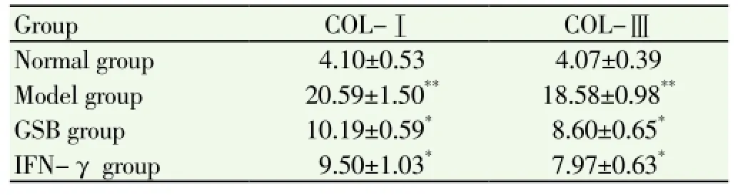
Table 1 Percentage of positive area in immunohistochemical image analysis (%).
4. Discussion
Gentiana scabra bage’s another name is gentian, also known as grass gentian. It is bitter in taste and cold in nature and the main active ingredients of it are base gentiana, gentiopicroside and gentiana trisaccharide. Gentiana scabra bage is a kind of Chinese herbs commonly used, which can be taken from many places and has small toxic side effects. It has such therapeutic effects as heat clearing and dampness eliminating, liver and gallbladder heat reducing, heat and humid of the lower abdominal organs and the role of appetite clearing. Also, it can be used to treat the heat in the liver caused by shock, red eyes by hot dysentery and swelling pharyngeal, jaundice, the loss of appetite, urinary tract infection, swelling of the scrotum, wet genital itch, etc. In clinic it is often used to treat infectious diseases, for example, colds, tonsillitis, bronchitis, acute dysentery and gastroenteritis[4,5].
We have found that gentiopicroside, gentiana scabra bage’s main active ingredient, dose have a protective effect on the liver[6-8]. However, many previous studies had focused on the therapeutic effect on the acute liver injury and there are few study on the chronic liver injury. Now there are few reports in China about the gentiana scabra bage’s effect on liver fibrosis caused by Paragonimus. A lot of remarkable achievements on the treatment of liver fibrosis have been reached, but a cheaper and more effective drug has not been found. There is no obvious liver and kidney toxicity after long-term use of gentiana scabra bage because of its small toxic side effects, so we used immunohistochemical technique to test the expression of hepatic type Ⅰ, Ⅲ
collagen proteins in rats on the pre-experiment basis with the purpose to explore new ways to treat liver fibrosis.
In this study, two mice in the model group were sacrificed randomly on the eighth week after the infection and it was found that the livers of these mice were darker and less smooth in general, with no significant changes in liver volume, the edge of the liver being blunting and its texture being solid. It was visible that circuitous cavernous lesions or sinus in the rat liver surface or facets, and the parasites were detected and it was found that more fibers were generated by pathological observation, which confirmed the formation of liver fibrosis and successful model. In order to simulate the treatment on human with liver fibrosis infected by Paragonimus skrjabini, we used gentiana scabra bage to treat hepatic fibrosis after insecticide with triclabendazole to the infected rats and the results showed that, compared with the model group, liver fibrous tissue proliferation was significantly reduced after the treatment of gentiana scabra bage in Paragonimus skrjabini rats with liver fibrosis and the swelling liver cells and the oppressed liver sinusoids also had been improved. It proves that gentiana scabra bage plays the role against hepatic fibrosis caused by Paragonimus skrjabin.
The study used immunohistochemical technique to test the content of hepatic type Ⅰ, Ⅲ collagen proteins in rats with liver fibrosis infected by Paragonimus skrjabini. The results showed that the expressions of hepatic type Ⅰ, Ⅲcollagen proteins were significantly decreased compared with the model group, but they still have not returned to the normal levels. It is consistent with the conclusion of Li et al[9], who found that GPS treatment could reduce the collagen production in rats with liver injury caused by carbon tetrachloride. IFN-γ was recommended to treat liver fibrosis by the United States institute of liver diseases in 1999 and it has been confirmed that IFN-γ has a positive effect on liver fibrosis through experiments and clinic[10-12]. This study once again confirmed that IFN-γ did have the exact effect on liver fibrosis. Taking IFN-γ as a positive control drug, we can make an objective assessment to gentiana scabra bage’s role against liver fibrosis. From the experimental results, we found that there were still some gaps between the effect of gentiana scabra bage and IFN-γ’s since the time was limited, the time to observe drug effect was short and the formation of fibrosis often was in the process during the evaluation of drug efficacy. So, more studies will be conducted to explore if the efficacy can be further increased while extending the treatment and if it can reserve completely the liver fibrosis.
The experimental results showed that gentiana scabra bage treatment could reduce the contents of hepatic type Ⅰ,Ⅲ collagen proteins significantly, thereby, it plays a role against hepatic fibrosis induced by Paragonimus skrjabini.
Conflict of interest statement
We declare that we have no conflict of interest.
[1] Liu T, Cai Q, Fu YQ,Yang SS. Research progress of traditional Chinese medicine gentian. Liaoning J Trad Chin Med 2004, 31(1): 85-86.
[2] Liu ZW, Chen CX. Research on gentiopicroside protective effect of liver. Herbs 2002; 33(1): 47.
[3] Wang GY, Huang CX, Jiang JX, Zhang Y. Pathology observation to Gentiopicroside’s experimental protective effect of liver cells in rat. Yunnan Med 1993; 14(4): 242-244.
[4] Liu YH, Zhou D, Chen YQ. The role of each component of okawa Dome pill to block calcium channel in the vascular endothelial cells. West Pharm J 2002; 17(1): 19-20.
[5] Zheng YY, Dai LY, Wang WB. Danshensu treatment of hepatic fibrosis and its mechanism study. China Liver Dis J 2003; 11(5): 288-290.
[6] Huang ZM, Zheng ZM, Cao WB. Pharmacological research onmixture’s role against hepatitis. Chin Pharm J 1992; 27(9): 555-556.
[7] Tong Li, Chen YY, Liu HH. The effects of gentiana injection on experimental hepatic injury. J First Military Med Univ 2001; 21(12): 906-907.
[8] Liu ZW, Chen CX, Jin RM, Shi GQ, Song CQ, Hu ZB. Research on gentiopicroside protective effect of liver. Herbs 2002; 33(1): 47-51.
[9] Li YQ, Zhao DH, Pan BR. Gentiopicroside’s role against liver injury in rats. J Fourth Military Med Univ 2001; 22(18): 1645-1649.
[10] Rockey DC, Chung JJ. Interferon gamma inhibits Lipocyte activation and extracellular matrix mRNA expression during experimental liver injury: Implications for treatment of hepatic fibrosis. J Investig Med 1994; 42: 660-670.
[11] Du X, Weng H, Cai W. Histological changes in 20 hepatic fibrosis patients with chronic hepatitis B after recombinant human interferon-gamma treatment. China Liver Dis J 2001; 9(5): 273-275.
[12] Cai WM. Effect of interferon-gamma on hepatic fibrosis in chronic hepatitis B virus infection. Clin Gastroenterol Hepatol 2005; 3(12): 1269.
ment heading
10.1016/S1995-7645(14)60188-7
*Corresponding author: Wen-Lin Wang, Professor, Department of Parasitology, Faculty of Basic Medicine, Kunming Medical University, Kunming 650500, China.
Tel: 0871-65920956
E-mail: wenlinwang331@163.com
ΔThese two authors contributed equally.
Foundation project: It is supported by National Natural Science Foundation of China (No. 81360252; 81360128); Natural Science Foundation of Yunnan Province (No. 2012FB025).
杂志排行
Asian Pacific Journal of Tropical Medicine的其它文章
- Inhibition of advanced glycation endproducts formation by Korean thistle, Cirsium maackii
- Anti-hypercholesterolemic effect of kenaf (Hibiscus cannabinus L.) seed on high-fat diet Sprague dawley rats
- Susceptibility to temephos, permethrin and deltamethrin of Aedes aegypti (Diptera: Culicidae) from Muang district, Phitsanulok Province, Thailand
- Cloning, expression, purification and bioinformatic analysis of 2-methylcitrate synthase from Mycobacterium tuberculosis
- Prevalence of shiga toxins (stx1, stx2), eaeA and hly genes of Escherichia coli O157:H7 strains among children with acute gastroenteritis in southern of Iran
- Larvicidal, ovicidal and repellent activities of marine sponge Cliona celata (Grant) extracts against Anopheles stephensi Liston (Diptera: Culicidae)