急性冠脉综合征患者mTOR活化与调节性T细胞及细胞因子的关系
2015-08-24张艳李志樑胡春玲
张艳,李志樑,胡春玲
急性冠脉综合征患者mTOR活化与调节性T细胞及细胞因子的关系
张艳1,2,李志樑1△,胡春玲2
目的 检测哺乳动物雷帕霉素靶蛋白(mTOR)在急性冠脉综合征(ACS)患者外周血T细胞中的活化状态,及其与调节性T细胞(Tregs)以及细胞因子的关系。方法 选取ACS患者32例(ACS组),同期因胸痛症状为主诉但心电图及冠脉造影正常,排除冠心病的胸痛综合征(CPS)患者28例为对照组;采用蛋白免疫印迹法检测2组外周血T细胞中mTOR、p70S6KT389磷酸化(p-p70S6KT389)及AKTS473磷酸化(p-AKTS473)水平;酶联免疫吸附测定法测T细胞亚群相关细胞因子干扰素-γ(IFN-γ)、白细胞介素(IL)-4、IL-17及转化生长因子-β(1TGF-β1)水平;流式细胞仪检测CD4+CD25+FoxP3+Tregs占CD4+T细胞的百分比;采用Spearman相关分析各指标之间的关联性。 结果 ACS 组mTOR及p-p70S6KT389表达量均高于对照组(均P<0.01);与对照组比较,ACS组IFN-γ、IL-17升高,Tregs百分比及TGF-β1减少(均P<0.05),而2组p-AKTS473、IL-4比较差异无统计学意义;相关分析显示p-p70S6KT389与IFN-γ及IL-17呈显著正相关(rs分别为0.91,0.92,均P<0.01),与Tregs及TGF-β1呈显著负相关(rs分别为-0.85,-0.80,均P<0.01)。结论 ACS患者外周血T细胞中mTORC1活化与Tregs减少及细胞因子失衡有关。
急性冠脉综合征;他克莫司结合蛋白质类;T淋巴细胞,调节性;细胞因子类;哺乳动物雷帕霉素靶蛋白
冠状动脉粥样硬化从发生、发展到斑块破裂、血栓形成最终引发急性冠脉综合征(ACS),是炎症和免疫交织的过程。调节性T细胞(Tregs)减少或功能缺陷、抗炎或促炎细胞因子平衡失调是ACS的重要发病机制之一。近来国外研究发现,抑制哺乳动物雷帕霉素靶蛋白(mTOR)或基因敲除后,可诱导产生较多的Tregs,提示mTOR在调节效应性T细胞与Tregs之间的平衡中扮演重要角色[1-3]。但目前尚鲜见有关ACS患者T细胞中mTOR与Tregs及细胞因子之间关系的研究。本研究通过检测ACS患者外周血T细胞中mTOR的活化状态、T细胞亚群相关细胞因子及Tregs百分比,并分析他们之间的关联,以期探讨mTOR在ACS免疫发病机制中的作用。
1 对象与方法
1.1 研究对象 选取2012年10月—2013年10月在南方医科大学附属珠江医院心内科住院的ACS患者共32例(ACS组),男20例,女12例,平均年龄(63.20±3.05)岁;其中不稳定型心绞痛(UAP)12例,非ST段抬高型心肌梗死(NSTEMI)10例,ST段抬高型心肌梗死(STEMI)10例。选择同期因胸痛症状为主诉但心电图及冠脉造影正常,排除冠心病的胸痛综合征(CPS)患者28例为对照组,男17例,女11例,平均年龄(63.35±2.74)岁。均排除合并感染、发热、肝肾功能不全、肿瘤和使用免疫抑制剂者。2组性别(χ2=0.876)、年龄(t=0.443)差异无统计学意义。
1.2 主要试剂及仪器 兔抗人mTOR抗体、兔抗人pp70S6K(Thr389)和p-Akt(Ser473)抗体(Cellsignal,美国);山羊抗兔二抗(southern biotech,美国);BCA法蛋白含量检测试剂盒(南京凯基生物发展有限公司);RosetteSep人T细胞富集试剂盒(StemCell,加拿大);人调节T细胞试剂盒(ebiosci⁃ence,美国);干扰素(IFN)-γ、白细胞介素(IL)-4、IL-17、转化生长因子(TGF)-β1酶联免疫吸附测定(ELISA)试剂盒(R&D,美国);Calibur型流式细胞仪(BD Bioscience,美国)。
1.3 血液标本采集 各组于入院后次日早晨空腹抽取静脉血20 mL,其中2 mL不抗凝,自然凝固20 min,2 000 r/min离心5~10 min,收集血清,ELISA法测细胞因子。余下的用肝素抗凝后,取其中部分用于流式细胞检测,其余用于提取T细胞。
1.4 T细胞的分离 按照RosetteSep人T细胞富集试剂盒说明提取T细胞,并用RPMI1640完全培养液调整细胞数为(2~4)×106/L。
1.5 mTOR、p70S6KT389磷酸化(p-p70S6KT389)和AKTS473磷酸化(p-AKTS473)水平检测 采用蛋白免疫印迹法:分离的T细胞离心,提取蛋白后BCA法测蛋白浓度;经电泳、转膜、室温封闭后,分别加入一抗(兔抗人)mTOR(1∶1 000)、p-p70S6K (Thr389,1∶1 000)、p-Akt(Ser473,1∶1 000)震荡过夜后洗涤并加入山羊抗兔二抗(1∶5 000),室温孵育,洗涤后暗胶片曝光,用Quantity One图像分析系统分析检测。
1.6 外周血CD4+CD25+FoxP3+Tregs水平检测 用流式细胞仪检测,收获各组抗凝血100 μL于试管,严格按照人调节T细胞试剂盒说明操作,最后以CD4+T细胞设门,分析荧光强度,记录CD4+CD25+FoxP3+Tregs百分率。
1.7 细胞因子的检测 血清IFN-γ、IL-4、IL-17、TGF-β1的测定均按照ELISA试剂盒说明操作进行。
2 结果
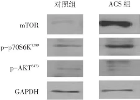
2.1 T细胞mTOR、p-p70S6KT389和p-AKTS473表达水平 ACS组的mTOR及p-p70S6KT389水平高于对照组(P<0.01);2组p-AKTS473表达水平差异无统计学意义,见图1、表1。

Fig.1 Expression of mTOR,p-p70S6KT389and p-AKTS473in peripheral T lymphocytes in two groups shown by Western blot图1 2组外周血T细胞mTOR、p-p70S6KT389和p-AKTS473表达水平
Tab.1 Comparison of mTOR,p-p70S6KT389and p-AKTS473expression in peripheral T lymphocytes in two groups表1 2组患者外周血T细胞mTOR、p-p70S6KT389和p-AKTS473表达水平比较 (±s)

Tab.1 Comparison of mTOR,p-p70S6KT389and p-AKTS473expression in peripheral T lymphocytes in two groups表1 2组患者外周血T细胞mTOR、p-p70S6KT389和p-AKTS473表达水平比较 (±s)
*P<0.05,**P<0.01;表2、3同
组别nmTOR p-AKTS473p-p70S6KT389对照组2850.12±5.5739.39±3.4484.76±4.93 ACS组32857.15±6.5642.53±4.21620.41±5.81 t 171.17**1.98141.63**
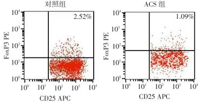
2.2 CD4+CD25+FoxP3+Tregs检测结果 ACS组患者CD4+CD25+FoxP3+Tregs占CD4+T细胞的比率为(1.09±0.16)%,显著低于对照组(2.52±0.10)%,差异有统计学意义(t=2.23,P<0.05),见图2。

Fig.2 CD4+CD25+FoxP3+Tregs/CD4+frequencies in two groups shown by FACS图2 流式细胞术测2组CD4+CD25+FoxP3+Tregs/CD4+T细胞比率
2.3 血清IFN-γ、IL-4、TGF-β1和IL-17细胞因子水平 与对照组比较,ACS组IFN-γ、IL-17水平显著升高,TGF-β1明显减少(均P<0.01),2组IL-4无明显差异(P>0.05),见表2。
Tab.2 Comparison of serum levels of cytokines in two groups表2 2组血清IFN-γ、IL-4、TGF-β1和IL-17细胞因子水平比较 (ng/L,±s)

Tab.2 Comparison of serum levels of cytokines in two groups表2 2组血清IFN-γ、IL-4、TGF-β1和IL-17细胞因子水平比较 (ng/L,±s)
组别对照组ACS组t n 28 32 IFN-γ 37.48±1.63 53.02±3.14 19.25**IL-4 18.74±2.19 21.03±2.20 1.31 TGF-β159.20±2.27 40.09±2.10 21.16**IL-17 14.46±1.42 59.02±2.05 23.62**
2.4 p-p70S6KT389、CD4+CD25+FoxP3+Tregs、细胞因子的相关分析 p-p70S6KT389与IFN-γ及IL-17均呈高度正相关,与TGF-β1及CD4+CD25+FoxP3+Tregs呈高度负相关(均P<0.01)。CD4+CD25+FoxP3+Tregs与TGF-β1水平呈高度正相关,与IFN-γ、IL-17均呈高度负相关(均P<0.01),见表3。

Tab.3 Correlation analysis on p-p70S6KT389,CD4+CD25+FoxP3+Tregs and cytokins表3 p-p70S6KT389、CD4+CD25+FoxP3+Tregs与细胞因子的相关分析 (rs)
3 讨论
目前研究已证实,大量存在于ACS患者循环及斑块中的活化CD4+T细胞及其分泌的炎症细胞因子,对粥样硬化进展起到极其重要的作用;而辅助性T细胞Th1、Th2、Th17及Tregs细胞亚群功能失衡,尤其是Tregs减少,是ACS的重要发病机制之一[4-5]。
mTOR是一种丝氨酸/苏氨酸激酶,在调节T细胞分化中起重要作用[6]。mTOR以mTORC1和mTORC2两种多蛋白复合物形式存在。而p70S6K (Thr389)及AKT(Ser473)磷酸化水平通常可分别作为评估mTORC1和mTORC2活性的指标[7]。近来国外学者们发现mTORC1在Th1和Th17亚群分化中起重要作用,而 Th2亚群则依赖于 mTORC2,mTORC1及mTORC2均影响FoxP3+Tregs的分化[1-3]。
本研究表明ACS组的mTOR及p-p70S6KT389水平高于对照组,但2组p-AKTS473表达水平无明显差异,提示mTORC1途径在ACS发病中可能发挥更重要的作用。本研究还观察到与对照组相比,ACS组外周血Treg比率及其主要细胞因子TGF-β1水平明显下降,Th1、Th17主要细胞因子IFN-γ和IL-17则升高,再次证实了Tregs表达下降,Th1、Th17型免疫反应亢进,同时引起抗炎及促炎因子平衡失调,促进了ACS发病进展过程[4-5],与既往研究结果相似[8]。2 组Th2细胞因子IL-4水平无明显差异,与Zhao等[9]研究结果相似,提示Th2型免疫反应在ACS病变中所起作用相对较小。相关分析结果提示Tregs在维持自身免疫稳定中的作用与TGF-β1有关,mTORC1活化可能与Th1、Th17、Tregs细胞因子失衡及Tregs减少有关。mTORC1活化使得T细胞向Th1和Th17亚群分化,Tregs细胞亚群减少,打破了机体维持外周自身免疫应答的稳态,同时导致T细胞亚群分泌的细胞因子失衡,最终促进ACS病变进展。在T细胞亚群的检测中,本研究主要集中于对ACS起潜在保护作用的Tregs细胞上,而对其他T细胞亚群只采用ELISA法检测其相关细胞因子,因此需要在下一步深入研究中进一步完善T细胞亚群的检测,这将会对本研究结果提供更充足的证据。
[1]Delgoffe GM,Kole TP,Zheng Y,et al.The mTOR kinase differen⁃tially regulates effector and regulatory T cell lineage commitment[J].Immunity,2009,30(6):832-844.
[2]Delgoffe GM,Pollizzi KN,Waickman AT,et al.The kinase mTOR regulates the differentiation of helper T cells through the selective activation of signaling by mTORC1 and mTORC2[J].Nat Immunol,2011,12(4):295-303.
[3]Soliman GA.The role of mechanistic target of rapamycin(mTOR)complexes signaling in the immune responses[J].Nutrients,2013,5 (6):2231-2257.
[4]Jiang HJ,Wang L,Li RZ,et al.Th1/Th2,Treg/Th17 drift in the re⁃search development of acute coronary syndrome[J].Chinese Journal of Clinicans,2013,7(7):3104-3106.[姜红菊,王蕾,李润智,等.Th1/ Th2、Treg/Th17漂移在急性冠状动脉综合征中的研究进展[J].中华临床医师杂志,2013,7(7):3104-3106].
[5]Sasaki N,Yamashita T,Takeda M,et al.Regulatory T cells in athero⁃genesis[J].J Atheroscler Thromb,2012,19(6):503-515.
[6]Lo YC,Lee CF,Powell JD.Insight into the role of mTOR and metab⁃olism in T cells reveals new potential approaches to preventing graft rejection[J].Curr Opin Organ Transplant,2014,19(4):363-371.
[7]Guertin DA,Stevens DM,Thoreen CC,et al.Ablation in mice of the mTORC components raptor,rictor,or mLST8 reveals that mTORC2 is required for signaling to Akt-FOXO and PKCalpha,but not S6K1 [J].Dev Cell,2006,11(6):859-871.
[8]Ma Y,Yuan X,Deng L,et al.Imbalanced frequencies of Th17 and Treg cells in acute coronary syndromes are mediated by IL-6-STAT3 signaling[J].PLoS One,2013,8(8):e72804.
[9]Zhao Z,Wu Y,Cheng M,et al.Activation of Th17/Th1 and Th1,but not Th17,is associated with the acute cardiac event in patients with acute coronary syndrome[J].Atherosclerosis,2011,217(2):518-524.
(2014-08-26收稿 2014-10-20修回)
(本文编辑 闫娟)
Relationship among mTOR levels,regulatory T cells and cytokins in patients with acute coronary syndrome
ZHANG Yan1,2,LI Zhiliang1△,HU Chunling2
1 Department of Cardiology,Zhujiang Hospital of Southern Medical University,Guangzhou 510282,China;2 Department of Internal Medicine,Guangdong Province Maternity and Child Care Hospital
△Corresponding Author E-mail:happydragon2012qq@126.com
Objective To assess whether mTOR was activated in peripheral T lymphocytes of acute coronary syndrome (ACS)patients and to investigate the relationship among mTOR,regulatory T cells and cytokins.Methods Patients with acute coronary syndrome(n=32)were selected in ACS group.Meanwhile,patients who complainted of chest pain but were proven to be normal in ECG and in coronary arteriography,were excluded as ACS patients but were diagnosed as Chest Pain Syndrome(CPS)and selected in CPS group(n=28).The expression levels of mTOR,phospho-p70S6KT389(indicative of mTORC1 activity)and phospho-AKTS473(indicative of mTORC2 activity)were investigated in T cells,which were isolated from peripheral blood of patients in ACS group or CPS group,using Western blot.The proportion of CD4+CD25+Foxp3+Tregs over CD4+cells were evaluated by FACS.And T cell subset related cytokines such as IFN-γ,IL-4,IL-17 and TGF-β1were exam⁃ined by ELISA.Then we investigated the relationship among mTOR,regulatory T cells and cytokins by Spearman analysis.Results mTOR and p-p70S6KT389expression were significantly enhanced in ACS group as compared with those in patients from CPS group(P<0.01).Higher levels of IFN-γ,IL-17 but lower level of TGF-β1cytokines as well as decreased propor⁃tion of Tregs were observed in ACS group than those in CPS group(P<0.05).There was no significant difference in the level of IL-4 and p-AKTS473between the two groups(P>0.05).By correlation analysis,p-p70S6KT389expression level positively correlated with IFN-γ,IL-17(rs=0.91,092,P<0.01)and negatively correlated with Tregs and TGF-β(1rs=-0.85,-0.80,re⁃spectively,P<0.01).Conclusion mTORC1 pathway was activated in peripheral T lymphocytes of ACS patients,and Tregs insufficiency and cytokins imbalance both contribute to the activation of mTORC1 pathway and pathological process of ACS.
acute coronary syndrome;tacrolimus binding proteins;T-lymphocytes,regulatory;cytokines;mTOR
R541.4
A
10.11958/j.issn.0253-9896.2015.04.022
1南方医科大学附属珠江医院心内科,广东广州(邮编510282);2广东省妇幼保健院内科
张艳(1979),女,主治医师,博士在读,主要从事冠心病防治的研究
△E-mail:happydragon2012qq@126.com
