Electroacupuncture down-regulates the expressions of colonic NGF and NGFR in visceral hypersensitivity rats
2015-06-19LiuYanan刘雅楠WuHuangan吴焕淦WangXiaomei王晓梅ShiZheng施征JiangYan江岩SongChunbin宋春斌ZhangYingying张英英TanLinying谭琳蓥
Liu Ya-nan (刘雅楠), Wu Huan-gan (吴焕淦), Wang Xiao-mei (王晓梅), Shi Zheng (施征), Jiang Yan (江岩), Song Chun-bin (宋春斌), Zhang Ying-ying (张英英), Tan Lin-ying (谭琳蓥)
1 Shanghai University of Traditional Chinese Medicine, Shanghai 201203, China
2 Shanghai Research Institute of Acupuncture and Meridian, Shanghai 200030, China
Basic Study
Electroacupuncture down-regulates the expressions of colonic NGF and NGFR in visceral hypersensitivity rats
Liu Ya-nan (刘雅楠)1, Wu Huan-gan (吴焕淦)2, Wang Xiao-mei (王晓梅)2, Shi Zheng (施征)2, Jiang Yan (江岩)2, Song Chun-bin (宋春斌)1, Zhang Ying-ying (张英英)2, Tan Lin-ying (谭琳蓥)2
1 Shanghai University of Traditional Chinese Medicine, Shanghai 201203, China
2 Shanghai Research Institute of Acupuncture and Meridian, Shanghai 200030, China
Objective:To observe the effect of electroacupuncture (EA) on the expressions of colonic nerve growth factor (NGF) and nerve growth factor receptor (NGFR) in visceral hypersensitivity rats, and investigate the mechanisms of EA on chronic visceral hypersensitivity.
Methods:A total of 24 neonatal rats were randomly divided into a normal group, a model group and an EA group. The models of visceral hypersensitivity rats were prepared according to the method of Al-Chaer. The rats in the EA group were treated with EA at Tianshu (ST 25) and Shangjuxu (ST 37), 20 min for each time, once a day, for 7 consecutive days. After the first treatment, the observation of abdominal withdrawal reflex (AWR) was used to evaluate the pain threshold of visceral hypersensitivity rats. After 7-day treatments, the colonic tissue of rat was collected to test the immunohistochemistry of NGF and NGFR.
Results:The score of AWR in the model group was higher than that in the normal group, but the score decreased after EA treatment. The positive expressions of colonic NGF and NGFR in the model group were increased than those in the normal group (P<0.05), while they were both decreased in the EA group after treatment (P<0.001).
Conclusion:The EA could up-regulate the pain threshold of visceral hypersensitivity rats, and down-regulate the expressions of colonic NGF and NGFR. The regulation of the expressions of colonic NGF and NGFR would be one of the peripheral mechanisms of EA in treating chronic visceral hypersensitivity.
Acupuncture Therapy; Electroacupuncture; Visceral Pain; Irritable Bowel Syndrome; Nerve Growth Factor; Receptors, Nerve Growth Factor; Hyperalgesia
Irritable bowel syndrome (IBS) is a common functional gastrointestinal disorder and characterized by abdominal pain or distention, and alteration of bowel habits. IBS appears to be related to mental factors, and visceral hypersensitivity is the main reason to cause IBS[1-2]. The incidence of typical IBS symptoms had up to 5%-15%[3]in the general population over the world. With the development of research in gastroenterology, the effect of enteric nervous system (ENS) in the pathogenesis of IBS has contracted more attention. Recent studies have found that ENS and intestinal local immunocyte could interact with each other, and releasevarious neurotransmitters, such as 5-hydroxytryptamine (5-HT), nerve growth factor (NGF), neuropeptide Y (NPY) and inflammatory factor, which are involved in the occurrence of visceral hypersensitivity[4].
NGF is a polypeptide that is critical for the growth and survival of nerve cells. It can promote the regeneration of cell functional synapses which are widely distributed in neuronal cell bodies and neurites[5-6]. Studies have shown that NGF was also an important substance intermediary for inflammatory pain, and it is also related to visceral hyperalgesia. NGF combines with specific nerve growth factor receptor (NGFR) on mast cell membrane, and then, the second messenger Ca2+stimulates and leads to mast cell activation itself, some allergic mediums and histamines would be released to the nerve endings to promote the release of neurotransmitter[7]. It’s been found higher expression of NGF in colonic mucosa of patients with IBS. While the positive NGF cells are mainly concentrated in the mucosa layer and myenteric plexus, and positive reaction is also visible in submucosal nervous plexus, while the expression is significantly higher than that in normal rats[8].
Electroacupuncture (EA) can reduce the visceral hypersensitivity of rats[9-10], and previous studies had found similar results[11-12]. It had been found that, exogenous injection of NGF could reduce the visceral pain threshold in rats, while the anti NGF antibody could reduce the visceral hypersensitivity induced by Trinitrobenzene Sulfonic Acid (TNBS)[13].
However it was still unknown whether colonic NGF and NGFR are involved in the occurrence of rats’ visceral hypersensitivity, and whether EA can regulate the expressions of colonic NGF and NGFR to induce the visceral hypersensitivity of rats. This study aims to explore the mechanism of EA for visceral hypersensitivity by observing the expressions of NGF and NGFR in colon.
1 Materials and Methods
1.1 Animal preparation
Twenty-four male newborn Sprague-Dawley rats (5 d) of clean grade, offered by the Experimental Animal Center of Shanghai University of Traditional Chinese Medicine [license number, SYXK (Shanghai) 2004-0005)] were used. Every 8 rats were bred with one adult female rat, room temperature: (22±2)℃, humidity: (60±5) %. The animal experiment was performed in the Animal Experimental Center of Shanghai University of Traditional Chinese Medicine from January 2007 to April 2007. All of the use of rats enforced strictly according to the rules of National Laboratory Animal Health. The experimental process obeyed the animal ethical standards.
1.2 Main reagents and instruments
Pentobarbital sodium, 0.4% paraformaldehyde, paraffin, xylene, ethanol, 0.01 mol/L citric acid buffer, PBS buffer, Envision reagent (Sigma), Olympus-CH optical microscope.
1.3 Modeling procedure and treatment
These 24 neonatal rats were randomly divided into a normal group, a model group and an EA group. The rats in the model group and the EA group were established as visceral hypersensitivity rats according to relevant literature[14]. At the beginning of 8 d from birth, the rats were stimulated by colorectal balloon stimulus in the waking state. The latex balloon which was inflated to be 0.2 mL (20 mm in length and 2 mm in diameter), gently inserted into the descending colon of rats, lasting for 1 min, and then the balloon gas was withdrawn, the stimulus balloon was removed slowly. The same stimulation was repeated after 30 min (to reduce pain or discomfort), and continued for 1 week. The abdominal withdrawal reflex (AWR) was used to check chronic visceral hypersensitivity in rats models. Two days after chronic visceral hypersensitivity model succeeded, the rats were divided into groups for different interventions.
1.3.1 Normal group
The rats in the normal group were not given any treatment, but only fixed in the same way as those in the EA group.
1.3.2 Model group
The rats in the model group were only modeled, without receiving any treatment, but only fixed in the same way as those in the EA group.
1.3.3 EA group
The rats in the EA group were treated with EA after modeled successfully.
Acupoints: Bilateral Tianshu (ST 25)[15]and Shangjuxu (ST 37)[11].
Method: Disposable acupuncture needles of 0.25 mm in diameter and 15 mm in length were selected. After routine disinfection of the skin, the needles were punctured into the acupoints. Then Han’s EA apparatus was used with square wave, 100 Hz for 3 s and 2 Hz for 3 s alternately in frequency, 0.2-0.6 ms in amplitude and 1 mA in intensity. The treatment retained for 30 min, once a day for 7 d in all.
1.4 Observation methods and measurements
1.4.1 AWR score
At the end of the first treatment of EA, the AWR score was used to assess pain threshold in rats with chronic visceral hypersensitivity. The AWR coring criteria were assessed according to the stimulation method of colorectal distention (CRD) of Al-Chaer ED, et al[16]. The balloon was taken into the anus of rats, and then it wasrapidly expanded under the pressure of 2.66 kPa, 5.32 kPa, 7.98 kPa and 10.64 kPa, each for 20 s. Each rat was taken for the next repeated assessment after 3 min. The same intensity of stimulation was repeated 3 times, and then the average value was taken as the final score.
1.4.2 Immunohistochemistry of colonic NGF and NGFR
On the second day after treatment, the rats were anesthetized with intraperitoneal injection of pentobarbital sodium [80 mg/(kg·BW)]. The colons were taken out quickly, about 1 cm colon tissues (starting at 5 cm away from the anus), rinsed by normal saline. Then 0.4% paraformaldehyde was used to fix the tissues. The expressions of NGF and NGFR were detected by immunohistochemical method.
The tissues were routinely dehydrated, hyalinized, embedded and sliced; the 4 μm paraffinized sections were posted on the glass slide coated with sliced adhesive, and baked as 58℃ for 24 h. After dewaxed water, the tissues were dewaxed by xylene I, II and III, each for 10 min, then washed by 95%, 85% and 75% ethanol, each for 2 min; then washed by 0.01 ml/L, pH 7.4 PBS 3 times, each for 3 min; 1% H2O2for 20 min; PBS 3 times, each for 3 min; antigen repairation for twice, each for 10 min. After the slices were cooled down to room temperature naturally, they were placed in 0.01 ml/L citric acid buffer (CB), pH 6, microwave III gear (98 ℃) twice, each for 10 min; PBS washing for 3 times, each for 3 min; closed with 1% normal serum for 20 min, at room temperature; no washing, added the appropriate dilution of primary anti (NGF 1:150 and NGFR 1:200, Sigma), 4 ℃ for 18 h; PBS washing for 3 times, each for 3 min; envision reagent (Sigma) for 30 min (37 ℃); PBS washing for 3 times, each for 3 min; 0.04% DAB + 0.03% H2O2colored for 8 min; water rinsing; hematoxylin stained for 30 s; water rinsing; acid ethanol blue for 2 s; water rinsing; microwave blue; conventional resin sheeted. Positive slide was known as positive control, and PBS buffer instead of the primary antibody as negative control. The Olympus-CH optical microscope was used to randomly continuously calculate 3 timesof optical density and the number of positive cells that expressed NGF and NGFR in the fields.
1.5 Statistical analysis

2 Results
2.1 Effect on AWR score of single EA
When the pressure of the colon balloon stimulation was 2.66 kPa, 5.32 kPa, 7.98 kPa and 10.64 kPa, the AWR score of visceral hypersensitivity rats was higher than those in the normal group (P<0.05). After one EA treatment, the AWR score in the EA group was significantly reduced, and the difference was significant compared with that in the model group (P<0.05), (Figure 1, Table 1).

Figure 1. The AWR score of single EA for chronic visceral hypersensitivity rats
Table 1. The AWR score comparison of single EA among the three groups

Table 1. The AWR score comparison of single EA among the three groups
Note: Compared with the normal group, 1) P<0.05; compared with the model group, 2) P<0.05
Group n 2.66 kPa 5.32 kPa 7.98 kPa 10.64 kPa Normal 8 0.0750±0.1389 1.0875±0.2475 2.8875±0.1553 3.0375±0.1923 Model 8 1.8875±0.15531)3.0750±0.55231)3.5375±0.36621)3.9625±0.10611)EA 8 0.1875±0.15532)1.2875±0.29002)2.9625±0.10612)3.5375±0.36622)
2.2 Effect on colonic NGF expression of EA
Positive target area and integral optical density of colonic NGF were significantly higher than those in the normal group, and the difference was statistically significant (P<0.001). After treatment, positive target area and integral optical density of colonic NGF in the EA group were significantly lower than those in the model group, and the differences were statistically significant (P<0.001), (Table 2, Figure 2-4).

Figure 2. Expression of NGF positive target total area
Table 2. Colonic NGF expression of EA for IBS rats

Table 2. Colonic NGF expression of EA for IBS rats
Note: Compared with the normal group, 1) P<0.001; compared with the model group, 2) P<0.001
Group nPositive target area (μm2) Integral optical density Normal8597.00±99.59 297.10±46.77 Model 8 1821.12±324.981)1349.68±247.121)EA 8 772.79±93.062)562.67±96.742)

Figure 3. Expression of NGF integral optical density
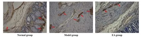
Figure 4. Effect on colonic NGF expression of EA for chronic visceral hypersensitivity rats (×200)
2.3 Effect on colonic NGFR expression of EA
Positive target area and integral optical density of colonic NGF were significantly higher than those in the normal group, and the differences were statistically significant (P<0.001). After treatment, both positive target area and integral optical density of colonic NGF in the EA group were significantly lower than those in the model group, and the differences were statistically significant (P<0.001), (Table 3, Figure 5-7).
Table 3. Colonic NGFR expression of EA for chronic visceral

Table 3. Colonic NGFR expression of EA for chronic visceral
Note: Compared with the normal group, 1) P<0.001; compared with the model group, 2) P<0.001
Group nPositive target area (μm2) Integral optical density Normal 8 889.08±113.54 555.97±63.76 Model 8 2393.63±368.741)1726.03±291.171)EA 81151.80±83.782)956.02±77.212)

Figure 5. Expression of NGFR positive target total area

Figure 6. Expression of NGFR integral optical density

Figure 7. Effect on colonic NGFR expression of EA for chronic visceral hypersensitivity rats (×200)
3 Discussion
Visceral hypersensitivity is a major cause of IBS[14,17]with chronic, repeated, lower abdominal pain and discomfort and altered bowel habits. Al-Chaer ED, et al[16]used a mechanical colorectal stimulation to neonatal rats to establish chronic visceral hypersensitivity model, a reliable animal model for research on the mechanism of EA on chronic visceral pain. The results of this study showed that the AWR scores of the rectus abdominis muscle at the pressure of 2.66 kPa, 5.32 kPa, 7.98 kPa and 10.64 kPa of colonballoon colonic stimulation were higher than those in the normal group. It could be indicated that the visceral hypersensitivity model has been established.
Tianshu (ST 25) is the Front-Mu point, while Shangjuxu (ST 37) is the lower He-Sea point of the large intestine. The combination of Tianshu (ST 25) and Shangjuxu (ST 37) could regulate the functions of the large intestine, spleen and stomach. Previous studies have shown that Tianshu (ST 25) and/or Shangjuxu (ST 37) can regulate the gastrointestinal function and treat gastrointestinal diseases[10-11,18-20]. Therefore, Shangjuxu (ST 37) and Tianshu (ST 25) were used to research the influence of EA for visceral hypersensitivity IBS rats in this experiment.
It was certified that EA can inhibit the chronic visceral hypersensitivity of rats. Cui KM, et al[9]reported that EA can significantly reduce the AWR scores of the rectus abdominis muscle at colonic distension pressure of 2.66 kPa, 5.32 kPa, 7.98 kPa and 10.64 kPa. This experiment also obtained the similar result. Some researchers[10-11]found that EA can significantly reduced chronic visceral hypersensitivity and the AWR scores of rats at the colonic distension pressure of 2.66 kPa, 5.32 kPa, 7.98 kPa and 10.64 kPa. However, the mechanism of EA is still unknown.
ENS could locally control the intestinal movement, secretion, blood flow, and transport water and electrolyte. It can also connect with the central nervous system via neurons, nerve fibers and neurotransmitters such as 5-HT[21-22], NGF[23]and NPY[24], to regulate gastrointestinal motility, pain, emotional reaction and immunity. Our previous study showed that NGF and NGFR were expressed in neural and non-neural tissues, and the expressions of both two were significantly enhanced in the intestinal mucosal epithelium and intestinal myenteric plexus of visceral hypersensitivity rats, suggesting that the expressions of NGF and NGFR maybe related to the enteric nervous and immune regulation. The result was same as the research of Barreau F, et al[25], suggesting that NGF and NGFRshould be associated with colonic notility and change of visceral hypersensitivity. After EA intervention, the positive expressions of NGF and NGFR in colon of rats decreased significantly, suggesting that EA can reduce the expressions of NGF and NGFR in colon.
In summary, this study showed that the pain threshold of chronic visceral hypersensitivity rats were lower, while the expressions of colonic NGF and NGFR increased significantly after colorectal stimulation. EA at Tianshu (ST 25) and Shangjuxu (ST 37) could enhance the pain threshold of the chronic visceral hypersensitivity rats, and down-regulate the expressions of colonic NGF and NGFR obviously. It indicates that EA can decrease the expressions of colonic NGF and NGFR after colorectal stimulation, which is possibly one of the peripheral mechanisms of EA in treatment of chronic visceral hypersensitivity.
Conflict of Interest
The authors declared that there was no conflict of interest in this article.
Acknowledgments
This work was supported by Project of Shanghai Natural Sciences Foundation (上海市自然科学基金项目, No.14ZR1438700); National Basic Research Program of China (973 Program) [国家重点基础研究发展计划 (973计划)项目, No. 2015CB554500]; The Second Period Xinglin Scholars Program of Shanghai University of Traditional Chinese Medicine (上海中医药大学第二期杏林学者项目); National Natural Sciences Foundation of China (国家自然科学基金项目, No.81202752).
Statement of Informed Consent
The treatment of animals conformed to the ethical criteria in this experiment.
[1] Mertz H, Naliboff B, Munakata J, Niazi N, Mayer EA. Altered rectal perception is a biological marker of patients with irritable bowel syndrome. Gastroenterology, 1995, 109(1): 40-52.
[2] Cervero F, Laird JM. Visceral pain. Lancet, 1999, 353(9170): 2145-2148.
[3] Xu XX, Li DG. Epidemiological characteristics of Irritable bowel syndrome. Zhonghua Liuxingbingxue Zazhi, 2003, 6(6): 523.
[4] Li YQ, Yu YB. Research progress of nerve immuneregulation on irritable bowel syndrome abdominal pain. Guoji Xiaohuabing Zazhi, 2012, 32(2): 65-67.
[5] Zhang YH, Nicol GD. NGF-mediated sensitization of the excitability of rat sensory neurons is prevented by a blocking antibody to the p75 neurotrophin receptor. Neurosci Lett, 2004, 366(2): 187-192.
[6] Pannese E, Procacd P. Ultrastructural localization of NGF receptors in satellite cells of the rat spinal ganglia. J Neurocytol, 2002, 31(8-9): 755-763.
[7] Metcalfe DD, Baram D, Mekori YA. Mast cells. Physiol Rev, 1997, 77(4): 1033-1079.
[8] Matricon J, Muller E, Accarie A, Meleine M, Etienne M, Voilley N, Busserolles J, Eschalier A, Lazdunski M, Bourdu S, Gelot A, Ardid D. Peripheral contribution of NGF and ASIC1a to colonic hypersensitivity in a rat model of irritable bowel syndrome. Neurogastroenterol motil, 2013, 25(11): e740-e754.
[9] Cui KM, Li WM, Gao X, Chung K, Chung JM, Wu GC. Electroacupuncture relieves chronic visceral hyperalgesia in rats. Neurosci Lett, 2005, 376(1): 20-23.
[10] Tian XY, Bian ZX, Hu XG, Zhang XJ, Liu L, Zhang H. Electro-acupuncture attenuates stress-induced defecation in rats with chronic visceral hypersensitivity via serotonergic pathway. Brain Res, 2006, 1088(1): 101-108.
[11] Tian SL, Wang XY, Ding GH. Repeated electroacupuncture attenuates chronic visceral hypersensitivity and spinal cord NMDA receptor phosphorylation in a rat irritable bowel syndrome model. Life Sci, 2008, 83(9-10): 356-363.
[12] Wu HG, Jiang B, Zhou EH, Shi Z, Shi DR, Cui YH, Kou ST, Liu HR. Regulatory mechanism of electroacupuncture in irritable bowel syndrome: preventing MC activation and decreasing SP VIP secretion. Dig Dis Sci, 2008, 53(6): 1644-1651.
[13] Delafoy L, Raymond F, Doherty AM, Eschalier A, Diop L. Role of nerve growth factor in the trinitrobenzene sulfonic acid-induced colonic hypersensitivity. Pain, 2003, 105(3): 489-497.
[14] Talley NJ, Boyce PM, Jones M. Predictors of health care seeking for irritable bowel syndrome: a population based study. Gut, 1997, 41(3): 394-398.
[15] Sugai GC, Freire Ade O, Tabosa A, Yamamura Y, Tufik S, Mello LE. Serotonin involvement in the electroacupuncture- and moxibustion-induced gastric emptying in rats. Physiol Behav, 2004, 82(5): 855-861.
[16] Al-Chaer ED, Kawasaki M, Pasricha PJ. A new model of chronic visceral hypersensitivity in adult rats induced by colon irritation during postnatal development. Gastroenterology, 2000, 119(5): 1276-1285.
[17] Drossman DA, Patrick DL, Whitehead WE, Toner BB, Diamant NE, Hu Y, Jia H, Bangdiwala SI. Further validation of the IBS-QOL: a disease-specific quality-oflife questionnaire. Am J Gastroenterol, 2000, 95(4): 999-1007.
[18] Wu HG, Zhao C, Shi Z, Chen HP, Liu Y, Liu SM. Clinical study on spleen-stomach-reinforcing moxibustion treatment of diarrhea-type irritable bowel syndrome. Shijie Zhenjiu Zazhi, 2002, 12(1): 10-15.
[19] Wang R, Bai HX, Ji LX, Jin XF, Yan LP. Study on the protective function of electric acupuncture at Tianshu (ST 25) with different accompanying points for colonic mucosa of rats with ulcerative colitis. Shijie Zhongxiyi Jiehe Zazhi, 2007, (11): 639-642.
[20] Wang W, Bai L, Gao ZX, Lü EJ. Clinical research on diarrhea-type irritable bowel syndrome treated by acupuncture. Zhongguo Wuzhenxue Zazhi, 2008, 18(26): 6335-6336.
[21] Sun G, Yang YS, Peng LH, Wang WF. Visceral sensitivity and expression of 5-hydroxytryptamine and c-fos in the spinal dorsal horn in a rat model with irritable bowel syndrome. Shijie Huaren Xiaohua Zazhi, 2007, 15(25): 2718-2722.
[22] Xiao DQ, Tang JS, Yuan B, Jia H. Blocking effects of 5-HT2receptor antagonist cyproheptadine applied to thalamic nucleus submedius on analgesia produced by high intensity electroacupuncture stimulation in rats. Clin J Neurosci, 2000, 16(4): 352-354
[23] Liu ZH, Pan PG, Qi YC, Zhao Y, Chai TQ, Tang CZ, Wang QY, Yang JJ, Lin JQ. Effect of electroacupuncture on neuronal apoptosis and protein expression of nerve growth factor in brain tissues of new-born rats with hypoxic-ischemic injury. Zhongguo Linchuang Kangfu, 2006, 10(23): 114-119.
[24] Wang ZY, Sun ZR, Liu RM. The role of neuropeptidecytokine network in the study on the correlation between acupuncture analgesia and acupuncture immunoregulation. Zhonghua Zhongyiyao Xuekan, 2010, 28(2): 297-299.
[25] Barreau F, Cartier C, Ferrier L, Fioramonti J, Bueno L. Nerve growth factor mediates alterations of colonic sensitivity and mucosal barrier induced by neonatal stress in rats. Gastroenterology, 2004, 127(2): 524-534.
Translator: Lan Tian-ying (兰天鹰)
电针下调内脏高敏感性大鼠结肠NGF和NGFR的表达
目的:观察电针对内脏高敏感大鼠结肠神经生长因子(nerve growth factor, NGF)和神经生长因子受体(nerve growth factor receptor, NGFR)表达的影响,探讨电针治疗慢性内脏高敏感性的作用机制。方法:将24只新生乳鼠随机分为正常组、模型组和电针组。参照Al-Chaer法制备内脏高敏感性大鼠模型。电针组予电针天枢、上巨虚治疗,每次20分钟,每天1次,连续7天。首次治疗后,通过观察大鼠腹壁撤回反射(abdominal withdrawal reflex, AWR)评分以评价内脏高敏感性大鼠的痛阈。治疗7天后,采集大鼠结肠组织进行NGF和NGFR免疫组织化学检测。结果:模型组大鼠的AWR评分均高于正常组,经电针治疗后均降低。模型组大鼠的结肠NGF和NGFR阳性表达较正常组显著增加(P<0.05),电针组治疗后NGF和NGFR阳性表达均显著降低(P<0.001)。结论:电针可使内脏高敏感性大鼠的痛阈升高,并降低结肠的NGF和NGFR表达。电针对结肠NGF和NGFR表达的调节可能是其治疗慢性内脏高敏感性的外周作用机制之一。
针刺疗法; 电针; 内脏痛; 肠易激综合征; 神经生长因子; 受体, 神经生长因子; 痛觉过敏
R2-03 【
】A
20 August 2014/Accepted: 15 October 2014
Author: Liu Ya-nan, master degree candidate
Wang Xiao-mei, post doctorate, vice professor, tutor of master degree candidate.
E-mail: wxm123@vip.sina.com
猜你喜欢
杂志排行

Journal of Acupuncture and Tuina Science的其它文章
- Therapeutic effect of tuina combined with Jin Gui Shen Qi Decoction on lumbar spinal stenosis
- Clinical observation of warm needling moxibustion for rheumatoid arthritis
- Summary of Professor Jin Yi-cheng’s academic thoughts on pediatric tuina therapy
- Triple needling plus moxibustion and Tanbo-plucking tender points for the third lumbar vertebra transverse process syndrome
- Effect of ginger-partitioned moxibustion on immunocytokines in patients with chronic nonbacterial prostatitis
- Effect of row needling in muscle regions combined with seven-star needle tapping on cognitive function and quality of life in patients with post-stroke upper limb spasticity