儿童股骨干骨折的规范化治疗
2015-04-25张立军
张立军
股骨干骨折是儿童骨折中较为常见的一种骨折。以男性多见,发生率接近女性的 3 倍[1-2]。骨折发生的两个高峰期是儿童期和青春期,儿童期骨折发生的原因多数为地面运动如滑板或摔倒等低能量损伤,所以多半是单纯性股骨干骨折。而青春期骨折常常是高能量损伤,如汽车与摩托车事故或高处坠落等,所以常伴有复合伤,如多处骨折、膝关节和韧带损伤、骨骺损伤及胸腹脏器损伤等。对于高能量损伤,需要常规检查头部 CT、腹部及盆腔超声等[3-8]。
简单有效就是最好的治疗儿童股骨干骨折的原则,恢复骨折的轴线与旋转畸形,并不强调解剖复位。年龄越小,骨质塑建的潜力越大,恢复正常的机会将会增加。按照这一准则,儿童股骨干骨折的治疗多以保守治疗为主。但随着 20 世纪 80 年代弹性髓内针的问世,无论国际还是国内,对于儿童股骨干骨折的治疗发生了较大的改变,即越来越趋向于手术干预。
在实际的临床工作中,股骨干骨折的治疗方法较多,包括悬吊与水平皮牵引、Pavlik 支具固定、单髋石膏固定或牵引后单髋石膏固定等保守治疗方式以及弹性与带锁髓内针固定、加压钢板与桥接钢板固定及外固定架固定等手术治疗方式。
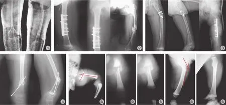
儿童股骨干骨折虽然多数可以获得较好的治疗结果,然而并发症也并非少见,包括骨延迟愈合和骨不连或感染性骨不连、肢体不等长、成角畸形以及血管和神经损伤等( 图 1~4 )。治疗方法的选择依据患者的年龄、体重、伴随的损伤情况、骨折的部位及类型等确定。按照年龄选择治疗方法得到多数学者的认同。
一、3 岁以内儿童股骨干骨折的治疗
新生儿产伤骨折和 3 个月以内小婴儿股骨干骨折,成角与短缩畸形程度有限,通过小夹板固定或应用 Pavlik 支具固定即可获得良好的疗效 ( 图 5,6 )。
临床上,多数病例治疗的经历提示,新生儿产伤骨折即便成角很大,也会随生长发育,逐渐获得塑建,多在1 年之内恢复轴线,一般不会留有残疾。
3 个月~3 岁的患儿通过悬吊皮牵引、水平皮牵引或不需要牵引,直接应用髋人字石膏固定。悬吊皮牵引的患儿很少发生肢体末端缺血性挛缩这一严重并发症,牵引中应注意观察。对于 2~3 岁的患儿,可以考虑水平皮肤牵引治疗股骨干骨折。牵引持续时间一般为 2~3 周,至骨折部位形成骨痂为止;也可以直接应用髋人字石膏固定治疗这一年龄段的股骨干骨折。牵引与直接髋人字石膏固定均有较好的疗效,但髋人字石膏固定的优点在于患儿住院时间短、疼痛时间短、费用低等。对于骨折重叠>2 cm 的患儿,应首先牵引,然后再行石膏固定[9-14]。

图1 股骨干骨折弹性髓内针治疗后发生骨不连。入针点位于髓腔:2 针 6 点固定作用减轻或消失;针尾留得过长,术后会出现疼痛、滑囊炎;外固定近端出现成角。纵向的微动有利于成骨,而不是横向的微动:骨不连发生的诱因图2 2 年共 6 次手术,每次手术均采用钢板固定,仍然骨不连。钢板选择得短;近端仅有 2枚螺丝;骨折线上不应再安置螺丝骨不连类型并非是萎缩型,说明骨折断端固定不稳图3 左股骨干骨折短段固定后发生骨不连。使用短段螺丝加钢丝固定是极其错误的图4 股骨远端骨折钢针固定后发生骨不连,骨折位置并非在骨骺区或接近骨骺的干骺端。选择“T”型钢板更加适宜,选择钢针固定,牢固、稳定性均不够图5 a:出生后当天发现左侧股骨干骨折,成角 80°;b:出生后 1 周摄片已经有明显的骨痂形成;c:出生后半个月,骨痂形成良好;d:出生后 4 个月,骨质已经完全愈合,但仍然有 42° 成角;e:出生后 8 个月,骨质愈合,成角消失Fig.1 Male, 6 years old. Nonunion occurred after using the elastic stablized intramedullary nail. The entry point was located in the marrow cavity.The fixed function with two nails and six points was decreasing or disappearing. The ends of nails were too long, and lead to pain and bursitis postoperatively. The angulation deformity occurred in the proximal part of external fixation. Slightly longitudinal motion, not horizontal motion,was beneficial for the formation of the new bone. The horizontal motion was one of nonunion.Fig.2 Female, 8 years old. She underwent six operations within two years. The plate fixations had been used in six operations, however, the nonunion occurred. The pitfalls of these plate fixation: plates were too short relative to the femoral fracture, only two screws proximal to the fracture line, screws should not be placed at the line of the fracture. It was not an atrophic nonunion, and this nonunion occurred from the unstable internal fixation.Fig.3 Male, 4 years old. Nonunion occurred after the short internal fixation with screws and wires for the proximal femur. The fixation was a huge pitfall.Fig.4 Male, 4 years old. Nonunion occurred after K wire fixation for the distal femoral shaft fracture. The fracture line was not located in the epiphyseal or metaphyseal region. The T figure plate may be the best choice. K wire was not stable and strong enough as internal fixator in this case Fig.5 a: Male, left femoral shaft fracture at birth with angulation of 80 degrees; b: The radiograph after one week showed obvious callus of new bone surrounding the fracture; c: The radiograph after two weeks showed abundant callus of new bone surrounding the fracture; d: The radiograph after four months showed complete healing of the fracture and the residual angulation of 42 degrees; e: The radiograph after eight months showed that the angulation disappeared
关于牵引中成角畸形的纠正和旋转畸形的预防。股骨干上 1 / 3 骨折端向前向外成角,因此牵引中应保持患肢屈髋外展位。远 1 / 3 骨折宜屈膝牵引,减少骨折远端向后倾。也可以采取股骨远端或胫骨近端骨牵引的方法治疗,但需要避开骨骺,避免损伤。
二、3 岁以上的儿童股骨干骨折的治疗
大龄儿童仍然可以选择牵引、石膏固定的方法治疗,并且也可以收到较好的疗效。但 20 世纪 80 年代初弹性髓内针的问世,对于传统的治疗方法有较大的冲击。欧美及国内文献资料报道,更多的家长愿意接受这一手术干预。与大龄儿童骨牵引相比,其区别仅在于大腿远端两侧的切口略大于骨穿针的切口,甚至可以做到很小的切口差别。而且弹性髓内针的应用术后可以不必外固定,术后短时间内甚至术后 7 天即可以负重,缩短了治疗时间,提高了生活质量,出现成角畸形及下肢不等长等并发症也少于保守治疗的患者。
弹性髓内针固定治疗股骨干骨折:这一年龄阶段应用弹性髓内针治疗股骨干骨折尤其是在欧洲已经成为主流。国内正在普及。骨折治疗的基本生物力学原则是保存骨的血液供应和维持骨的生理、力学环境。弹性髓内钉治疗股骨干骨折,骨折处不需要切口,这样就避免了剥离外骨膜;非扩髓的小直径髓内针产热量低,破坏骨内膜小,增加了骨质愈合的潜在能力;手术不剥离外骨膜,不扩髓对骨折处血运影响较小,故骨折愈合快。弹性髓内针固定骨折后能产生骨折端的纵向微动,不产生应力遮挡,加速了骨折愈合速度;其仅在骨质的一端两侧作 3 cm 以内的较小切口,软组织损伤很小,外观满意。整个手术操作过程简便,瘢痕小,失血少,视为微创手术,患儿家属容易接受。
弹性髓内针是利用 2 根弹性钛针或钢针在髓腔内通过双弓形在髓腔内产生弹性内卡和针的两端 6 点支撑原理来固定骨折 (图 7)。髓腔内通过限制骨折端的旋转、移位,较好地维持了骨折端的解剖对位。其弹性强度可以通过弓形幅度来调整。与带锁髓内针相比较,弹性髓内针并不损伤骨骺与骺板。适应于骨干的横行骨折和短斜行骨折。较大的斜行骨折、伴有分离碎骨片的粉碎性骨折以及体重过大,超过 45 kg 的患儿应慎行或并非完全适应。
具体操作:术前选择 2 根直径相同的弹性髓内针,选针的原则是弹性髓内针的直径是髓腔直径的 1 / 3 或2 / 5。弹性髓内针直径选得大一些,将有利于骨折的固定与稳定,但操作的难度会增加。直径选得小一些,操作起来会更加容易。预弯弹性髓内针,预弯弧的高度为髓腔直径的 3 倍,弧弓的顶点应恰好固定在骨折处,弧形的方向与髓内针钉头的喙状结构方向一致。在固定后达到交叉应力,髓内成“X”形的 6 点固定。
术前常规消毒铺敷,常规麻醉,手术在 C 型臂透视机下进行。首先在离患肢骨骺板约 2 cm 处定位,肢体两侧经皮小切口。切口长度 1~3 cm,在套管保护下用开孔器分别对骨皮质打孔,逆骨骺板方向操作,与骨质纵轴呈斜向 30°~45° 开路。将选择好 2 根直径相同的、已经预弯好的髓内针分别由开孔处置入至骨折处。在 C 型臂透视机监测下进行骨折复位,较小的儿童单纯手法整复,纵向牵引患肢, 横向挤压骨折端可以有效地使骨折复位。闭合复位后可以通过髓内针的旋转使骨折复位更加满意。较大的儿童可以应用牵引床复位。如果复位十分困难,通过骨折处小切口辅助复位,持骨钳将骨折端固定,然后将髓内针穿过骨折向骨折近端推进。髓内针钉头直至固定到转子之间,两侧插入固定到松质骨内。髓内针针尾保留的长度0.5 cm 为佳,短段保留针尾不会并发针尾处疼痛或针尾滑囊炎。拔出困难时可以选用三关节咬骨钳便于取出。针尾埋于皮下。皮内缝合,闭合切口。
文献报道术后不必外固定,术后 1 周即可以逐渐负重行走。但笔者在实际临床工作中更愿意外固定 3~4 周,这样有利于患儿的恢复,减少骨折处成角畸形等并发症的发生。术后根据骨折愈合情况,一般 6 个月~1 年可以取出弹性髓内针。由于损伤小,取出钢针甚至可以在门诊局麻下完成。
保证弹性髓内针固定与治疗成功的关键点在于入针点的位置与固定、弹性髓内针弧形顶点的固定和针头顶点喙状突的固定。每一位点固定到位对于骨折的复位与稳定均十分重要。入针点偏离干骺端而接近髓腔,髓内针顶点喙状突固定的不够远,未达到大小转子处,即髓内针两端接近髓腔或就在髓腔内,针的弧形顶点也不在骨折处,这样就达不到 6 点固定这一目的,骨折一定会移位、成角或出现并发症[15-26]。
钢针、钢板及外固定架治疗股骨干骨折:股骨干骨折切开复位传统的治疗方法即为钢板内固定,钢板进行内固定治疗股骨干骨折报道很多。其优点是不需要 C 型臂透视机,骨折对位对线理想。其适用于小转子下骨折或干骺端骨折,而对于接近于骺板的干骺端骨折 ( 3 cm 左右 ),应用“T”形钢板更加适宜。
对于<3 cm 的或股骨远端骨骺部骨折,钢板已不适用,需要应用直径 ≤2 mm 的钢针进行骨骺、干骺端内固定,并且需要不少于 3~4 根的钢针固定,应用 2 根钢针固定达不到牢固、稳定的目的,增加发生骨折处的成角畸形愈合的机会,甚而会发生骨不连接。
钢板固定的不足之处是手术切口大,骨膜剥离范围广,骨折端因此减少或失去了血液供应,少部分骨折会出现延迟愈合,甚而出现骨不连接。取出钢板后,由于螺丝孔和钢板的应力遮挡效应,致板源性骨吸收作用又容易发生再骨折,并且有需要二次手术、切口瘢痕影响外观等不足。所以,对于儿童股骨干骨折,应用钢板进行内固定有逐渐减少的趋势。
对于干骺端或骨干粉碎性骨折,应用肌肉下桥接钢板( submuscular bridge plating ) 可以减少普通钢板很多的弊病。尤其适应于大的斜折性和粉碎性骨折 ( 图 8 )。桥接钢板属于微创技术,微创是外科手术治疗的发展趋势。目前,微创、长桥接钢板的应用已经逐渐得到医生的认同,是粉碎性骨折首选,是钢板治疗长骨骨折的又一较好的选择。
选择 4.5 mm 厚、10~16 孔的长加压或加压锁定钢板;钢板的长度应在 C 型臂透视机的检测下决定,并按股骨侧位外观塑形,钢板尽可能长,固定稳定,而且切口长度不变;牵引床牵引、人为牵引或股骨固定器、外固定器牵引,使骨折尽可能复位;股骨远端外侧作 3 cm 长切口,经阔筋膜、股外侧肌进入到肌肉下方、骨膜外;放入已经预弯或塑形后的钢板,沿骨膜外缓慢向近端穿行,如果操作困难,近端可以作另一开口;到位后两端及中间应用钢针临时固定,保持轴线,并用 C 型臂透视机正侧位检测,侧位钢板与股骨可以不完全一致相贴;近端和远端各为 3枚螺丝,尽可能远离骨折处固定,是为桥接。也可均匀或不规则分布;经皮安置螺丝在 C 型臂透视机检测下进行,侧位片是一个标准的卵圆孔 ( 先打入钢针作为导针,再顺钢针放入保护器,之后钻头钻孔 )。术后根据骨折稳定情况,考虑是否需要石膏或支具外固定。
由于是肌肉下、骨膜外安置钢板,骨折断端髓内外血液供应均未遭到破坏,所以,骨折愈合快,而且切口小、瘢痕少,家属容易接受。

图6 患儿,2 个月,左侧股骨干骨折,Pavlik 吊带固定之后摄片图 7 6 点固定髓内针两侧弓形顶点固定骨折处,使骨折复位。两端干骺端固定使骨折稳定,避免旋转和移位图 8 患者,女,7 岁,左股骨近段长斜形骨折,桥接钢板固定图 9 患者,男,12 岁,体重 75 kg,带锁髓内针固定Fig.6 Female, 2 months old. The radiograph showed left femoral shaft fracture after the fixation with pavlik harnessFig.7 Male, 5 years old. Elastic stablized intramedullary nails were performed in this case. The fracture was immobilized by six points. The apex of the nail bow was located at the fracture line. The metaphyseal nails avoided the rotation and displacementFig.8 Female, 7 years old. Long oblique fracture of the left proximal femur and the bridge plating fixationFig.9 Male, 12 years old. The weight of this case is 75 kg. The rigid locked intramedullary nail was performed
对于开放性骨折、粉碎性骨折以及伴有软组织损伤严重的骨折可以选用外固定架治疗。外固定支架治疗儿童股骨骨折,创伤小于钢板内固定,对软组织覆盖干扰少,对骨的血供破坏少,不影响儿童的生长发育;而且骨外固定器在术后骨折发生移位后具有可调性等优点。外固定架在骨折固定早期可提供牢固的骨折断端稳定,骨折接近愈合的后期可以轻微放松锁钮,使骨折端能够相互接触,承受压力。放松锁钮消除了支架固定所产生的应力遮挡作用,有利于骨痂的生成以及骨折的进一步骨化、愈合。外固定架的不足有:钉道感染,多半不会引起骨质感染;膝关节僵硬,因钢针固定骨折周围肌肉所引起,通过功能锻炼或去除外固定架后一般均能恢复;神经血管损伤,并不多见;再骨折,由于外固定架固定时间有限,去除外固定架后由于没有支具的保护或意外损伤,出现再骨折的几率增大。减少发生再骨折的方法是,去架前松动锁钮,负重后 1 个月再去架,去除外固定架后应用支具保护将会更加有效[27-36]。
带锁髓内针固定治疗股骨干骨折同样属于微创手术,该手术的优点是骨折处无手术切口;较好的复位与轴线;带锁可以控制旋转;术后不用外固定;术后早期负重;不影响临近关节的功能等 ( 图 9 )。由于术后可以早期负重,所以患者及家属容易接受。带锁髓内针的不足是手术需要扩髓,破坏了骨髓内结构,带锁本身有骨折处应力遮挡,同样可以产生骨延迟愈合与骨不连,而影响带锁髓内针广泛应用最为严重的并发症是股骨头缺血性坏死,虽然发生率低,但这是影响其普及或广泛应用的主要弊端,也是医生和患者担心的最严重的并发症。有文献报道,由梨状窝入针,股骨头坏死发生率为 2%,大转子顶点入针,股骨头坏死率为 1.4%。在临床工作中实际遇上股骨头缺血性坏死的病例很少。而大转子外侧入针,未发现股骨头坏死的报道。另外,目前应用于儿童的带锁髓内针最细的直径是 8.5 mm 左右,仍然较粗,而且所设计的带锁髓内针仅适用于大转子和梨状窝进针,属于左右侧通用。因此,目前带锁髓内针仅在国内很少几家医院应用。现今,发达国家已经出现了直径 7 mm、左右分侧、适应于大转子外侧进针的带锁髓内针,这样将会使更小的患者,甚而是7~8 岁的股骨干骨折的患者能够接受带锁髓内针的治疗,既可以达到微创的目的,又可以极大地降低或避免术后股骨头缺血性坏死和股骨近端生长紊乱,同时又可以术后即时获得负重行走。是儿童股骨干骨折治疗的又一合适的选择[37-41]。
三、小结
3 岁以内股骨干骨折以保守治疗为主,Pavlik 吊带或全麻牵引下髋人字石膏固定效果较好;3~10 岁:稳定性骨折适宜弹性髓内针治疗;干骺端骨折可以考虑钢板或钢针内固定;长斜型、螺旋型,尤其是粉碎性骨折适宜桥接钢板固定;而感染、开放性骨折宜选用外固定架治疗;10 岁 ( 甚至更小 ) 以上,尤其是体重超过 50 kg 的患者可以考虑带锁髓内针固定。
[1] Beaty JH. Operative treatment of femoral shaft fractures in children and adolescents. Clin Orthop Relat Res, 2005,(434):114-122.
[2] Heideken JV, Svensson T, Blomqvist P, et al. Incidence and trends in femur shaft fractures in Swedish children between 1987 and 2005. J Pediatr Orthop, 2011, 31(5):512-519.
[3] Rewers A, Hedegaard H, Lezotte D, et al. Childhood femur fractures, associated injuries, and sociodemographic risk factors: a population-based study. Pediatrics, 2005, 115(5):e543-552.
[4] Bridgman S, Wilson R. Epidemiology of femoral fractures in children in the West Midlands region of England 1991 to 2001.J Bone Joint Surg Br, 2004, 86(8):1152-1157.
[5] Loder RT, O’Donnell PW, Feinberg JR. Epidemiology and mechanisms of femur fractures in children. J Pediatr Orthop,2006, 26(5):561-566.
[6] Kanlic E, Cruz M. Current concepts in pediatric femur fracture treatment. Orthopedics, 2007, 30(12):1015-1019.
[7] Anglen JO, Choi L. Treatment options in pediatric femoral shaft fractures. J Orthop Trauma, 2005, 19(10):724-733.
[8] Flynn JM, Luedtke LM, Ganley TJ, et al. Comparison of titanium elastic nails with traction and a spica cast to treat femoral fractures in children. J Bone Joint Surg Am, 2004, 86-A(4):770-777.
[9] Epps HR, Molenaar E, O’connor DP. Immediate single-leg spica cast for pediatric femoral diaphysis fractures. J Pediatr Orthop, 2006, 26(4):491-496.
[10] d’Ollonne T, Rubio A, Leroux J, et al. Early reduction versus skin traction in the orthopaedic treatment of femoral shaft fractures in children under 6 years old. J Child Orthop, 2009,3(3):209-215.
[11] Cassinelli EH, Young B, Vogt M, et al. Spica cast application in the emergency room for select pediatric femur fractures.J Orthop Trauma, 2005, 19(10):709-716.
[12] Czertak DJ, Hennrikus WL. The treatment of pediatric femur fractures with early 90-90 spica casting. J Pediatr Orthop, 1999,19(2):229-232.
[13] Infante AF Jr, Albert MC, Jennings WB, et al. Immediate hip spica casting for femur fractures in pediatric patients. A review of 175 patients. Clin Orthop Relat Res, 2000, (376):106-112.
[14] Jauquier N, Doerfler M, Haecker FM, et al. Immediate hip spica is as effective as, but more efficient than, flexible intramedullary nailing for femoral shaft fractures in pre-school children. J Child Orthop, 2010, 4(5):461-465.
[15] Simanovsky N, Porat S, Simanovsky N, et al. Close reduction and intramedullary flexible titanium nails fixation of femoral shaft fractures in children under 5 years of age. J Pediatr Orthop B, 2006, 15(4):293-297.
[16] Bopst L, Reinberg O, Lutz N. Femur fracture in preschool children: experience with flexible intramedullary nailing in 72 children. J Pediatr Orthop, 2007, 27(3):299-303.
[17] Sink EL, Faro F, Polousky J, et al. Decreased complications of pediatric femur fractures with a change in management.J Pediatr Orthop, 2010, 30(7):633-637.
[18] Ellis HB, Ho CA, Podeszwa DA, et al. A comparison of locked versus nonlocked Enders rods for length unstable pediatric femoral shaft fractures. J Pediatr Orthop, 2011, 31(8):825-833.
[19] Wall EJ, Jain V, Vora V, et al. Complications of titanium and stainless steel elastic nail fixation of pediatric femoral fractures.J Bone Joint Surg Am, 2008, 90(6):1305-1313.
[20] Ellis HB, Ho CA, Podeszwa DA, et al. A comparison of locked versus nonlocked Enders rods for length unstable pediatric femoral shaft fractures. J Pediatr Orthop, 2011, 31(8):825-833.
[21] Ho CA, Skaggs DL, Tang CW, et al. Use of flexible intramedullary nails in pediatric femur fractures. J Pediatr Orthop,2006, 26(4):497-504.
[22] Jencikova-Celerin L, Phillips JH, Werk LN, et al. Flexible interlocked nailing of pediatric femoral fractures: experience with a new flexible interlocking intramedullary nail compared with other fixation procedures. J Pediatr Orthop, 2008,28(8):864-873.
[23] 翁刘其, 李明, 许益文. 闭合复位经皮弹性髓内针治疗儿童四肢骨折. 重庆医科大学学报, 2011, 36(2):218-223.
[24] 张敏刚, 王恒冰, 王延宙, 等. 弹性髓内针固定治疗儿童长骨骨折. 临床骨科杂志, 2009, 12(5):535-537.
[25] 张中发, 刘刚, 李茂东, 等. 弹性髓内针在儿童股骨干骨折中的应用. 中国骨与关节损伤杂志, 2012, 27(4):338-339.
[26] 庄云强, 徐荣明, 仲肇平, 等. 弹性髓内针固定与钢板固定治疗儿童股骨干骨折疗效比较. 中国骨与关节损伤杂志, 2009,24(4):336-337.
[27] Jarvis J, Davidson D, Letts M. Management of subtrochanteric fractures in skeletally immature adolescents. J Trauma, 2006,60(3):613-619.
[28] Hedequist D, Bishop J, Hresko T. Locking plate fixation for pediatric femur fractures. J Pediatr Orthop, 2008, 28(1):6-9.
[29] Hedequist DJ, Sink E. Technical aspects of bridge plating for pediatric femur fractures. J Orthop Trauma, 2005, 19(4):276-279.
[30] Eidelman M, Ghrayeb N, Katzman A, et al. Submuscular plating of femoral fractures in children: the importance of anatomic plate precontouring. J Pediatr Orthop B, 2010, 19(5):424-427.
[31] Abbott MD, Loder RT, Anglen JO. Comparison of submuscular and open plating of pediatric femur fractures: a retrospective review. J Pediatr Orthop, 2013, 33(5):519-523.
[32] Samora WP, Guerriero M, Willis L, et al. Submuscular bridge plating for length-unstable, pediatric femur fractures. J Pediatr Orthop, 2013, 33(8):797-802.
[33] Matzkin EG, Smith EL, Wilson A, et al. External fixation of pediatric femur fractures with cortical contact. External fixation of pediatric femur fractures with cortical contact. Am J Orthop,2006, 35(11):498-501.
[34] Hedin H, Hjorth K, Rehnberg L, et al. External fixation of displaced femoral shaft fractures in children: a consecutive study of 98 fractures. J Orthop Trauma, 2003, 17(4):250-256.
[35] Miner T, Carroll KL. Outcomes of external fixation of pediatric femoral shaft fractures. J Pediatr Orthop, 2000, 20(3):405-510.
[36] Sabharwal S. Role of Ilizarov external fixator in the management of proximal/distal metadiaphyseal pediatric femur fractures. J Orthop Trauma, 2005, 19(8):563-569.
[37] MacNeil JA, Francis A, El-Hawary R. A systematic review of rigid, locked, intramedullary nail insertion sites and avascular necrosis of the femoral head in the skeletally immature.J Pediatr Orthop, 2011, 31(4):377-380.
[38] Beebe KS, Sabharwal S, Behrens F. Femoral shaft fractures: is rigid intramedullary nailing safe for adolescents? Am J Orthop,2006, 35(4):172-174.
[39] Kanellopoulos AD, Yiannakopoulos CK, Soucacos PN. Closed,locked intramedullary nailing of pediatric femoral shaft fractures through the tip of the greater trochanter. J Trauma,2006, 60(1):217-223.
[40] Gordon JE, Khanna N, Luhmann SJ, et al. Intramedullary nailing of femoral fractures in children through the lateral aspect of the greater trochanter using a modified rigid humeral intramedullary nail: preliminary results of a new technique in 15 children. J Orthop Trauma, 2004, 18(7):416-424.
[41] Keeler KA, Dart B, Luhmann SJ, et al . Antegrade intramedullary nailing of pediatric femoral fractures using an interlocking pediatric femoral nail and a lateral trochanteric entry point. J Pediatr Orthop, 2009, 29(4):345-351.
