强直性脊柱炎合并下颈椎骨折脱位患者的手术治疗
2014-02-14宋跃明刘立岷曾建成孔清泉
汪 雷 宋跃明 刘 浩 刘立岷 龚 全 李 涛 曾建成 孔清泉
. 强直性脊柱炎 Ankylosing spondylitis .
强直性脊柱炎合并下颈椎骨折脱位患者的手术治疗
汪 雷 宋跃明 刘 浩 刘立岷 龚 全 李 涛 曾建成 孔清泉
目的探讨强直性脊柱炎 ( ankylosing spondylitis,AS ) 合并下颈椎骨折脱位的临床特点及手术治疗要点。方法2010 年 2 月至 2013 年 12 月,我科共收治 21 例 AS 合并脊柱骨折患者,其中下颈椎骨折7 例 ( C5~6椎骨折脱位 1 例,C6~7骨折脱位 6 例 ):男 6 例,女 1 例,年龄平均 47.8 ( 40~52 ) 岁。对 7 例均行手术治疗,采用单纯前路植骨融合内固定手术治疗 1 例,单纯后路手术长节段固定融合术治疗 1 例,前后联合入路手术治疗 5 例。结果7 例术后获得平均 18.2 ( 6~34 ) 个月随访。术中术后均未出现神经损害症状加重情况,手术切口均 I 期愈合;术后脑脊液漏 1 例,换药对症处理 2 周后愈合。1 例 C5~6椎骨折脱位患者行前路手术后第 2 天出现内固定移位,急诊行前后联合入路翻修术;6 例术后神经功能较术前明显改善,1 例无明显改善 ( 术前为 Frankel B 级 )。随访期间均达骨性融合,未出现内固定松动断裂移位现象。结论AS 脊柱骨折好发于颈胸交界区,大多合并有脊髓损伤,手术方式上宜行前后联合入路复位固定或后路长节段固定植骨融合术,术中应先充分减压后再精细复位,避免加重神经损伤。
脊柱炎,强直性;脊柱骨折;矫形外科手术;外科手术;颈椎
强直性脊柱炎 ( ankylosing spondylitis,AS ) 是一 种以椎间关节和韧带骨化及全身骨骼骨质疏松为特征的慢性炎症性疾病,其结果导致脊柱强直后韧性减弱并脆性增加[1-2]。因此,在遭受外力作用甚至无明显外力情况下均可引起脊柱骨折[3-4],临床研究发现下颈椎及颈胸交界区是这类骨折的高发部位[5-7],且多为经椎间隙的三柱骨折[8-9],因骨折端极不稳定而使得其发生脊髓损伤的风险约为普通颈椎骨折的3 倍[5]。回顾近 4 年来,我科收治的 21 例 AS 合并脊柱骨折的患者资料,其中 7 例发生于下颈椎。分析此 7 例下颈椎骨折患者的临床特点,并探讨其手术要点。
资料与方法
一、一般资料
2010 年 2 月至 2013 年 12 月,我科收治 21 例AS 合并脊柱骨折患者,其中 7 例发生在下颈椎( C5~6椎骨折脱位 1 例,C6~7椎骨折脱位 6 例 )。男6 例,女 1 例。年龄平均 47.8 ( 40~52 ) 岁。4 例伤前已确诊为 AS 并进行药物治疗,3 例因创伤入院后发现。受伤机制为车祸伤 3 例,跌倒伤 3 例,重物砸伤 1 例。影像资料分析均为累及三柱的骨折脱位,术前神经功能 Frankel 分级:B 级 2 例,C 级2 例,D 级 2 例,E 级 1 例;其中 1 例合并左锁骨骨折,1 例合并有胸腰段脊柱骨折;其中 3 例因胸椎重度后凸而无法在平卧状态下进行颅骨牵引 ( 予以颈胸支具固定 ),其余 4 例均在术前予以颅骨牵引制动。
二、临床诊断
所有患者入院后均常规行以下实验室检查:ESR、CRP、抗“O”、HLA-B27。术前均完善了以下影像学检查:颈椎正侧位 X 线、颈椎三维 CT重建、颈椎 MRI、腰椎和骨盆及其它可疑损伤部位的 X 线检查。最后根据患者的病史、症状和体征、以上辅助检查结果,采用纽约标准[10]( New York criteria ) 进行诊断。
三、手术方式
对于经椎间隙的无明显脱位或者脱位程度较轻的骨折采用单纯前路单间隙减压及植骨融合内固定术 ( 本组仅 1 例 ) ;对于明显骨折脱位者均采用前后联合入路:先行后路切开减压、复位及多节段侧块螺钉或椎弓根螺钉固定 ( 4~6 个运动节段 ),自体骨后外侧植骨融合,再行前路椎间自体髂骨块植骨或cage 支撑植骨及钛板螺钉内固定术,或者根据具体情况先行前路手术后再行后路固定融合,其中 1 例因术中生命征不稳定而仅行后路减压复位,长节段固定融合。
四、术后处理
所有患者术后常规预防感染、糖皮质激素、抗骨质疏松 ( 降钙素、钙剂、阿仑磷酸钠 )、保护胃黏膜等治疗。术后第 2 天开始根据患者神经功能情况分别行主动或被动踝关节背伸及直腿抬高锻炼。切口引流管于术后 48 h 拔除,严格卧床休息后 4 周根据复查情况佩戴头颈胸支具坐起或下地行走锻炼,头颈胸支具保护 3~6 个月。术后 6 周、3 个月、6 个月、1 年复查 X 线片及三维 CT 以了解骨折愈合情况及内固定位置。7 例术后平均随访 18.2 ( 6~34 ) 个月。
结 果
颈椎前后联合入路手术时间 180~280 min,出血量 400~650 ml。术后 7 例均未出现切口感染,切口一期愈合 ( 图 1,2 )。其中 1 例因术中损伤硬膜导致术后脑脊液漏,经积极换药对症处理后 2 周愈合。1 例 C5~6椎骨折脱位患者入院后急诊行前路手术后第 2 天复查 X 线片发现钛网移位,伤椎脱位复发而急诊行前后联合入路翻修手术。术后 6 个月复查,6 例有脊髓损伤的患者中 5 例神经功能有明显恢复:2 例由术前 Frankel B 级恢复至 D 级,2 例由 C 级分别恢复至 D 和 E 级,1 例由 D 级恢复至E 级,1 例术前为 B 级,术后肌力未见明显恢复。随访期间 7 例均达骨性融合,未出现内固定松动断裂移位现象。
讨 论
一、AS 合并下颈椎骨折的临床特点
AS 的病理特点是脊柱椎间盘、韧带、关节突关节的广泛炎症和骨化,椎体骨质疏松[1-2]。脊柱活动度丢失、弹性下降及脆性增加使得轻微的外力即可导致脊柱骨折[11-13],甚至会发生医源性骨折[14]。文献报道下颈椎及颈胸交界区更是高发部位,发生于此处的骨折约占 AS 脊柱骨折的 73%[15-16]。就损伤机制而言,过伸性损伤较过屈位损伤更为常见[17]。本组 21 例中有 7 例发生于下颈椎,与文献报道一致。强直的脊柱发生骨折犹如长骨干横断骨折,骨折端极不稳定,因而有极高的神经损伤风险[18-20],尤其在伤后运送及麻醉后体位变动过程中极易加重神经损伤[18]。本组 7 例中 6 例均有不同程度的脊髓损伤。治疗方法上在 20 世纪 80~90 年代多主张保守治疗,主要有颈围制动、颈椎牵引、头颈胸石膏、Halo-Vest 架固定等,但远期随访发现这些固定方式不够牢固,骨折愈合欠佳导致骨折端的移位,且须长期卧床制动,常发生褥疮、肺部感染、深静脉血栓形成等并发症[15,21-22],因此,保守治疗近年已较少采用,仅应用于有绝对手术禁忌的患者。目前多主张早期手术治疗,通过复位固定来重建脊柱稳定性并达到骨折部位的骨性融合[17,23]。
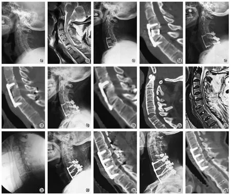
图1 a:患者,男,40 岁,因跌倒伤入院。颈椎侧位 X 线提示典型 AS;b:颈椎 MRI 可见 C5~6椎骨折部位脊髓有明显受压;c:前路手术后复查 X 线可见内固定移位,骨折处再次出现脱位;d:前路手术后复查三维 CT 可见内固定移位,骨折处再次出现脱位;e:前后联合入路翻修术后 1 周复查颈椎 X 线见复位理想,内固定位置良好;f:前后联合入路翻修术后 1 周复查颈椎三维 CT 见复位理想,内固定位置良好;g:术后 1 年复查颈椎 X 线见内固定位置较前无变化,未见松动移位下沉;h:术后 1 年复查颈椎三维 CT 见内固定位置较前无变化,未见松动移位下沉,骨折处已达骨性融合图 2 a:患者,男,52 岁,因车祸伤入院。颈椎三维 CT 提示 C6~7椎骨折脱位;b:颈椎 MRI 可见骨折部位脊髓有明显受压;c:术中摆放体位后 C 型臂透视见骨折脱位已基本复位;d:前后联合入路术后 1 周复查颈椎 X 线见复位理想,内固定位置良好;e:前后联合入路术后 1 周复查颈椎三维 CT 见复位理想,内固定位置良好;f:术后 1 年复查颈椎正侧位 X 线见内固定位置较前无变化,未见松动移位下沉;g:术后 1 年复查颈椎三维 CT 见内固定位置未见松动移位下沉,骨折处已达骨性融合Fig.1 a: The lateral X-ray of the cervical spine of a 40-year-old male patient who fell and hurt showed classic AS; b: The MRI of the cervical spine showed obvious spinal cord compression after C5-6fractures; c: The X-ray of the cervical spine after the anterior operation showed displacement of internal fixation and recurrence of dislocation at C5-6; d: The 3D CT of the cervical spine after the anterior operation showed displacement of internal fixation and recurrence of dislocation at C5-6; e: The X-ray of the cervical spine at 1 week after the combined anterior-posterior revision showed good restoration and satisfactory location of internal fixation; f: The 3D CT of the cervical spine at 1 week after the anterior-posterior revision showed good restoration and satisfactory location of internal fixation; g: The X-ray of the cervical spine at 1 year after the operation showed satisfactory location of internal fixation without looseness, shift or sinking; h: The 3D CT of the cervical spine at 1 year after the operation showed satisfactory bone fusion and location of internal fixation without looseness, shift or sinkingFig.2 a: The 3D CT of the cervical spine of a 52-year-old male patient who was injured in a car accident showed fractures and dislocations at C6-7; b: The MRI of the cervical spine showed spinal cord compression after C6-7fractures and dislocations; c: The C-arm X-ray after the body postition was set during the operation showed acceptable restoration at C6-7; d: The X-ray of the cervical spine at 1 week after the anterior-posterior operation showed good restoration and satisfactory location of internal fixation; e: The 3D CT of the cervical spine at 1 week after the anteriorposterior operation showed good restoration and satisfactory location of internal fixation; f: The anteroposterior and lateral X-ray of the cervical spine at 1 year after the operation showed satisfactory location of internal fixation without looseness, shift or sinking; g: The 3D CT of the cervical spine at 1 year after the operation showed satisfactory bone fusion and location of internal fixation without looseness, shift or sinking
二、手术时机和手术方式的选择
AS 下颈椎骨折后骨折端极不稳定,且多数患者就诊时已发生脊髓损伤,尽早手术减压固定不仅可以促进残存脊髓功能恢复,还可一定程度上限制脊髓继发性损伤和减少脊髓再次发生机械性损伤的几率。有回顾性研究结果[24]显示接受早期手术 ( 伤后<24 h ) 治疗者运动功能改善程度明显优于晚期手术( 伤后>24 h ) 治疗者。Kanter 等[25]研究指出,对尚无脊髓损伤或仅为不完全性脊髓损伤患者应早期手术减压;对完全性或中央型脊髓损伤者可等病情充分稳定后再行手术。对于骨折脱位者是否进行牵引存在争议。有学者认为 AS 颈椎骨折脱位在术前牵引复位有可能加重脊髓损伤,其理论基础是这类患者行闭合牵引治疗时所有的应力在损伤部位过于集中。我们的经验提示,若三维 CT 及 MRI 显示在损伤节段椎管有明显狭窄,牵引复位可能造成脊髓挫伤和挤压加重,可考虑支具临时固定,完善术前准备后尽快手术,若椎管无明显狭窄,脊髓无显著压迫, 术前进行颅骨牵引还是很有必要的,其作用一是颈部制动,二是一定程度上完成复位 ( 复位本身就是一种理想的减压方式 )。
过去不少学者采用单纯前路减压及固定融合术[26],其优点是创伤较小、减压彻底、植骨融合率高等,Kouyoumdjian 等[27]的研究获得了较好的临床效果,但随访报道易发生内固定物松动,包括钢板螺钉及 cage 的松动移位,甚至有报道钢板螺钉脱出而导致食道漏[28-30]。分析其原因,可能由于骨折端的局部应力集中,当颈部屈曲时导致后方骨折端易分离,加之继发有骨质疏松,故内固定器易松动和移位。因此,我们认为单纯前路手术仅适用于骨质条件较好且骨折无明显移位的患者,且手术固定节段应较常规手术适当延长。
后来有学者主张后路长节段固定融合术[31],生物力学证实对于完全失稳者采用后路固定术的稳定性优于单纯前路手术,但仍有报道此术式仍不能获得理想的稳定性,有前柱张开、假关节形成等并发症发生[32-33]。从我们的临床经验来看,后路长节段固定融合仍是一个可选择的手术方式。C3~6因椎弓根体积较小而侧块较大可采用侧块螺钉固定,C7至上胸椎椎弓根体积相对较大可采用椎弓根螺钉固定,固定节段数应选择在骨折端上各 2~3 个节段。这一点和 Shen[34]、Cornefjord[31]等的研究结论相一致。单纯后路手术较适用于骨质尚好、前柱无减压要求及前柱无明显骨质缺损的患者。
对于骨质疏松严重、前柱骨质缺损、前后柱均有减压要求、颈部有明显后凸畸形者则需选择前后联合入路手术,前后路联合减压/固定融合术分别针对前柱和后柱进行减压固定,可使脊柱得到 360°融合,故相比较于单纯前路或后路手术具有稳定性更好、减压更彻底、早期下床活动的优点,目前被大多数学者采用[17,23,26,28,35-36],本组中 6 例均采用此术式均获得了良好的融合效果。至于先行前路还是后路手术,我们认为如果术前颅骨牵引可将骨折端复位,可先行前路手术,反之则先采用后路手术。但该术式存在手术时间长、创伤大、出血多的缺点,发生围手术期并发症的风险也会增加,我们认为对于心肺功能不理想、不能耐受长时间麻醉的患者不宜采用此术式,可采用单纯后路手术。因此,采用前后路联合术式前必须对患者全身情况进行充分评估。
三、麻醉注意事项及手术相关并发症
AS 下颈椎骨折的骨折端极不稳定且常存在颈椎后凸畸形,采用常规的气管插管方式易导致神经损伤加重[6]。因此,对此类患者插管最好在不搬动颈椎的情况下进行。若条件允许采用经鼻盲插管[37]或光学纤维喉镜引导下插管[38]比较安全。由于 AS下颈椎骨折患者肺功能往往较差,且往往手术麻醉时间较长,术后拔管需慎重,拔管前应充分评估患者的呼吸功能,必要时术后早期可留置气管插管。手术时的体位摆放十分重要:首先搬动时应特别小心,最好能在支具保护下搬动,并采用神经诱发电位监测装置进行监测,固定头架时须使颈椎曲线与伤前颈椎曲线保持一致,同时应根据伤前颈椎曲线,对手术床作出相应调整,以免麻醉下造成医源性的脊髓损伤。因 AS 患者韧带广泛骨化,骨质退变严重,椎管容积减小及缓冲性降低,在椎管减压前就进行骨折脱位的复位操作很容易导致神经损伤加重,所以在手术操作过程中,无论采用何种手术方式,都应遵循先充分减压后在直视下进行精细复位的原则,避免盲目复位而加重神经损伤。
这类患者一般术后卧床时间相对较长,卧床期间易发生褥疮、坠积性肺炎、深静脉血栓形成等并发症[39],而这些并发症尤其是肺部感染是导致患者死亡的主要原因,所以术后应加强相应的护理和治疗以积极预防此类并发症的发生。
[1]Foo D, Sarkarati M, Marcelino V. Cervical spinal cord injury complicating ankylosing spondylitis. Paraplegia, 1985, 23(6):358-363.
[2]Pedersen W, Clausen S, Kriegbaum NJ. Spinal lesions in patients with ankylosing spondylitis. Scand J Rheumatol, 1987, 16(5):381-382.
[3]Detwiler KN, Loftus CM, Godersky JC, et al. Management of cervical spine injuries in patients with ankylosing spondylitis. J Neurosurg, 1990, 72(2):210-215.
[4]Rowed DW. Management of cervical spinal cord injury in ankylosing spondylitis: the intervertebral disc as a cause of cord compression. J Neurosurg, 1992, 77(2):241-246.
[5]Tico N, Ramon S, Garcia-Ortun F, et al. Traumatic spinal cord injury complicating ankylosing spondylitis. Spinal Cord, 1998, 36(5):349-352.
[6]Fox MW, Onofrio BM, Kilgore JE. Neurological complications of ankylosing spondylitis. J Neurosurg, 1993, 78(6):871-878.
[7]Vosse D, Feldtkeller E, Erlendsson J, et al. Clinical vertebral fractures in patients withs ankylosing spondylitis. J Rheumatol, 2004, 31(10):1981-1985.
[8]刘海春, 陈允震, 张剑锋, 等. 强直性脊柱炎颈椎骨折诊断及后路内固定治疗. 中国矫形外科杂志, 2004, 12(18): 1369-1372.
[9]郭昭庆, 党耕町, 陈仲强, 等. 强直性脊柱炎脊柱骨折的特点及诊断. 中华骨科杂志, 2003, 23(10):577-580.
[10]Eastmond CJ, Robertson EM. A prospective study of early diagnostic investigations in the diagnosis of ankylosing spondylitis. Scott Med J, 2003, 48(1):21-23.
[11]Hitchon PW, From AM, Brenton MD, et al. Fractures of thoracolumbar spine complicating ankylosing spondylitis. Neurosurg, 2002, 97(2):218-222.
[12]Thumbikat P, Hariharan RP, Ravichandran G, et al. Spinal cord injury in patients with ankylosing spondylitis: a 10-year review. Spine, 2007, 32(26):2989-2995.
[13]Altenbernd J, Bitu S, Lemburg S, et al. Vertebral fractures in patients with ankylosing spondylitis: a retrospective analysis of 66 patients. Rofo, 2009, 181(1):45-53.
[14]Kim KT, Lee SH, Suk KS. Spinal pseudarthrosis in advanced ankylosing spondylitis with sagittal plane deformity: clinical characteristics and outcome analysis. Spine, 2007, 2(15): 1641-1647.
[15]Westerveld LA, Verlaan JJ, Oner FC. Spinal fractures in patients with ankylosing spinal disorders: a systematic review of the literature on treatment, neurological status and complications. Eur Spine J, 2009, 18(2):145-156.
[16]Calin A. Ankylosing spondylitis. Medicine, 2006, 34(10): 396-400.
[17]May PJ, Raunest J, Herdmann J, et al. Treatment of spinal fracture in ankylosing spondylitis. Unfallchirurg, 2002, 105(2):165-169.
[18]Jacobs WB, Fehlings MG. Ankylosing spondylitis and spinal cord injury: origin,incidence, management, and avoidance. Neurosurg Focus, 2008, 24(1):E12.
[19]Grisolia A, Bell R, Peltier L. Fractures and dislocations of the spine complicating ankylosing spondylitis: a report of six cases. Clin Orthop, 2004, 42(2):129-134.
[20]Machado P, Gawronski J, Gall A. Ankylosing spondylitis and spinal cord injury. Acta Reumatol Port, 2008, 33(2):231-237.
[21]Einsiedel T, Schmelz A, Arand M, et al. Injuries of the cervical spine in patients with ankylosing spondylitis: experience at two trauma centers. J Neurosurg Spine, 2006, 5(1):33-45.
[22]Bessant R, Keat A. How should clinicians manage osteoporosis in ankylosing spondylitis? J Rheumatol, 2002, 29(7): 1511-1519.
[23]Payer M. Surgical management of cervical fractures in ankylosing spondylitis using a combined posterior-anterior approach. J Clin Neurosci, 2006, 13(1):73-77.
[24]Guest J, Eleraky MA, Apostolides PJ, et al. Traumatic central cord syndrome: results of surgical management. J Neurosurg, 2002, 97(1 Suppl):S25-S32.
[25]Kanter AS, Wang MY, Mummanent PV. A treatment algorithm for the management of cervical spine fractures and deformity in patients with ankylosing spondylitis. Neurosurg Focus, 2008, 24(1):E11.
[26]Metz-Stavenhagen P, Krebs S, Meier O. Cervical fractures in ankylosing spondylitis. Orthopade, 2001, 30(12): 925-931.
[27]Kouyoumdjian P, Guerin P, Schaelderle C, et al. Fracture of the lower cervical spine in patients with ankylosing spondylitis: Retrospective study of 19 cases. Orthop Traumatol Surg Res, 2012, 98(5):543-551.
[28]Lu GH, Wang B, Kang YJ, et al. Combined anterior and posterior surgery for treatment of cervical fracture-dislocation in patients with ankylosing spondylitis. Chin J Traumatol, 2009, 12(3):148-152.
[29]康意军, 陈飞, 吕国华, 等. 强直性脊柱炎颈椎骨折的治疗. 中国脊柱脊髓杂志, 2006, 16(6):424-428.
[30]Zdichavsky M, Blauth M, Bosch U, et al. Late esophageal perforation complicating anterior cervical plate fixation in ankylosing spondylitis: a case report and review of the literature. Arch Orthop Trauma Surg, 2004, 124(5):349-353.
[31]Cornefjord M, Alemany M, Olerud C. Posterior fixation of subaxial cervical spine fractures in patients with ankylosing spondylitis. Eur Spine J, 2005, 14(4):401-408.
[32]Olerud C, Frost A, Bring J. Spinal fractures in patients withankylosing spondylitis. Eur Spine J, 1996, 5(1):51-55.
[33]Cooper PR, Cohen A, Rosiello A, et al. Posterior stabilization of cervical spine fractures and subluxations using plates and screws. Neurosurgery, 1988, 23(3):300-306.
[34]Shen FH, Samartzis D. Surgical management of lower cervical fracture in ankylosing spondylitis. J Trauma, 2006, 61(4): 1005-1009.
[35]EL-Masry MA, Badawy WS, Chan D. Combined anterior and posterior stabilization for treating an unstable cervical spine fracture in a patient with long standing ankylosing spondylitis. Injury, 2004, 35(10):1064-1067.
[36]Kuroiwa T, Yoshii T, Sakaki K, et al. Vertebral locking lesion following cervical spine fracture in ankylosing spondylitis. Orthopedics, 2012, 35(6):1005-1008.
[37]Lu PP, Brimacombe J, Ho AC, et al. The intubating laryngeal mask airway in severe ankylosing spondylitis. Can J Anaesth, 2001, 48(10):1015-1019.
[38]Langford RA, Leslie K. Awake fibreoptic intubation in neurosurgery. J Clin Neurosci, 2009, 16(3):366-372.
[39]张凤山, 刘忠军, 陈仲强, 等. 强直性脊柱炎颈椎骨折的手术治疗. 中华创伤骨科杂志, 2006, 12(8):1139-1143.
( 本文编辑:王永刚 )
Surgical management of ankylosing spondylitis combined with lower cervical spine fractures and dislocations
WANG Lei, SONG Yue-ming, LIU Hao, LIU Li-min, GONG Quan, LI Tao, ZENG Jian-cheng, KONG Qing-quan. Department of Orthopedics, West China Hospital, Sichuan University, Chengdu, Sichuan, 610041, PRC
ObjectiveTo explore the clinical characteristics and surgical treatment of lower cervical spine fractures and dislocations combined with ankylosing spondylitis ( AS ).MethodsFrom February 2010 to December 2013, 21 patients with AS combined with spinal fractures were adopted, including 7 patients with lower cervical spine fractures. There were 6 males and 1 female, whose average age was 47.8 years old ( range: 40-52 years ). All the 7 patients underwent surgical treatment, including 1 patient with C5-6fractures and dislocations and 6 patients with C6-7fractures and dislocations. Simple anterior interbody fusion and internal fxation was performed on 1 patient, simple posterior long-segmental fxation and fusion on 1 patient, and a combined anterior-posterior approach on the other 5 patients.ResultsAll the 7 patients were followed up for a mean period of 18.2 months ( range: 6-34 months ). There was no aggravation of neuronal damage during and after the operation, and primary healing of surgical incisions was achieved in all the patients. Cerebrospinal fuid leakage was noticed in 1 patient, who recovered after 2 weeks of changing dressing. Internal fxation loosening was found in 1 patient with C5-6fractures and dislocations at the 2nd day after the anterior surgery, and a combined anterior-posterior revisional operation was performed immediately. The remarkable improvement of the neuronal function was obtained in 6 patients after the operation, and no obvious improvement in 1 patient ( Frankel grade B preopertively ). Bone fusion was achieved in all the patients, without loosening, breakage or displacement of internal fixation during the follow-up.ConclusionsThe lower cervical vertebrae is the common location of AS and spinal fractures, usually combined with spinal cord injury ( SCI ). A combined anterior-posterior reduction and posterior long-segmental fxation and bone graft fusion are 2 satisfactory surgical methods. The aggravation of neuronal damage can be effectively avoided, with complete decompression before fne reduction.
Spondylitis, ankylosing; Spinal fractures; Orthopedic procedures; Surgical procedures, Operative; Cervical vertebrae
10.3969/j.issn.2095-252X.2014.10.006
R681.5, R682.3
610041 成都,四川大学华西医院骨科
宋跃明,Email: hx_sym@163.com
2014-07-16 )