What Moved into the Lung? An Unusual Case of Foreign Body Migration
2013-11-18YantingWangXiaodongYangShaohuaLiuandYanhuaHuang
Yan-ting Wang,Xiao-dong Yang*,Shao-hua Liu,and Yan-hua Huang
Department of Respiratory Medicine,West China Hospital of Sichuan University,Chengdu 610041,China
FOREIGN body migration into the lung is a rare complication that can cause various symptoms.In adults,however,foreign body deposited in the lung can be tolerated and remain undetected for a long time.Delayed diagnosis and subsequent delayed treatment is usually associated with atypical complications.In this article,we describe a case of foreign body migration into the tracheobronchial tree.After a long time delay diagnosis,the foreign body was ultimately removed by operation.
CASE DESCRIPTION
On August 1th,2012,a 52-year-old woman was admitted for the chief complaint of refractory pneumonia and intermittent hemoptysis of twenty years’s duration.She had been repeatedly hospitalized due to several undefined respiratory tract infections with recurrent hemoptysis and productive cough.The patient had been received antibiotic treatment each time but still complained of a recurrent massive hemoptysis.
A physical examination mainly revealed a frail woman in moderate respiratory distress with rales in her right lung.Her respiratory rate,23 times per minute and oxygen saturation,95% on her baseline 2 L of oxygen by nasal cannula.The heart rate and rhythm were regular.The abdomen was soft and non-tender.The extremity examination revealed no joint swelling,purpura,or clubbing.
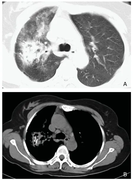
Blood routine examination revealed white blood cell count at 15.2×10⁹/L,neutrophils 0.83,hemoglobin 86.0 g/L,and platelets 230×109/L.The serum electrolytes,liver function tests,coagulation studies,and urinalysis were normal.Chest computed tomography (CT) revealed the right upper lung infiltrates and bronchiectasis (Fig.1).There were two 2 cm-long needle-like high density shadows distinguished from the surrounding tissue (Fig.1).Bronchoscopic biopsy found nothing but chronic inflammation.
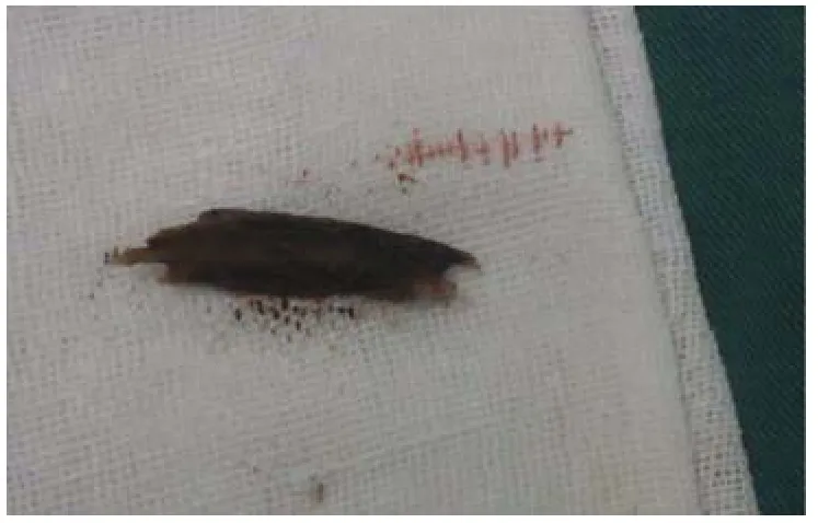
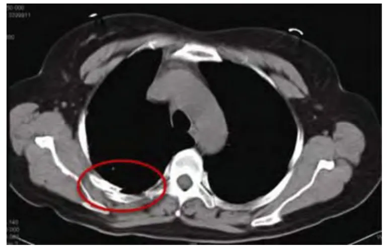
The patient failed to provide the reliable history of trauma or aspiration.She could not offer any past imaging data either.It is difficult for us to explain what the high-density image was.The patient preferred surgery as first-line treatment rather than other interventions because she had suffered from hemoptysis and refractory pneumonia for a long time.Right upper lobectomy was performed which revealed that a bone fragment was embedded in the resected specimen (Fig.2).Then after carefully reviewed our patient's chest CT,we found that there is an old fracture site at the trailing edge of the right fourth rib (Fig.3).We considered that the period after her injuring was too long for her to remember.The patient’s own fractured rib had been moved and finally settled in the lung to cause various respiratory symptoms.Her condition improved within three days after the removal of the foreign body.The postoperative course and follow-up was unremarkable.

Figure 1.Chest computed tomography scan imagines obtained with lung window settings at the level of the carina (A) and mediastinal window settings at the level of the carina (B).

Figure 2.A fragment of the fractured rib.

Figure 3.An old fracture site at the trailing edge of the right fourth rib.
DISCUSSION
Tracheobronchial foreign body (TFB) is defined as the presence of any foreign object below the vocal cords.1This condition is often clinically encountered due to the aspiration.Lone and Lateef2analyzed the infinite variety of foreign bodies had been inhaled in different ways and posed different problems.But,TFB derived from autologous tissue migration had never been reported.In our case report,the key what made it unusual was the migration of such a foreign object which came from her own fractured rib and settled in the lung.Even more striking,she could not remember she had suffered a trauma.Some scholars hypothesized that this migration may have occurred due to the negative intrathoracic pressures and continuous respiratory excursions.3
Diagnosing of TFB is challenging,because the airway foreign body causes various symptoms,ranging from coughing and wheezing to hemoptysis and dyspnea.These symptoms mimick other diseases,such as asthma,bronchiectasis or pneumonia.So sometimes leads to the delay in diagnosis.4The radiological findings are often non-specific and inadequate to exclude the diagnosis of other diseases.Chest radiographs were obtained in 59 patients with foreign body aspiration and TFB was seen on the chest radiograph in merely 19 patients (32%),even though 92% of the patients had some abnormalities on chest radiographs.1CT is a powerful noninvasive examine for guiding diagnosis and assessing the need for bronchoscopy or thoracic surgeon.Bronchoscopy may provide some help,but it can only deal with the main bronchus foreign body.In this case,open-lung biopsy was the final,definitive and therapeutic option.The thoracic surgeon is of paramount importance in the management of TFBs which cannot be extracted by bronchoscopy.Thus,in every patient with persistent cough and hemoptysis,with or without a history of foreign body aspiration,with or without the typical imaging,the possibility of retained foreign body should be kept in mind.
1.Swanson KL,Prakash UBS,McDougall JC,et al.Airway foreign bodies in adults.J Bronchol Interv Pulmonol 2003;10:107-11.
2.Lone SA,Lateef M.Foreign body in tracheobronchial tree.JK Sci 2004;6:77-80.
3.Venissac N,Alifano M,Dahan M,et al.Intrathoracic migration of Kirschner pins.Ann Thorac Surg 2000;69:1953-5.
4.Kogure Y,Oki M,Saka H.Endobronchial foreign body removed by rigid bronchoscopy after 39 years.Interact Cardiovasc Thorac Surg 2010;11:866-8.
杂志排行
Chinese Medical Sciences Journal的其它文章
- Primary Endodermal Sinus Tumor in the Posterior Cranial Fossa:Clinical Analysis of 7 Cases
- Coronary Artery Perforation Complicated With Acute Aortic Valve Regurgitation During Percutaneous Coronary Intervention:Report of Two Cases
- CORRECTION
- Mechanical Stimulus Inhibits the Growth of a Bone Tissue Model Cultured In Vitro△
- Recurrent Seizures Manifestations in a Case of Congenital Hypoparathyroidism:a Case Report
- Reconstruction of the Medial Patellofemoral Ligament Using Hamstring Tendon Graft With Different Methods: a Biomechanical Study
